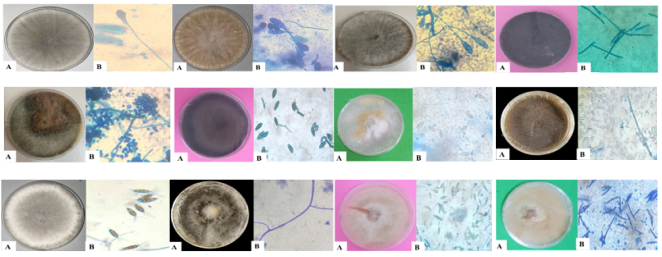
Sesame (Sesamum indicum L.) is an important oil seed crop in Chad, but its productivity is constrained by seed-borne fungal pathogens affecting seed quality, germination, and seedling establishment. This study examined the different seed-borne fungi associated with sesame varieties using two International Seed Testing Association (ISTA)-recommended methods: the blotter paper method and agar plate method. The results show that 22 fungal species were identified, with notable differences between the two methods. The blotter paper method detected 13 species, while the agar plate method revealed 22 species, with 3 375 isolates detected using the agar plate method compared to 1 070 isolates on blotter paper method. Using the blotter paper method, Alternaria sesami (13.28%), A. brassicicola (12.51%) and Macrophomina phaseolina (10.69%) were the most frequent species and Bipolaris spicifera (3.86%) and Nigrospora sphaerica (4.02%) were the least frequent. With the agar plate method, the most frequent species were A. sesami (16.92%), Aspergillus niger (12.57%) and Cercospora sesami (12.5%). Pestalotia guepini and Verticillium dahliae were the least frequent species, with respective frequencies of 0.41 and 0.61%. These results revealed a wide diversity of seed-borne fungi of sesame, highlighting the need to develop effective strategies to control these seed-borne fungi.
| Published in | Journal of Plant Sciences (Volume 14, Issue 2) |
| DOI | 10.11648/j.jps.20261402.13 |
| Page(s) | 93-103 |
| Creative Commons |
This is an Open Access article, distributed under the terms of the Creative Commons Attribution 4.0 International License (http://creativecommons.org/licenses/by/4.0/), which permits unrestricted use, distribution and reproduction in any medium or format, provided the original work is properly cited. |
| Copyright |
Copyright © The Author(s), 2026. Published by Science Publishing Group |
Sesamum indicum, Seed-borne Fungi, Agar Plate Method, Blotter Paper Method, Chad
Name | Origin | Seed color | Growth cycle (days) | Yield (Kg/ha) |
|---|---|---|---|---|
32-15 | Burkina Faso | White | 80-100 | 400-1000 |
DLS1 | Chad | Cream | 100-130 | 500-1000 |
Ker manne | Chad/Local | White | 100-120 | 500-1000 |
Ker Ndaa | Chad/Local | White | 110-135 | 600-1000 |
Guera Local | Chad/Local | Black | 100-120 | 600-1200 |
Makaye | India | White | 90-115 | 500-1100 |
Pachequeno sel | Chad | White | 80-100 | 500-1000 |
S42 -Burkina | Burkina Faso | White | 85-90 | 500-1200 |
S42 ITRAD | Chad | White | 80-90 | 500-1200 |
Sesame varieties | |||||
|---|---|---|---|---|---|
Fungal specie | S42-ITRAD | S42-Burkina | Makaye | Pachequeno sel | 3215 |
Alternaria brassicicola | 9.23 (12) | 10.27 (15) | 31.91 (30) | 18.05 (24) | 13.6 (17) |
Alternaria raphani | / | 11.64 (17) | 19.15 (18) | 6.02 (8) | 6.4 (8) |
Alternaria sesami | 15.38 (20) | 10.27 (15) | 0 (0) | 27.07 (36) | 19.2 (24) |
Aspergillus flavus | 9.23 (12) | 4.79 (7) | 8.51 (8) | 9.02 (12) | 8 (10) |
Aspergillus niger | 16.92 (22) | 10.96 (16) | / | 9.77 (13) | 4 (5) |
Bipolaris spicifera | / | 6.85 (10) | / | 3.01 (4) | / |
Cercospora sesami | 10.77 (14) | 17.81 (26) | 8.51 (8) | 3.76 (5) | 11.2 (14) |
Fusarium fujikuroi | 0 (0) | 0 (0) | 7.45 (7) | 6.77 (9) | / |
Fusarium oxysporum | 18.46 (24) | 5.48 (8) | / | / | 16 (20) |
Fusarium roseum | 6.15 (8) | 6.85 (10) | / | 6.02 (8) | 4.8 (6) |
Macrophomina phaseolina | 7.69 (10) | 9.59 (14) | 10.64 (10) | 0.75 (1) | 10.4 (13) |
Nigrospora sphaerica | / | / | 8.51 (8) | 0 (0) | 0 (0) |
Fusarium equiseti | 6.15 (8) | 5.48 (8) | 5.32 (5) | 9.77 (13) | 6.4 (8) |
(Total number) | (130) | (146) | (94) | (133) | (125) |
Sesame varieties | |||||
|---|---|---|---|---|---|
Fungal specie | DLS1 | Keur manne | Keur ndaa | Guera Local | Mean (Total) |
Alternaria brassicicola | 3.05 (4) | 0 (0) | 13.43 (9) | 9.84 (12) | 12.15 (123) |
Alternaria raphani | 7.63 (10) | 6.56 (8) | 1.49 (1) | 9.84 (12) | 7.64 (82) |
Alternaria sesami | 18.32 (24) | 9.84 (12) | 10.45 (7) | 9.02 (11) | 13.28 (149) |
Aspergillus flavus | 6.87 (9) | / | / | 7.38 (9) | 5.98 (67) |
Aspergillus niger | 12.98 (17) | 12.3 (15) | / | 18.85 (23) | 9.53 (111) |
Bipolaris spicifera | 7.63 (10) | 17.21 (21) | / | / | 3.86 (45) |
Cercospora sesami | 8.4 (11) | 4.1 (5) | / | / | 7.17 (83) |
Fusarium fujikuroi | 6.11 (8) | 10.66 (13) | 13.43 (9) | / | 4.94 (46) |
Fusarium oxysporum | 7.63 (10) | 13.11 (16) | 10.45 (7) | 8.2 (10) | 8.81 (95) |
Fusarium roseum | 5.34 (7) | 4.92 (6) | 0 (0) | 22.13 (27) | 6.25 (72) |
Macrophomina phaseolina | 10.69 (14) | 10.66 (13) | 35.82 (24) | 0 (0) | 10.69 (99) |
Nigrospora sphaerica | 5.34 (7) | 5.74 (7) | 14.93 (10) | 1.64 (2) | 4.02 (34) |
Fusarium equiseti | / | 4.92 (6) | / | 13.11 (16) | 5.68 (64) |
(Total number) | (131) | (122) | (67) | (122) | (1070) |
Sesame varieties | |||||
|---|---|---|---|---|---|
Fungal specie | S42-ITRAD | S42-Burkina | Makaye | Pachequeno sel | 3215 |
Alternaria brassicicola | 4.41 (18) | 6.17 (25) | 8.4 (21) | 4.97 (22) | 6.15 (23) |
Alternaria raphani | 5.39 (22) | 4.94 (20) | 0 (0) | 4.51 (20) | 5.35 (20) |
Alternaria sesami | 18.38 (75) | 18.52 (75) | 20.4 (51) | 19.41 (86) | 16.84 (63) |
Aspergillus flavus | 2.94 (12) | 1.73 (7) | 3.2 (8) | 2.71 (12) | 2.67 (10) |
Aspergillus niger | 13.97 (57) | 10.86 (44) | 5.2 (13) | 18.51 (82) | 10.43 (39) |
Bipolaris spicifera | 2.7 (11) | 3.46 (14) | 2.8 (7) | 2.03 (9) | 2.67 (10) |
Cercospora sesami | 8.33 (34) | 9.14 (37) | 14 (35) | 7.9 (35) | 10.43 (39) |
Cladosporium herbarum | 1.96 (8) | 2.72 (11) | 2.8 (7) | 2.26 (10) | 2.94 (11) |
Colletotrichum gloeosporioides | 2.45 (10) | 0.99 (4) | / | / | / |
Curvularia lunata | 2.94 (12) | 2.22 (9) | 2.8 (7) | 1.35 (6) | 2.41 (9) |
Fusarium fujikuroi | 10.29 (42) | 10.86 (44) | 17.6 (44) | 9.71 (43) | 9.36 (35) |
Fusarium oxysporum | 6.86 (28) | 7.65 (31) | 6 (15) | 6.77 (30) | 8.56 (32) |
Fusarium roseum | 3.92 (16) | 3.95 (16) | 5.2 (13) | 5.42 (24) | 5.61 (21) |
Macrophomina phaseolina | 2.7 (11) | 2.22 (9) | 3.2 (8) | 2.03 (9) | 3.21 (12) |
Nigrospora sphaerica | 1.47 (6) | 1.73 (7) | / | 1.81 (8) | 2.41 (9) |
Penicillium sp. | 2.45 (10) | 2.22 (9) | / | / | 3.48 (13) |
Pestalotia guepini | / | / | / | 1.81 (8) | 1.87 (7) |
Phoma sorghina | 1.72 (7) | 2.96 (12) | / | 2.03 (9) | / |
Fusarium equiseti | 2.7 (11) | 1.98 (8) | 4 (10) | 2.48 (11) | 2.94 (11) |
Rhizopus sp. | 2.21 (9) | 2.96 (12) | 2 (5) | 3.39 (15) | / |
Trichoderma harzianum | 2.21 (9) | 2.72 (11) | 2.4 (6) | 0.9 (4) | 2.67 (10) |
Verticillium dahliae | / | / | / | / | / |
(Total number) | (408) | (405) | (250) | (443) | (374) |
Sesame varieties | |||||
|---|---|---|---|---|---|
Fungal specie | DLS1 | Keur manne | Keur ndaa | Guera Local | Mean (Total) |
Alternaria brassicicola | 5.93 (23) | 5.71 (19) | 5.41 (18) | 10.43 (46) | 6.4 (215) |
Alternaria raphani | 5.41 (21) | 6.01 (20) | 6.91 (23) | 4.99 (22) | 4.83 (168) |
Alternaria sesami | 23.71 (92) | 12.61 (42) | 7.21 (24) | 15.19 (67) | 16.92 (575) |
Aspergillus flavus | 2.32 (9) | 2.7 (9) | 4.5 (15) | 2.04 (9) | 2.76 (91) |
Aspergillus niger | 12.11 (47) | 11.71 (39) | 12.61 (42) | 17.69 (78) | 12.57 (441) |
Bipolaris spicifera | 3.87 (15) | 2.1 (7) | 3.3 (11) | 2.04 (9) | 2.77 (93) |
Cercospora sesami | 9.54 (37) | 15.92 (53) | 17.72 (59) | 19.5 (86) | 12.5 (415) |
Cladosporium herbarum | 3.87 (15) | 3.9 (13) | 3.3 (11) | 0.23 (1) | 2.66 (87) |
Colletotrichum gloeosporioides | 3.35 (13) | 2.4 (8) | 2.4 (8) | 1.81 (8) | 1.49 (51) |
Curvularia lunata | 1.29 (5) | 2.7 (9) | 2.4 (8) | 1.36 (6) | 2.16 (71) |
Fusarium fujikuroi | 11.6 (45) | 10.81 (36) | 12.01 (40) | 10.2 (45) | 11.38 (374) |
Fusarium oxysporum | 7.47 (29) | 12.31 (41) | 11.71 (39) | / | 7.48 (245) |
Fusarium roseum | / | 3.6 (12) | 6.01 (20) | 4.31 (19) | 4.22 (141) |
Macrophomina phaseolina | 1.8 (7) | 2.4 (8) | 2.4 (8) | 2.27 (10) | 2.47 (82) |
Nigrospora sphaerica | 2.06 (8) | / | / | / | 1.05 (38) |
Penicillium sp. | / | / | / | / | 0.91 (32) |
Pestalotia guepini | / | / | / | / | 0.41 (15) |
Phoma sorghina | 1.8 (7) | / | / | / | 0.95 (35) |
Fusarium equiseti | / | / | / | 2.72 (12) | 1.87 (63) |
Rhizopus sp. | / | 2.4 (8) | / | / | 1.44 (49) |
Trichoderma harzianum | 1.8 (7) | 2.7 (9) | 2.1 (7) | 1.81 (8) | 2.15 (71) |
Verticillium dahliae | 2.06 (8) | / | / | 3.4 (15) | 0.61 (23) |
(Total number) | (388) | (333) | (333) | (441) | (3375) |
CRD | Completely Randomized Design |
F | Frequency of Each Fungus |
IMPM | Institute of Medical Research and Medicinal Plants Studies |
ITRAD | Chadian Institute of Agronomic Research for Development |
PDA | Potato Dextrose Agar |
URBOA | Research Unit of Applied Botany |
| [1] | Noureldin, S. & Butovchenko, A. (2019). Cultivation technology of sesame seeds and its production in the world and in Egypt. Earth and Environmental Science, 403(1): 1755–1763. |
| [2] | Aondona, M. M., Kundam, D. N., Nyinjo, M. E., Bunde-Tsebga, C. M., Famuwagun, A. A., Aluko, R. E. & Girgih, A. T. (2025). Chemical composition, functional and phytochemical properties of black, brown and white varieties of Nigerian sesame seeds. The International Journal of Engineering and Science, 14(6): 5-13. |
| [3] |
ANSES. (2025). Table de composition nutritionnelle des aliments (Ciqual) [Table of nutritional composition of foods (Ciqual)]. Available at:
https://ciqual.anses.fr/ on 17/10/2025. |
| [4] | Gopinath, K. A., Venkateswarlu, B., Venkateswarlu, S., Yadav, S. K., Balloli, S. S., Srinivasa Rao, Ch., Prasad, Y. G. & Maheswari, M. (2011). Organic Sesame Production, Technical Bulletin, Central Research Institute for Dryland Agriculture, Santoshnagar, Hyderabad, Andhra Pradesh, India. 34 p. |
| [5] | Manjeet, R., Kumar, A., Sheoran, R. K. & Verma, P. K. (2020). Screening of sesame (Sesamum indicum L.) genotypes for major diseases. Electronic Journal of Plant Breeding, 11(4): 1227-1232. |
| [6] |
Food and Agriculture Organization of the United Nations (2025). FAOSTAT Statistical Database. FAO, Rome. Available at:
http://www.fao.org/faostat/en/#data/QC on 02/12/ 2025. |
| [7] | Ngarbelem D. H., Keuete K. E., Serferbe, S., Njimona, I. & Tsopmbeng, N. G. (2025). Effect of plant extracts on seed germination and fungal infections of sesame varieties cultivated in Chad. African Crop Science Journal, 33(2): 151-163. |
| [8] | Langham, D. R. & Cochran A. K. (2021). Fungi, Oomycetes, bacteria, and viruses associated with sesame (Sesamum indicum L.). Sesame Research, LLCR&D, Texas, USA. 743 p. |
| [9] | Soalla, W. R., Zida, P. E., Neya, B. J. & Koita, K. (2023). Morphological and molecular identification of fungi associated with sesame diseased plants of the three agroclimatic zones of Burkina Faso. American Journal of Plant Sciences, 14: 290-307. |
| [10] | Arun, A. T. & Singh, M. (2023). Seed mycoflora of sesame (Sesamum indicum L.) and their phytopathogenic effect. International Journal of Current Microbiology and Applied Sciences, 12(8): 49-64. |
| [11] | Nayyar, B. G., Akram, A., Arshad, M., Mughal, S. M., Akhund, S. & Mushtaq, S. (2013). Mycoflora detected from seeds of Sesamum indicum L. in Sialkot, Pakistan. Journal of Pharmacy and Biological Sciences, 7(3): 99-103. |
| [12] | Pravallika, P. L., Bhattiprolu, S. L., Radhika, K. & Raghavendra, M. (2018). Standardization of detection methods for seed borne fungi in sesame. International Journal of Chemical Studies, 6(5): 2261-2265. |
| [13] | Ouali, D. P., Zida, P. E., Soalla, W. R. & Guissou, K. M. L. (2023). Morphological identification of the main fungi associated with sesame in Burkina Faso. American Journal of Plant Sciences, 14: 882-895. |
| [14] | Umbrey, Y., Divya, M., Das, T., Das, S. & Mahapatra, S. (2021). Isolation and identification of seed borne mycoflora associated with popular rice cultivars in North East India. Journal of Cereal Research, 13(1): 43-50. |
| [15] | Serferbe, S., Tsopmbeng, N. G., & Kuiate, J. R. (2016). Seed-borne fungi associated with rice seeds varieties in Bongor, Chad Republic. International Journal of Current Microbiology and Applied Sciences, 5(12): 161–170. |
| [16] | ISTA. (2001). International Rules for Seed Testing. Seed Science and Technology, 335 p. |
| [17] | Champion, R. (1997). Identifier les champignons transmis par les semences. Institut National de la Recherche Agronomique [Identifying seed-transmitted fungi. National Institut of Agronomic Research], 398 p. |
| [18] | Mathur, S. B. & Kongsdal, O. (2003). Common laboratory seed health testing methods for detecting fungi. Danish Government Institute of Seed Pathology for Developing Countries, 425 p. |
| [19] | Warham, E. J., Butler, L. D. & Sutton, B. C. (2008). Contrôle des semences de maïs et de blé. Guide de laboratoire [Control of maize and wheat seeds. Laboratory guide]. International Maize and Wheat Improvement Center, Lisboa, Mexico, 86 p. |
| [20] | Bankole, S. A., Schollenberger, M. & Drochner, W. (2006). Mycotoxins in food systems in sub-Saharan Africa: A review. Mycotoxin Research, 22(3): 163-169. |
| [21] | Pitt, J. I. & Hocking, A. D. (2009). Fungi and food spoilage (3rd ed.). Springer, 189 p. |
| [22] | Anand T. & Senthilraja G. (2025). Detection of seedborne pathogens in sesame and their management through seed biopriming. Plant Science Today (Early Access). 1: 1-8 |
| [23] | Mahlakshmi, P. & Devi, P. A. (2021). Integrated management of root rot of sesame (Sesamum indicum L.) caused by Macrophomina phaseolina. Journal of Entomology and Zoology Studies, 9: 1006-1009. |
| [24] | Amusa, N. A., Kehinde, I. A., & Oladapo, M. O. (2010). Seed-borne fungal pathogens of some Nigerian crops. African Journal of Food, Agriculture, Nutrition and Development, 10(9): 4103–4116. |
| [25] | Leslie, J. F., Summerell, B. A. & Bullock, S. (2006). The Fusarium Laboratory Manual. Blackwell Publishing, Iowa, USA. |
| [26] | Rachel, B., Gupta, K. N., Salbarde, A. & Bisen, R. (2022). Detection of seed associated mycoflora of sesame. The Pharma Innovation Journal, 11(10): 1707-1713. |
| [27] | Tollenaere, C., Susi, H. & Laine, A. L. (2016). Evolutionary and epidemiological implications of multiple infection in plants. Trends in Plant Science, 21(1): 80-90. |
| [28] | Abdullah, A. S., Moffat, C. S., Lopez-Ruiz, F. J., Gibberd, M. R., Hamblin, J. & Zerihun, A. (2017). Host–multi-pathogen warfare: Pathogen interactions in co-infected plants. Frontiers in Plant Science, 8: 1806-1821. |
| [29] | Fang, X., Zhang, C., Wang, Z., Duan, T., Yu, B., Jia, X. & Nan, Z. (2021). Co-infection by soil-borne fungal pathogens alters disease responses among diverse alfalfa varieties. Frontiers in Microbiology, 12: 664-683. |
| [30] | Akhund, S. (2018). Identification and pathogenicity of Fusarium species associated with sesame (Sesamum indicum L.) seeds from the Punjab, Pakistan. Physiological and Molecular Plant Pathology, 102: 128-135. |
| [31] | Ouali, P. D., Zida, E. P. & Guissou, L. M. K. (2024). Évaluation du pouvoir pathogène de 42 isolats de Fusarium associés aux semences de sésame (Sesamum indicum L.) au Burkina Faso [Evaluation of pathogenic potential of 42 Fusarium isolates associated with sesame seeds (Sesamun indicum L.) in Burkina Faso]. International Journal of Biological and Chemical Sciences, 18(3): 1046-1061. |
| [32] | Zhang, P., Li, F., Wang, D., Tian, Y., Rong, Y., Wu, Y., Zheng, F., Qin, H., Teng, Z., Gao, T. & Zhang, H. (2025). Soil bacteria and fungi diversity analysis reveals effects of sesame (Sesamum indicum L.) under waterlogging stress. BMC Microbiology, 25(1): 620-638. |
APA Style
Ngarbelem, H. D., Njimona, I., Kamdoum, E. K., Serferbe, S., Songmi, S. G., et al. (2026). Seed-borne Fungi Associated with Sesame Seeds Cultivated in Chad Republic. Journal of Plant Sciences, 14(2), 93-103. https://doi.org/10.11648/j.jps.20261402.13
ACS Style
Ngarbelem, H. D.; Njimona, I.; Kamdoum, E. K.; Serferbe, S.; Songmi, S. G., et al. Seed-borne Fungi Associated with Sesame Seeds Cultivated in Chad Republic. J. Plant Sci. 2026, 14(2), 93-103. doi: 10.11648/j.jps.20261402.13
@article{10.11648/j.jps.20261402.13,
  author = {Hermann Djelassem Ngarbelem and Ibrahim Njimona and Elie Keuete Kamdoum and Signaboubo Serferbe and Sedric Gerryco Songmi and Natacha Ngamveng Nkotto and Gaston Tsopmbeng Noumbo},
  title = {Seed-borne Fungi Associated with Sesame Seeds Cultivated in Chad Republic},
  journal = {Journal of Plant Sciences},
  volume = {14},
  number = {2},
  pages = {93-103},
  doi = {10.11648/j.jps.20261402.13},
  url = {https://doi.org/10.11648/j.jps.20261402.13},
  eprint = {https://article.sciencepublishinggroup.com/pdf/10.11648.j.jps.20261402.13},
  abstract = {Sesame (Sesamum indicum L.) is an important oil seed crop in Chad, but its productivity is constrained by seed-borne fungal pathogens affecting seed quality, germination, and seedling establishment. This study examined the different seed-borne fungi associated with sesame varieties using two International Seed Testing Association (ISTA)-recommended methods: the blotter paper method and agar plate method. The results show that 22 fungal species were identified, with notable differences between the two methods. The blotter paper method detected 13 species, while the agar plate method revealed 22 species, with 3 375 isolates detected using the agar plate method compared to 1 070 isolates on blotter paper method. Using the blotter paper method, Alternaria sesami (13.28%), A. brassicicola (12.51%) and Macrophomina phaseolina (10.69%) were the most frequent species and Bipolaris spicifera (3.86%) and Nigrospora sphaerica (4.02%) were the least frequent. With the agar plate method, the most frequent species were A. sesami (16.92%), Aspergillus niger (12.57%) and Cercospora sesami (12.5%). Pestalotia guepini and Verticillium dahliae were the least frequent species, with respective frequencies of 0.41 and 0.61%. These results revealed a wide diversity of seed-borne fungi of sesame, highlighting the need to develop effective strategies to control these seed-borne fungi.},
 year = {2026}
}
TY  - JOUR
T1  - Seed-borne Fungi Associated with Sesame Seeds Cultivated in Chad Republic
AU  - Hermann Djelassem Ngarbelem
AU  - Ibrahim Njimona
AU  - Elie Keuete Kamdoum
AU  - Signaboubo Serferbe
AU  - Sedric Gerryco Songmi
AU  - Natacha Ngamveng Nkotto
AU  - Gaston Tsopmbeng Noumbo
Y1  - 2026/04/16
PY  - 2026
N1  - https://doi.org/10.11648/j.jps.20261402.13
DO  - 10.11648/j.jps.20261402.13
T2  - Journal of Plant Sciences
JF  - Journal of Plant Sciences
JO  - Journal of Plant Sciences
SP  - 93
EP  - 103
PB  - Science Publishing Group
SN  - 2331-0731
UR  - https://doi.org/10.11648/j.jps.20261402.13
AB  - Sesame (Sesamum indicum L.) is an important oil seed crop in Chad, but its productivity is constrained by seed-borne fungal pathogens affecting seed quality, germination, and seedling establishment. This study examined the different seed-borne fungi associated with sesame varieties using two International Seed Testing Association (ISTA)-recommended methods: the blotter paper method and agar plate method. The results show that 22 fungal species were identified, with notable differences between the two methods. The blotter paper method detected 13 species, while the agar plate method revealed 22 species, with 3 375 isolates detected using the agar plate method compared to 1 070 isolates on blotter paper method. Using the blotter paper method, Alternaria sesami (13.28%), A. brassicicola (12.51%) and Macrophomina phaseolina (10.69%) were the most frequent species and Bipolaris spicifera (3.86%) and Nigrospora sphaerica (4.02%) were the least frequent. With the agar plate method, the most frequent species were A. sesami (16.92%), Aspergillus niger (12.57%) and Cercospora sesami (12.5%). Pestalotia guepini and Verticillium dahliae were the least frequent species, with respective frequencies of 0.41 and 0.61%. These results revealed a wide diversity of seed-borne fungi of sesame, highlighting the need to develop effective strategies to control these seed-borne fungi.
VL  - 14
IS  - 2
ER  -
Department of Plant Biology, University of Dschang, Dschang, Cameroon
Institute of Medical Research and Medicinal Plants Studies (IMPM), Yaounde, Cameroon
Department of Plant Biology, University of Dschang, Dschang, Cameroon
Department of Plant Biology, University of Dschang, Dschang, Cameroon
Department of Plant Biology, University of Dschang, Dschang, Cameroon
Department of Plant Biology, University of Dschang, Dschang, Cameroon
Information