Case Report | | Peer-Reviewed

Psittacosis with Abnormal Mental Behavior: A Rare Case Report and Literature Review

Received: 5 January 2025     Accepted: 2 February 2025     Published: 17 February 2025
Views:       Downloads:
Abstract

Psittacosis, an infectious disease caused by Chlamydia psittaci, has clinical manifestations ranging from asymptomatic infection to severe atypical pneumonia, and even rare fatal meningitis. Psittacosis is difficult to identify early due to its nonspecific clinical presentation. In this report, we describe a male patient with atypical severe pneumonia and meningitis caused by Chlamydia psittacosis. The patient presented to the Department of Neurology with a fever for 5 days, abnormal mental behavior for 5 h, and no history of avian or poultry exposure. Following empirical antimicrobial therapy, the patient’s condition deteriorated rapidly, and he suffered respiratory failure, shock, and psychiatric disorders. The patient was rapidly transferred to ICU, where he received antishock, invasive mechanical ventilation therapy, and organ support therapy. Sputum and cerebrospinal fluid metagenomic secondary sequencing (mNGS) identified sequence reads related to Chlamydia thermosum. The patient was administered targeted drugs, as well as fluoroquinolone antimicrobial agents. As a result, the patient improved and left the ICU after 25 days, before returning to a near-premorbid condition after discharge. Psittacosis and meningitis should be considered in patients suffering from atypical pneumonia with fever, headache, and neuropsychiatric symptoms, and mNGS is a useful test for etiological screening. The case reports in this study hopefully help to provide some references for earlier diagnosis of psittacosis and development of life-saving treatment plan.

Published in International Journal of Medical Case Reports (Volume 4, Issue 1)
DOI 10.11648/j.ijmcr.20250401.12
Page(s) 7-16
Creative Commons

This is an Open Access article, distributed under the terms of the Creative Commons Attribution 4.0 International License (http://creativecommons.org/licenses/by/4.0/), which permits unrestricted use, distribution and reproduction in any medium or format, provided the original work is properly cited.

Copyright

Copyright © The Author(s), 2025. Published by Science Publishing Group

Keywords

Psittacosis, Chlamydia Psittaci, Severe Pneumonia, Meningitis, Metagenomic Sequencing

1. Introduction
Psittacosis, also known as guano disease, is transferred via natural hosts, including the parrot and many other birds and poultry. The infectious psittaci chlamydia can survive for months after shedding from the host. Humans become infected with chlamydia mainly by inhaling aerosols with bacteria or by making contact with bird urine, feces, or other excrement . Psittacosis is a zoonotic disease that mainly manifests as community-acquired pneumonia. A previous a meta-analysis showed that Chlamydia psittacosis constitutes only 1% of community-acquired pneumonia pathogens worldwide, with few reports . Due to the low incidence rate, clinicians have insufficient awareness of the disease, which, combined with the nonspecific nature of the disease and the rapid development to respiratory failure and multiple organ dysfunction, increases the risk of clinical misdiagnosis, mistreatment, and improper use of antibiotics. Case reports on psittacosis have increased with improved detection methods and improved understanding of the disease. Chlamydia psittacosis mainly manifests as atypical pneumonia, while intracranial infection is rare. We successfully treated and cured a case of Chlamydia psittaci, severe pneumonia, and meningitis. In this case, the disease was diagnosed by metagenomic sequencing (meta genomics next generation sequencing [mNGS]), which has improve accuracy and reduce the delay in diagnosis of psittacosis . The patient presented with abnormal mental behavior caused by intracranial infection with Chlamydia psittaci thermoplasma, but showed a good prognosis owing to timely detection by mNGS and rapid treatment. Such a case has rarely been reported in the existing literature.
2. Case Report
2.1. Medical History and Clinical Manifestations
The patient was a young 29-year-old male who attended Wuming Hospital of Guangxi Medical University for fever for 5 days and abnormal mental behavior for 5 h on January 23, 2022. The patient experienced hallucinations, involuntary shaking, headache, nausea, vomiting, and speech disorder. The patient was a fruit-farmer who frequently worked in an orchard, but reported no apparent contact with wild birds, no history of raising domestic birds, no recent history of poultry slaughter and foreign tourism, and good drinking habits.
Physical examination showed a body temperature of 40.0°C, pulse at 131 beats/min, respiratory rate of 20 breaths/min, blood pressure of 133/81 mmHg, poor mental state, no rash and no eschar on the skin, and significant wet rales in the lower left lung. Cardiac and abdominal examination showed no obvious abnormalities, with no edema in the lower limbs. Neurological examination showed that the patient was conscious, with clear and fluent speech. The patient suffered hallucinations, but had good computing, understanding, and memory. The bilateral ocular movements were normal, and the diameters of the left and right pupils were approximately 3.0 mm and 2.5 mm, respectively. The patient showed sensitivity to light and a symmetrical frontal grain eye crack. The nasolabial groove was not changed to shallow, with no skew quarrel, an extended tongue in the middle, no hoarseness, normal pharyngeal reflex and muscle tone of the extremities. The patient also had a muscle strength level of 5, knee reflex, ankle reflex (+ +), negative pathological signs, no neck resistance, Brudzinski’s sign (−), Kernig sign (−), limbs felt normal, skin scratch test (−), and no abnormal sweating occurred. A novel coronavirus nucleic acid test and a human influenza A and B virus RNA test were both negative.
On the day of admission, lumbar puncture showed that the cerebrospinal fluid (CSF) was colorless and transparent, the CSF drip rate was 90 drops/min, and the CSF biochemistry, measurement, and neococcal capsule antigens were normal.
2.2. Laboratory and Medical Technical Examination
The results of the complete blood cell count were as follows: red blood cell (RBC) count, 3.59×10¹²/L; hemoglobin (HGB), 122 g/L; white blood cell (WBC) count, 2.58×10⁹/L; neutrophil (NEUT) ratio, 93.3%; and platelet (PLT) count, 148 × 109/ L. The concentration of C-reactive protein (CRP) was >200.00 mg/L, that of serum amyloid A protein (SAA) was >600.0 mg/L, and that of procalcitonin (PCT) was >100.0 ng/ml. The results of the emergency head and chest computed tomography (CT) were as follows: lower lobe inflammation in both lungs (Figure 1A2), with review recommended after treatment review; and the plain brain CT scan showed no obvious abnormalities (Figure 1A1).
The patient was admitted to the neurology department for mental behavior abnormalities. Early the next morning, the patient suffered an epileptic seizure, respiratory failure, and infectious shock, and was transferred to the intensive care unit (ICU) for rescue treatment. The diameters of the left and right pupils were approximately 2.0 mm and 2.5 mm, respectively, and neither pupil responded to light. The patient had an acute physiology and chronic health evaluation (APACHE-II) score of 22, a sepsis related organ failure assessment (SOFA) score of 10, and a predicted mortality risk of 42.40%. Lumbar puncture again still showed colorless clear CSF on January 28, 2022, at which point the following measurements were taken (Table 1). According to the above characteristics, a preliminary diagnosis was made: 1. central nervous system infection (such as viral encephalitis or autoimmune encephalitis); 2. septic shock; 3. severe pneumonia; 4. acute respiratory failure; 5. liver insufficiency; 6. metabolic acidosis; and 7. myocardial damage. (Table 1)
Endotracheal intubation, ventilator-assisted ventilation, anti-infective treatment and supportive treatment was carried out. The patient was initially treated with empirical broad-spectrum antibacterial and antifungal drugs, anti-infection (imipenem 2.0 g q8h, vancomycin 1 g q12h + voriconazole 0.24 g 12 h, azithromycin 0.5 g qd orally [from January 28, 2022], methylprednisolone 80 mgq12h [from January 27, 2022]). Following treatment, the patient was still febrile, with no significant improvement in clinical symptoms. The results of the two CSFs, bronchoalveolar lavage fluid (BALF), and peripheral blood cultures were all negative.
The results of mNGS were as follows: sputum test on January 27 and CSF mNGS DNA test on January 28: 2 days later, sputum identified 1,823 of 2,750 sequences corresponding to Chlamydia parasitus, with a relative abundance of 39.40% and coverage of 9.5611%, as well as 11 Haemophilus influenzae in 15 bacteria, and sequence reads from other suspected pathogenic microorganisms. The relative abundance of 1,630 of the 1,644 sequence reads in CSF was 59.90%, with a coverage of 9.5818%; furthermore, eight cytomegalovirus type 5 were detected in eight of the viral sequences, and the microbe sequences were negative for CSF RNA.
Table 1. Clinical and laboratory findings.

Laboratory projects

Result

Normal range of value

RBC

3.59×10^12/L;

(4.0-5.5)×10^12

HGB

122 g/L

120-160

WBC

2.58×10^9/L

(4.0-10.0)×10^9

NEUT ratio

93.30%

50%-70%

PLT

148 × 10^9/ L

(100-300)×10^9

CRP

>200.0 mg/L

0.0-10.0

SAA

>600.0 mg/L

0.0-10.0

PCT

>100.0 ng/ml

0.0-0.05

intracranial pressure

450 mmH2O

80-180

CSF biochemical protein quantification

134 mg/L

150-450

CSF glucose

5.23 mmol/L

2.5-4.4

CSF chloride

144.1 mmol/L

120-130

CSF of ADA

1 U/L

0-8.0

CSF of LDH

38 U/L

0-4.0

CSF of complement C3

0.01 g/L

0.005-0.009

CSF of complement C4

0 g/L

0.005-0.009

CSF of IgG

0.09 g/L

0.01-004

CSF of RBC

20×10^6/L

0-0.0

CSF of WBC

2×10^6/L

(0-8.0)×10^6

CSF of blood serum bacterial endotoxin

0.031 EU/mL

0-0.11

G test

<37.500 pg/mL

0-70.0

total bilirubin

23.6 umol/L

0.0-21.0

direct bilirubin

14 umol/L

1.70-6.80

ALT

70 U/L

7.0-40.0

AST

346 U/L

13.0-35.0

ALB

26.6 g/L

40.0-55.0

creatinine

86.3 umol/L

44.0-115.0

CK

9874.0 U/L

40.0-200.0

CK-MB

105 U/L

0.0-24.0

HBD

693.5 U/L

76.0-195.0

LDH

1084 U/L

120.0-250.0

pH

7.227

7.35-7.45

PCO2

33.9 mmHg

35-45

PO2

145 mmHg

80-100

lactate

8.9 mmol/L

0.5-2.0

According to the above characteristics, a preliminary diagnosis was made: 1. central nervous system infection (such as viral encephalitis or autoimmune encephalitis); 2. septic shock; 3. severe pneumonia; 4. acute respiratory failure; 5. liver insufficiency; 6. metabolic acidosis; and 7. myocardial damage.
2.3. Treatment and Outcome
According to the mNGS results, doxycycline was added to fight chlamydia infection, and voriconazole, vancomycin and imipenem cilastatin were changed to moxifloxacin. As a result, the oxygenation index (PO2/FiO2) gradually improved after admission (Figure 2). Additionally, the WBC count increased first after the decline, the CRP and PCT levels gradually returned to normal (Figures 3-5), the chest CT lesions showed gradual absorption, and subdural effusion decreased after treatment (Figures 6-8). Regarding the patient’s mental state, 3-week treatment resulted in a gradual improvement in consciousness, with slightly vague speech and slow walking persisting, but no cough, no fever, no black stool, no blood, no nausea, vomiting, or other discomfort. Physical examination showed stable vital signs, fair spirit, rough breathing in both lungs, no wet rales, no wheezing, and no obvious abnormalities. The patient left the ICU on February 17, 2022, and was successfully discharged from the hospital on March 16, 2022, following which the patient returned to near premorbid status.
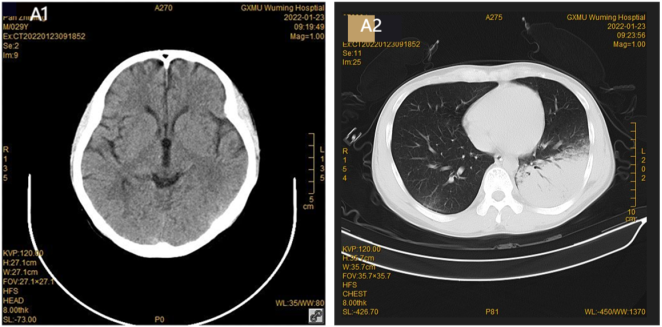
Figure 1. No A1 abnormal findings of cranial CT on admission. Sheet and plaque changes of the lower lobe of A2 lungs, and tracheal shadow on the left side.
Figure 2. Patient PO2/FiO2 trend chart of treatment changes in the ICU.
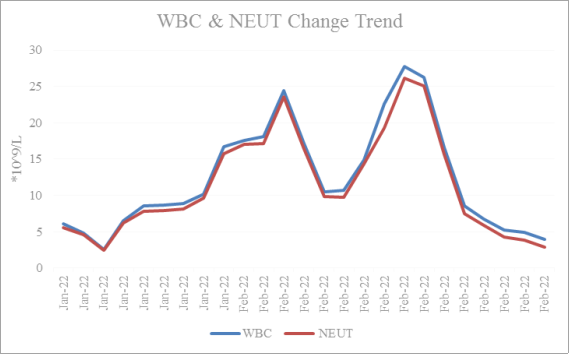
Figure 3. Trend ends of leukocytes (WBC) and neutrophils (NEUT).
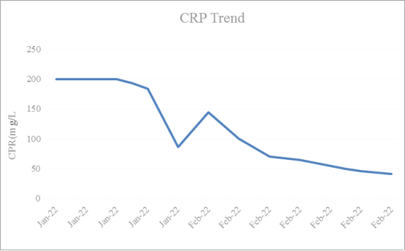
Figure 4. Trend chart of patients C-reactive protein (CRP).
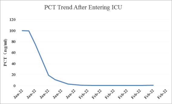
Figure 5. Patient change trend diagram of procalcitonin (PCT).
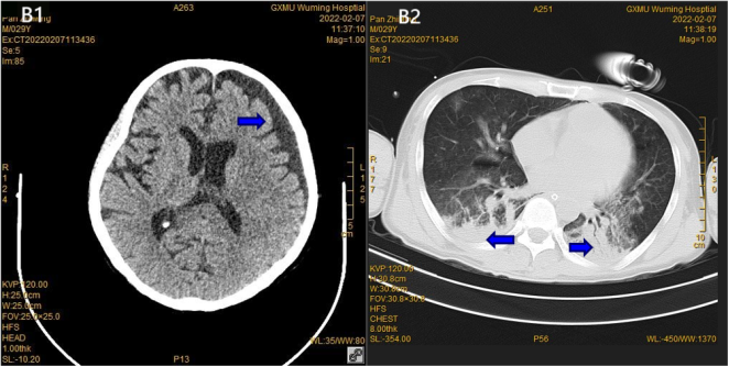
Figure 6. Review of B1 cranial CT after 15 days showed a small amount of subdural effusion in the left frontotemporal region. The corresponding brain parenchyma was shifted medially, the brain parenchyma density was normal, the ventricle, cisterna and sulcus morphology were normal, and the B2 pulmonary lesions increased on the right side and decreased on the left side.
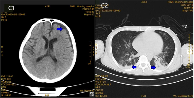
Figure 7. Left frontal and temporal subdural effusion of C1 showed a decrease after 23 days, and the C2 lung inflammation was improved compared to the anterior absorption.
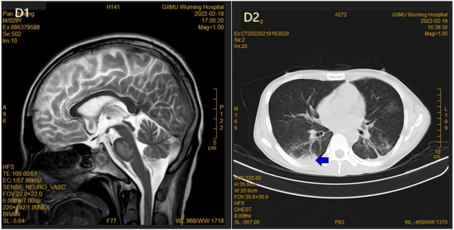
Figure 8. D1 MR subdural effusion was absorbed after 26 days; subacute cerebral infarction was suspected, and D2 inflammation of lungs was also absorbed.
3. Discussion
Psittacosis is caused by Chlamydia psittaci, that uses the energy of the host cells to complete its metabolism and reproduction. Chlamydia psittaci is a strict obligate eukaryotic cell parasitic pathogen with a unique two-phase development cycle that can form an infective protomer and a reproductive reticulate body . Chlamydia psittaci has 10 genotypes, of which types A and E are pathogenic in humans . The ability to cause disease mainly depends on bacterial body lipopolysaccharide, membrane proteins, plasmid encoding proteins, and secreted effectors. Chlamydia adheres and invades the host to cause inflammation and pathological damage, regulates the host cell function to facilitate intracellular growth, and inhibits host immune cells to escape immune-mediated killing . However, the pathogenic factors, pathways, and modes and targets of action of Chlamydia psittaci are still not fully elucidated and need further study .
Although contact with birds or birds is a major risk factor for psittacosis , some patients, including the current case, have no history of direct contact with epidemic birds or poultry.
The clinical manifestations of psittacosis are diverse and nonspecific; the incubation period is usually 5-19 days, but can be up to 28 days . The initial asymptomatic infection can lead to multiple organ failure, including atypical severe pneumonia, liver injury, and respiratory failure. Almost all infected patients have febrile manifestations and generally show sudden-onset symptoms, including high fever, headaches, chills, muscle soreness, and mild dry cough, while severe cases may include dyspnea . More than one-third of patients develop severe headaches, which may be an indicator of the occurrence and severity of meningitis. Fatal meningitis is a common serious complication that mainly manifests as intense headache and psychiatric disorders . In addition to fever, the current patient had no respiratory symptoms such as cough, and instead presented with rare mental behavior abnormalities. Some patients also have gastrointestinal symptoms , such as nausea and vomiting, which was also observed in this case. The high APACHE-II score, SOFA score, and mortality risk rate indicated the severity of severe pneumonia and meningitis. However, it is difficult to determine the cause solely based on clinical manifestations .
Most patients with psittacosis have a normal or mildly elevated leukocyte count, mainly with an elevated proportion of neutrophils, and a normal or slight elevation in inflammatory indices, including CRP, abnormal elevation, and PCT . The inflammatory indices of the current patient were largely consistent with the literature reports, although the abnormal increase in PCT was > 100 ng/ml, which was likely due to the presence of other pathogenic bacteria, such as Haemophilus influenzae. However, this finding also suggested a particularly severe inflammatory response, which was gradually reduced to < 1.0 ng/ml after 8 days of treatment, together with CRP, indicating effective inflammatory control. Additionally, this patient had multiple organ injuries, including in the brain, lung, myocardium, and liver, which is consistent with previous literature reports . However, cases of meningitis are rare in the literature. Imaging manifestations of the lungs show different degrees of exudation and consolidation, of which patchy shadows and mesh infiltrations are the most common; in some severe cases, lung lobe large shadow and extensive bilateral pneumonia may also appear, presenting from the hilum outward fan or subpleural wedge patch and occasionally with pleural effusion, while chest lesions can be absorbed within 2-4 weeks after treatment . The lung lesions reported in this case and in the previous literature were generally consistent. Brain lesions have not been reported previously, and no significant abnormalities were observed in the brain parenchyma in this case; however, the meningeal inflammation reaction was intense, which led to subdural effusion.
Regarding the diagnosis of Chlamydia psittaci, conventional culture, complement binding test, immunofluorescence, enzyme-linked immunosorbent test and other methods are difficult to test the problems , which cause difficulties in determining the clinical etiology. Although polymerase chain reaction (PCR) improves the detection sensitivity and shortens the detection time and has replaced conventional culture methods, it is more sensitive in the acute stage of the onset . Additionally, the PCR conditions in technical laboratories are highly demanding, and many hospitals do not perform routine examinations. Recently, the use of mNGS technology has provided a new detection means for diagnosis in patients with an unexplained infection. This technology provides high throughput sequencing of all nucleic acid sequences in clinical samples, and can be used to more comprehensively and accurately detect various pathogens, especially for cross-species transmission and new and rare pathogens. Additionally, mNGS technology is less time-consuming than the conventional pathogen detection, has a low positive rate, high positive diagnosis and negative exclusion diagnostic value, and has been used in clinical diagnosis . Under normal circumstances, as CSF is sterile and psittacosis does not colonize humans, no coexisting or opportunistic bacteria are present in CSF. Therefore, contamination of CSF by psittacosis such as chlamydia and by other pathogens can be eliminated as long as the sampling, storage, and transport processes strictly follow aseptic procedures. Certainly, qualified the sample helps mNGS in accurate etiological diagnosis . In the Compendium of Measures to Control Chlamydia psittaci Infection Among Humans (Psittacosis), the use of additional diagnostic techniques, such as genome sequencing, is encouraged . In this case, the nucleic acid sequence of Chlamydia psittaci was detected in CSF and sputum specimens via mNGS, combined with clinical manifestations and related examination, and finally confirmed as Chlamydia psittaci infectious pneumonia and meningitis.
Chlamydia psittaci is an intracellular parasitic bacterium against which β lactam antibiotics are ineffective due to its lack of a cell wall. Antibiotics with high intracellular activity should be selected in clinical practice, including tetracyclines, fluoroquinolones, and macrolides, though tetracyclines are preferred . These antibiotics’ main mechanism of action is via specific binding to the A position of the 30S subunit of the bacterial ribosome, which prevents the association of aminoacyl-tRNA at this position, thereby inhibiting the growth of the peptide chain and affecting the bacterial protein synthesis . In the current case, after treatment remained febrile with routinely used ultra-broad-spectrum antimicrobial coverage, and no significant improvement in oxygenation index and brain function was observed. However, according to the mNGS results, targeted treatment with tetracycline and intravenous quinolones and macrolides achieved good results, clinically supporting the diagnosis of Chlamydia psittacosis.
4. Conclusion
The incidence of psittacosis is low and the rate of misdiagnosis and mistreatment is high, which is complicated by rapid disease development and poor prognosis. When clinicians encounter patients with unexplained fever, shortness of breath, headache or neuropsychiatric disorder symptoms of meningitis, normal or slightly elevated WBC count, combined with multiple organ function damage, and chest CT showing large sheet lung consolidation or ground glass pattern, especially with a history of bird contact history, atypical pathogen infection should be suspected. In particular, the possibility of psittacosis should be considered in cases with serious infection but poor response to conventional anti-infection treatment. Clinicians should execute mNGS testing as early as possible to clarify the cause of disease and ensure that the targeted anti-infection treatment is started in time .
Abbreviations

mNGS

Metagenomic Secondary Sequencing

CSF

Cerebrospinal Fluid

RBC

Red Blood Cell

HGB

Hemoglobin

WBC

White Blood Cell

PLT

Platelet

CRP

C-reactive Protein

SAA

Serum Amyloid A Protein

PCT

Procalcitonin

CT

Computed Tomography

ICU

Intensive Care Unit

APACHE-II

Acute Physiology and Chronic Health Evaluation

SOFA

Sepsis Related Organ Failure Assessment

ADA

Adenosine Deaminase

LDH

Lactate Dehydrogenase

G test

Fungal D-glucan Detection

ALT

Alanine Transaminase

AST

Aspartate Aminotransferase

ALB

Albumin

HBD

Hydroxybutyric Dehydrogenase

BALF

Bronchoalveolar Lavage Fluid

PCR

Polymerase Chain Reaction

Acknowledgments
The authors have no competing interests to declare.
Funding
Guangxi medical and health appropriate technology development and promotion and application project (contract No: S023100).
Data Availability Statement
All data generated or analysed during this study are included in this published article.
Conflicts of Interest
The authors of this manuscript declare that there are no conflicts of interest associated with this work.
References
[1] Kozuki E, Arima Y, Matsui T, et al. Human psittacosis in Japan: notification trends and differences in infection source and age distribution by gender, 2007 to 2016. Ann Epidemiol. 2020; 44: 60-63.
[2] Hogerwerf L, DE Gier B, Baan B, VAN DER Hoek W. Chlamydia psittaci (psittacosis) as a cause of community-acquired pneumonia: a systematic review and meta-analysis. Epidemiol Infect. 2017; 145(15): 3096-3105.
[3] Cheng Y-J, Lin K-Y, Chen C-C, Huang Y-L, Liu C-E, Li S-Y. Zoonotic atypical pneumonia due to Chlamydophila psittaci: first reported psittacosis case in Taiwan. J Formos Med Assoc. 2013; 112(7): 430-3.
[4] Shaw KA, Szablewski CM, Kellner S, Kornegay L, Bair P, Brennan S, et al. Psittacosis outbreak among workers at chicken slaughter plants, Virginia and Shi et al. BMC Infectious Diseases (2021) 21: 621Page 7 of 8Georgia, USA, 2018. Emerg Infect Dis. 2019; 25(11): 2143-5.
[5] Branley JM, Weston KM, England J, Dwyer DE, Sorrell TC. Clinical features of endemic community-acquired psittacosis. New Microbe New Infect. 2014; 2(1): 7-12.
[6] Yung AP, Lindsay GM. Psittacosis - a review of 135 cases. Med J. 1988; 148: 228-33.
[7] Chen X, Cao K, Wei Y, et al. Metagenomic next-generation sequencing in the diagnosis of severe pneumonias caused by Chlamydia psittaci. Infection. 2020; 48(4): 535-542.
[8] Peng Li, Lihua Song, Jinhua Miao. Progress in the pathogenic mechanism of Chlamydia psittaci. International Journal of Laboratory Medicine, 2016, 37(06): 780-782.
[9] Zeidler H, Kuipers J, Köhler L. Chlamydia-induced arthritis. Curr Opin Rheumatol. 2004; 16(4): 380-392.
[10] Mingyi Shu, Zhongyu Li. Progress in studying the pathogenic mechanism of Chlamydia. The Chinese Journal of Zoonosis, 2021, 37(06): 542-547.
[11] Elwell C, Mirrashidi K, Engel J. Chlamydia cell biology and pathogenesis. Nat Rev Microbiol. 2016; 14(6): 385-400.
[12] Radomski N, Einenkel R, Müller A, Knittler MR. Chlamydia-host cell interaction not only from a bird's eye view: some lessons from Chlamydia psittaci. FEBS Lett. 2016; 590(21): 3920-3940.
[13] Balsamo G, Maxted AM, Midla JW, et al. Compendium of Measures to Control Chlamydia psittaci Infection Among Humans (Psittacosis) and Pet Birds (Avian Chlamydiosis), 2017. J Avian Med Surg. 2017; 31(3): 262-282.
[14] Spoorenberg SM, Bos WJ, van Hannen EJ, et al. Chlamydia psittaci: a relevant cause of community-acquired pneumonia in two Dutch hospitals. Neth J Med. 2016; 74(2): 75-81.
[15] Stewardson AJ, Grayson ML. Psittacosis. Infect Dis Clin N Am. 2010; 24(1): 7-25.
[16] Katsura D, Tsuji S, Kimura F, Tanaka T, Eguchi Y, Murakami T. Gestational psittacosis: a case report and literature review. J Obstet Gynaecol Research. 2020; 46(5): 673-7.
[17] Jin W, Liang R, Tian X, Cheng Y, Kong X, He F, Zhang C, Wang G, Li S, Lu H, Sun S, Shen L. Clinical features of psittacosis in 46 Chinese patients. Enferm Infecc Microbiol Clin (Engl Ed). 2023 Nov; 41(9): 545-548.
[18] Stidham RA, Richmond-Haygood M. Case report: Possible psittacosis in a military family member-clinical and public health management issues in military settings. MSMR. 2019; 26(7): 2-7.
[19] Fraeyman A, Boel A, Van Vaerenbergh K, De Beenhouwer H. Atypical pneumonia due to Chlamydophila psittaci: 3 case reports and review of literature. Acta Clin Belg. 2010; 65(3): 192-196.
[20] Cunha BA. The atypical pneumonias: clinical diagnosis and importance. Clin Microbiol Infect. 2006; 12 Suppl 3: 12-24.
[21] Corsaro D, Greub G. Pathogenic potential of novel Chlamydiae and diagnostic approaches to infections due to these obligate intracellular bacteria. Clin Microbiol Rev. 2006; 19(2): 283-297.
[22] Nieuwenhuizen AA, Dijkstra F, Notermans DW, van der Hoek W. Laboratory methods for case finding in human psittacosis outbreaks: a systematic review. BMC Infect Dis. 2018; 18(1): 442. Published 2018 Aug 30.
[23] Gu W, Miller S, Chiu CY. Clinical Metagenomic Next-Generation Sequencing for Pathogen Detection. Annu Rev Pathol. 2019; 14: 319-338.
[24] Brown JR, Bharucha T, Breuer J. Encephalitis diagnosis using metagenomics: application of next generation sequencing for undiagnosed cases. J Infect. 2018; 76(3): 225-240.
[25] Gu L, Liu W, Ru M, et al. The application of metagenomic next-generation sequencing in diagnosing Chlamydia psittaci pneumonia: a report of five cases. BMC Pulm Med. 2020; 20(1): 65. Published 2020 Mar 17.
[26] Langelier C, Kalantar KL, Moazed F, et al. Integrating host response and unbiased microbe detection for lower respiratory tract infection diagnosis in critically ill adults. Proc Natl Acad Sci U S A. 2018; 115(52): E12353-E12362.
[27] Kohlhoff SA, Hammerschlag MR. Treatment of Chlamydial infections: 2014 update. Expert Opin Pharmacother. 2015; 16(2): 205-212.
[28] Drysdale MJ, Lentzen G, Matassova N, Murchie AI, Aboul-Ela F, Afshar M. RNA as a drug target. Prog Med Chem. 2002; 39: 73-119.
[29] NI Y, ZHONG H, GU Y, et al. Clinical Features, Treatment, and Outcome of Psittacosis Pneumonia: A Multicenter Study [J]. Open Forum Infect Dis, 2023, 10(2): ofac518.
Cite This Article
  • APA Style

    Sheng, S., Lin, Z., Li, L., Zhuang, Z., Yang, C., et al. (2025). Psittacosis with Abnormal Mental Behavior: A Rare Case Report and Literature Review. International Journal of Medical Case Reports, 4(1), 7-16. https://doi.org/10.11648/j.ijmcr.20250401.12

    Copy | Download

    ACS Style

    Sheng, S.; Lin, Z.; Li, L.; Zhuang, Z.; Yang, C., et al. Psittacosis with Abnormal Mental Behavior: A Rare Case Report and Literature Review. Int. J. Med. Case Rep. 2025, 4(1), 7-16. doi: 10.11648/j.ijmcr.20250401.12

    Copy | Download

    AMA Style

    Sheng S, Lin Z, Li L, Zhuang Z, Yang C, et al. Psittacosis with Abnormal Mental Behavior: A Rare Case Report and Literature Review. Int J Med Case Rep. 2025;4(1):7-16. doi: 10.11648/j.ijmcr.20250401.12

    Copy | Download

  • @article{10.11648/j.ijmcr.20250401.12,
      author = {Shanfeng Sheng and Zhaohua Lin and Lianmao Li and Zhiquan Zhuang and Chunqiong Yang and Zheng Li},
      title = {Psittacosis with Abnormal Mental Behavior: A Rare Case Report and Literature Review},
      journal = {International Journal of Medical Case Reports},
      volume = {4},
      number = {1},
      pages = {7-16},
      doi = {10.11648/j.ijmcr.20250401.12},
      url = {https://doi.org/10.11648/j.ijmcr.20250401.12},
      eprint = {https://article.sciencepublishinggroup.com/pdf/10.11648.j.ijmcr.20250401.12},
      abstract = {Psittacosis, an infectious disease caused by Chlamydia psittaci, has clinical manifestations ranging from asymptomatic infection to severe atypical pneumonia, and even rare fatal meningitis. Psittacosis is difficult to identify early due to its nonspecific clinical presentation. In this report, we describe a male patient with atypical severe pneumonia and meningitis caused by Chlamydia psittacosis. The patient presented to the Department of Neurology with a fever for 5 days, abnormal mental behavior for 5 h, and no history of avian or poultry exposure. Following empirical antimicrobial therapy, the patient’s condition deteriorated rapidly, and he suffered respiratory failure, shock, and psychiatric disorders. The patient was rapidly transferred to ICU, where he received antishock, invasive mechanical ventilation therapy, and organ support therapy. Sputum and cerebrospinal fluid metagenomic secondary sequencing (mNGS) identified sequence reads related to Chlamydia thermosum. The patient was administered targeted drugs, as well as fluoroquinolone antimicrobial agents. As a result, the patient improved and left the ICU after 25 days, before returning to a near-premorbid condition after discharge. Psittacosis and meningitis should be considered in patients suffering from atypical pneumonia with fever, headache, and neuropsychiatric symptoms, and mNGS is a useful test for etiological screening. The case reports in this study hopefully help to provide some references for earlier diagnosis of psittacosis and development of life-saving treatment plan.},
     year = {2025}
    }
    

    Copy | Download

  • TY  - JOUR
    T1  - Psittacosis with Abnormal Mental Behavior: A Rare Case Report and Literature Review
    AU  - Shanfeng Sheng
    AU  - Zhaohua Lin
    AU  - Lianmao Li
    AU  - Zhiquan Zhuang
    AU  - Chunqiong Yang
    AU  - Zheng Li
    Y1  - 2025/02/17
    PY  - 2025
    N1  - https://doi.org/10.11648/j.ijmcr.20250401.12
    DO  - 10.11648/j.ijmcr.20250401.12
    T2  - International Journal of Medical Case Reports
    JF  - International Journal of Medical Case Reports
    JO  - International Journal of Medical Case Reports
    SP  - 7
    EP  - 16
    PB  - Science Publishing Group
    SN  - 2994-7049
    UR  - https://doi.org/10.11648/j.ijmcr.20250401.12
    AB  - Psittacosis, an infectious disease caused by Chlamydia psittaci, has clinical manifestations ranging from asymptomatic infection to severe atypical pneumonia, and even rare fatal meningitis. Psittacosis is difficult to identify early due to its nonspecific clinical presentation. In this report, we describe a male patient with atypical severe pneumonia and meningitis caused by Chlamydia psittacosis. The patient presented to the Department of Neurology with a fever for 5 days, abnormal mental behavior for 5 h, and no history of avian or poultry exposure. Following empirical antimicrobial therapy, the patient’s condition deteriorated rapidly, and he suffered respiratory failure, shock, and psychiatric disorders. The patient was rapidly transferred to ICU, where he received antishock, invasive mechanical ventilation therapy, and organ support therapy. Sputum and cerebrospinal fluid metagenomic secondary sequencing (mNGS) identified sequence reads related to Chlamydia thermosum. The patient was administered targeted drugs, as well as fluoroquinolone antimicrobial agents. As a result, the patient improved and left the ICU after 25 days, before returning to a near-premorbid condition after discharge. Psittacosis and meningitis should be considered in patients suffering from atypical pneumonia with fever, headache, and neuropsychiatric symptoms, and mNGS is a useful test for etiological screening. The case reports in this study hopefully help to provide some references for earlier diagnosis of psittacosis and development of life-saving treatment plan.
    VL  - 4
    IS  - 1
    ER  - 

    Copy | Download

Author Information