Controlling the microbiological quality of water in hemodialysis centers is essential to avoid complications in hemodialysis patients that may be caused by microorganisms. The aim of this study was to determine the microbiological quality of water after the installation of a new water treatment system in the hemodialysis department of the Yaoundé University Hospital Center. A total of sixteen (16) samples were taken every two weeks at sites A (network inlet), B (filter outlet/osmosis inlet), C (osmosis outlet) and D (loop return) between May and July 2023. Microorganisms were isolated after filtration of 100 ml of water through a nitrocellulose membrane, microporosity 0.22 µm, then deposited on Tryptone Glucose Extract Agar (TGEA) medium and incubated at room temperature between 17 and 22°C for 7 days. After subculturing on different media, the pure microorganisms were identified by their cultural characteristics and marketed biochemical galleries. The compliance threshold was below 100CFU/ml. Of the samples analyzed, 56% (9/16) were declared non-compliant (>100UFC/ml) versus 43% (7/16) compliant (<100CFU/ml). Only samples from the fourth series were all compliant at points A, B, C and D. Of the microorganisms identified, five (5) species were Gram-negative bacilli, including Acinetobacter baumanii, Pseudomonas luteola, Burkholderia cepacia, Pseudomonas aeruginosa and Stenotrophomonas maltophilia. Gram-positive cocci were all coagulase-negative Staphylococcus and yeasts were Candida spp. The most frequently isolated bacterial genera were Pseudomonas (29.17%), Staphylococcus (25%), Acinetobacter (16.67%), Stenotrophomonas (12.50%), Candida (12.50%) and Burkholderia (4.17%). In this study, although the samples from the fourth series of sampling were all compliant at the various sampling points, the high rate of non-compliance and the detection of a variety of microorganisms demonstrate the need to reinforce the disinfection system in the hemodialysis water treatment circuit.
This is an Open Access article, distributed under the terms of the Creative Commons Attribution 4.0 International License (http://creativecommons.org/licenses/by/4.0/), which permits unrestricted use, distribution and reproduction in any medium or format, provided the original work is properly cited.
The most common modality of renal replacement therapy worldwide is hemodialysis (89%)
[1]
Pecoits-Filho R, Okpechi IG, Donner JA, Harris DCH, Aljubori HM, Bello AK, Bellorin-Font E, Caskey FJ, Collins A, Cueto-Manzano AM, Feehally J, Goh BL, Jager KJ, Nangaku M, Rahman M, Sahay M, Saleh A, Sola L, Turan Kazancioglu R, Walker RC, Walker R, Yao Q, Yu X, Zhao MH, Johnson DW: Capturing and monitoring global differences in untreated and treated end-stage renal disease, replacement therapy modality and outcomes. Kidney Int Suppl (2011) 10: e3-e9, 2020.
. This is a procedure in which a dialysis machine and a special filter called an artificial kidney or dialyzer are used to cleanse the blood of impurities
In the course of an average week of hemodialysis, a patient may be exposed to 300-600 liters of water, providing multiple opportunities for exposure to waterborne pathogens
[3]
Kohn, Orly F.; Plascencia, Miguel; Taylor, Yolanda; Koyner, Jay L.. Novel Use of Premixed Dialysate Bags during Water Supply Interruption in Acute Hospital Setting. Kidney360 February 2021, 2(2): p 339-343.
. The water used for treatment comes from the mains and is therefore not sterile
[4]
Skarupskiene I, Bumblyte IA, Kuzminskis V. Endotoksinu kiekis hemodializei naudojamame vandenyje ir dializes tirpale [The level of endotoxins in hemodialysis water and dialysate]. Medicina (Kaunas). 2007; 43 Suppl 1: 81-84.
[4]
. Patients undergoing hemodialysis are vulnerable because of their weakened immunity, and contaminants such as microorganisms, endotoxins, peptidoglycans and bacterial DNA fragments present in the water and resulting dialysate can cross the dialyzer membrane, stimulate cytokine production and trigger a rise in acute-phase reagents. These can give rise to acute intra-dialytic complications such as fever, acute respiratory distress syndrome (ARDS), disseminated intravascular coagulation (DIC), chills, hypotension, headaches, nausea and cramps , 6]. Controlling the quality of the water used in hemodialysis is therefore a major concern, due to the various contaminations of microbiological origin that can have serious repercussions for hemodialysis patients. Despite advances in extrarenal purification techniques, the quality of water and associated dialysis solutions have been implicated in adverse patient outcomes
[7]
Coulliette AD, Arduino MJ. Hemodialysis and water quality. Semin Dial. 2013; 26(4): 427-438.
In Europe, a microbiological quality control of dialysate from several hemodialysis centers in Germany showed a high concentration of microorganisms and endotoxins above the standard (<100 CFU/ml and <0.25EU/ml) set by the Association for Advancement of Medical Instrumentation (AAMI)
[8]
Pisani B, Simões M, Prandi MAG, Rocha MMM, Gonçalves CR, Vaz TMI, et al. Surto de bacteriemia por Pseudomonas aeruginosa na unidade de hemodiálise de um hospital de Campinas, São Paulo, Brasil. Rev. Inst. Adolfo Lutz. 2000; 59(1/2): 51-56.
. They have set standards to avoid any risk of contamination, as in 1996, when an epidemic of bacteremia occurred in a hemodialysis center in Campinas, in the Brazilian state of São Paulo
[9]
R Bambauer, M Schauer, WK Jung, V Daum, J Vienken ASAIO Journal (American Society for Artificial Internal Organs: 1992) 40 (4), 1012-1016, 1994.
. Following this episode, water and dialysate samples were collected at various sites in the hemodialysis system for monitoring. At the first collection, 80% of samples showed Pseudomonas aeruginosa and Burkholderia cepacia, two Gram-negative bacteria, while at the second collection, 100% of samples showed identification with both bacteria
[9]
R Bambauer, M Schauer, WK Jung, V Daum, J Vienken ASAIO Journal (American Society for Artificial Internal Organs: 1992) 40 (4), 1012-1016, 1994.
. In Cameroon, studies carried out in 2015 by Gueguim et al on-hemodialysis water showed microbiological contamination, with 83.3% non-compliance at the Centre Hospitalier Universitaire de Yaoundé
[10]
Cédric Gueguim, Nnanga Nga, François Kaze Folefack, Alain Ragon, Hortense Gonsu Kamga. Microbiological Analysis of Hemodialysis Water at the University Teaching Hospital of Yaounde, Cameroon. American Journal of Biomedical and Life Sciences. Vol. 4, No. 6, 2016, pp. 81-86.
. These results led to the installation of a new hemodialysis water treatment system in the CHUY hemodialysis unit. The aim of this study was to investigate the microbiological quality of hemodialysis water after installation of the new system.
2. Materials and Methods
2.1. Sample Collection and Transport
2.1.1. Collection
Water sampling was carried out under rigorous aseptic conditions. After disinfecting the sampling sites with a hydro-alcoholic solution, we let the water run for 1 to 2 minutes to ensure that the sample was not contaminated by traces of the disinfectant, then collected it in sterile single-use bottles
[11]
French standard. (2008). Water treatment and distribution system for dilution of concentrated hemodialysis solutions - Design, operation, performance and safety requirements. S93-315: (2008).
The vials were clearly labelled and transported immediately to the laboratory, accompanied by a form containing all the necessary information (date, time and place of collection) for immediate analysis.
2.2. Microbiological Analysis
2.2.1. Isolation and Quantification of Microorganisms
To count the number of microorganisms suspended in the water sample, a 100 ml volume of water sample was filtered through a nitrocellulose membrane with microporosity (0.22 µm). The filter was then placed on Tryptone Glucose Extract Agar (TGEA) and incubated at room temperature (17-22ºC) for 7 days
[11]
French standard. (2008). Water treatment and distribution system for dilution of concentrated hemodialysis solutions - Design, operation, performance and safety requirements. S93-315: (2008).
. The number of colonies found was expressed as the mean colony-forming units (CFU/mL)
[11]
French standard. (2008). Water treatment and distribution system for dilution of concentrated hemodialysis solutions - Design, operation, performance and safety requirements. S93-315: (2008).
. Sterile water for injection (SWFI) was used as a control.
2.2.2. Microorganism Identification
After Gram staining, pure isolates were transferred to Chapman, Mueller Hinton, MacConkey and Sabouraud agars and incubated at 37°C for 24 to 48 hours.
Microorganisms were identified using commercially available biochemical strips: API 20NE strip for identification of bacteria classified as non-fermentative, API 20E strip for identification of enterobacteria, mannitol, catalase, coagulase and DNase tests for identification of staphylococcus species.
2.3. Data Processing and Analysis
Data were collected, processed and analyzed using Excel (version 2010). Results were presented in the form of tables, graphs or narratives.
3. Results
3.1. Colony Count After Culture
To check whether the number of germs present in CHUY's hemodialysis water met international standards, colonies isolated on membrane filters were counted at each sampling point and expressed as CFU/ml.
Table 1. Colony counts after membrane filtration.
Number of colonies UFC/ml for each sample after 7 days
Sample 1 May 11, 2023
Sample 2 May 29, 2023
Sample 3 June 12, 2023
Sample 4 June 26, 2023
Point A
PC
PC*.
PC*.
PC*.
Point B
PC
PC
PC*.
PC*.
Point C
PC
PC
PC
PC*.
Point D
PC
PC
PC
PC*.
Point A: Network inlet; Point B: Filter outlet/Osmosis inlet; Point C: Osmosis outlet; Point D: Loop return;
PC: Presence of colonies >100CFU/ml (non-compliant); PC*: Absence of colonies <100CFU/ml (compliant)
The results for the first sample were above the standard on all points (>100UFC/ml); Two weeks later, a second sample was taken and only non-conformity was observed on points B, C and D (>100UFC/ml); The analysis of the third sample showed non-conformity on points C and D (>100UFC/ml); And the fourth sample had all points conforming to the standards in force (<100UFC/ml).
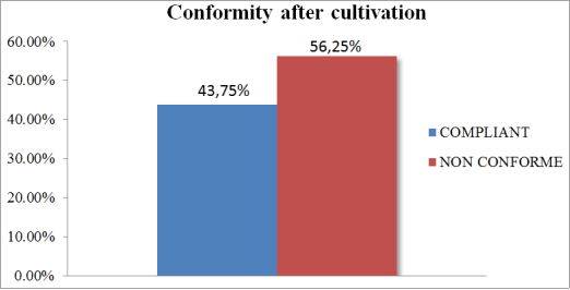
3.2. Compliance After Cultivation
According to Figure 1, 56.25% of samples were non-compliant, with colony counts well above the norm (˃100 CFU / mL), while only 43.75% of samples were compliant (˂100 CFU / mL).
Gram staining and fresh state observation were carried out to determine the type of germ present at each sampling point. Table 2 below shows the results obtained. Our results show that we mainly isolated gram-negative bacilli, then gram-positive cocci and finally yeasts.
Table 2. Type of bacteria at each sampling point.
Type of germs
Levy 1/ May 11, 2023
Sampling 2/ May 29, 2023
Sampling 3/ June 12, 2023
Sampling 4/ June 26, 2023
A B C D
A B C D
A B C D
A B C D
Gram- bacilli
+ + + +
+ + + +
+ + + +
+ + + +
Gram+ cocci
- - - -
- - - -
+ + - -
+ + + +
Yeast
- - - -
- - - +
+ - - -
+ - - -
(+) = present (-) = absent
3.4. Microorganisms Isolated in Hemodialysis Water
The results show a variation in the germs isolated, depending on the type of sample taken. In the first sampling, Acinetobacter baumanii was more frequently isolated, followed by Pseudomonas aeruginosa in the second sampling. On the other hand, Staphylococcus coagulas negativa was most frequently isolated in the third sampling.
Table 3. Microorganism species isolated from hemodialysis water.
Type of isolated germs
Levy 1/ May 11, 2023
Sampling 2/ May 29, 2023
Sampling 3/ June 12, 2023
Sampling 4/ June 26, 2023
A B C D
A B C D
A B C D
A B C D
Acinetobacter baumanii
+ + + +
- - - -
- - - -
- - - -
Pseudomonas luteola
+ + - -
- + - -
- - - -
- - - -
Burkholderiacepacia
- - + -
- - - -
- - - -
- - - -
Pseudomonas aeruginosa
- - - -
- + + +
- - - +
- - - -
Stenotrophomonasmaltophilia
- - - -
- - + -
- - - -
- + - +
Coagulase-negative Staphylococcus
- - - -
- - - -
+ + - -
+ + + +
Candida sp
- - - -
- - - +
+ - - -
+ - - -
(+) =Presence (-) = Absence Point A: Network inlet; Point B: Filter outlet/Osmosis inlet; Point C: Osmosis outlet; Point D: Loop return
3.5. Distribution of Bacterial Genera
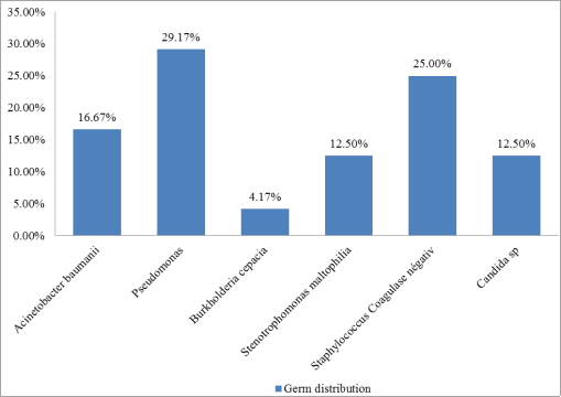
The figure below shows the representation of the germs isolated. The genus Pseudomonas accounts for 29.17% of germs isolated, Staphylococcus 25%, Acinetobacter baumanii 16.67%, Sténotrophomonas maltophilia 12.50%, Candida spp 12.50% and Burholderia cepacia 4.17%.
Every week, hemodialysis patients are exposed to around 400 l of water used for the production of dialysis fluids which, albeit with the interposition of an artificial semi-permeable membrane, come into direct contact with the bloodstream
[12]
Pontoriero G, Pozzoni P, Andrulli S, Locatelli F. The quality of dialysis water. Nephrol Dial Transplant. 2003; 18 Suppl 7: vii21-vii56.
. The best possible treatment of water intended for dialysis is therefore of paramount importance, and recurrent maintenance of treatment devices is essential to reduce or eliminate the load of possible contaminants. Disinfection should be carried out at least once a week, using appropriate and effective products. During our expedition to the CHUY hemodialysis department, the system was treated weekly with a solution of hydrogen peroxide and citric acid, added at the system inlet. This solution is used as a water disinfectant because of its effectiveness in combating impurities and micro-organisms.
Contamination of dialysis water has been a problem in hemodialysis departments for several years, due to the presence of contaminants such as bacteria and endotoxins that can come into contact with blood and present risks for hemodialysis patients. Indeed, contamination by microorganisms >100UFC/ml can lead to septicemia and even endotoxemia.
The study showed bacterial loads ranging from 10 to 1000UFC/ml, which is higher than international standards (<100UFC/ml). These results differ from those of Oumokhtar et al
[13]
O Oumokhtar B, Lalami Ael O, Mahmoud M, Berrada S, Arrayhani M, Houssaini TS. Prevent infection linked to the dialysis water in a hemodialysis center in Fez city (Morocco). Pan Afr Med J. 2013; 16: 122. Published 2013 Nov 28.
published in 2013, carried out in a hemodialysis center in the city of Fez, Morocco, with a bacterial load meeting standards of 8.74±5.52 CFU/ml. But similar to the results of Heidarieh et al
[14]
Heidarieh P, Hashemi Shahraki A, Yaghoubfar R, Hajehasani A, Mirsaeidi M. Microbiological Analysis of Hemodialysis Water in a Developing Country. ASAIO J. 2016 May-Jun; 62(3): 332-9.
announced in 2016 and carried out in five hospitals in a developing country (one in Tehran, and the others in Alborz), which reported variant loads of 50 to 1000 CFU/ml.
The sampling points were all non-compliant at the first sampling, but compliance was observed as early as the second sampling for point A, at the third sampling for points A and B, and compliance for all points at the fourth sampling. The low microbial load at point A (network inlet) can be explained by the use of a disinfectant (hydrogen peroxide) and a descaler (citric acid) at this site by the maintainer of the CHUY hemodialysis water treatment room, with a treatment frequency of once a week. Point B (filter outlet/Osmosis inlet) has a filter designed to retain particles suspended in the water. To block microorganisms, this filter had a porosity of 1 µm. It is recommended to have a filter with a porosity of 0.2µm, so it would be possible that it acts much more as a support for bacterial growth, hence the high contamination observed.
The high level of contamination at point C (Osmosis outlet) indicates the installation of a biofilm, which explains the results at point D (loop return), which also show the development of a biofilm. This situation could reflect a failure in the maintenance of the disinfection system used to purify the water distributed in the hemodialysis circuit. In fact, it has been shown that the presence of biofilm on pipes leads to rapid bacterial growth after just a few hours of disinfecting the water system
[15]
Nystrand R. Thoughts about biofilm in dialysis water systems. EDTNA ERCA J. 2003 Jul-Sep; 29(3): 127-30.
Microbiological analysis of the water revealed the presence of various types of micro-organisms. Among these, a number of Gram-negative bacilli (64%), Gram-positive cocci (24%) and yeasts (12%) were isolated. These results are very similar to those of Gueguim et al
[10]
Cédric Gueguim, Nnanga Nga, François Kaze Folefack, Alain Ragon, Hortense Gonsu Kamga. Microbiological Analysis of Hemodialysis Water at the University Teaching Hospital of Yaounde, Cameroon. American Journal of Biomedical and Life Sciences. Vol. 4, No. 6, 2016, pp. 81-86.
performed in the hemodialysis department of CHUY and published in 2016 with the presence of Gram-negative Bacilli (50%) and Gram-positive Cocci (33.3%). Similarly, those of Al-Haik et al
[16]
MUKALLA - HADHRAMAUT - YEMEN: CONTAMINATION RATES AND ANTIBIOTIC SUSCEPTIBILITY OF BACTERIAL ISOLATES". Universal journal of pharmaceutical research, vol. 4, no. 6, January 2020,
performed at the Artificial Kidney Center in the town of Mukalla published in 2020 with (85.7%) Gram-negative Bacilli and (14.2%) Gram-positive Cocci. Several Gram-negative Bacilli were isolated from the water distribution system, with a clear predominance of the Pseudomonas genus. As the Pseudomonas genus is able to multiply rapidly even in sterile water, they can reach high concentrations (>100,000 CFU/ml) in less than 48 hours in dialysis solutions, and this bacterial growth could be even more rapid due to the presence of glucose and bicarbonate in dialysis solutions, producing high levels of toxin responsible for infections in hemodialysis patients
[17]
Santos, F.; Santos, A. G.; Biernat, J. C.; Souza, M. E. L.; Raubach, A.; Aguirre, A.; Antoniazzi, V. R.; Scorsatto, K.; Seibel, I.; Demin, S. S.; Hickmann, A. C.; Oliveira, F. P.; Borba, E. V.; Simon, S.; Santos, S. S. Detecção de endotoxina pelo teste do limulus amoebocyte lysate (LAL) em unidades de hemodiálise. Rev. Virt. Med, 1, 2000.
[17]
.
The high concentration of bacteria found in hemodialysis water also suggests a high level of endotoxins. Endotoxins are low-molecular-weight proteins (2,000 to 20,000 Da) that can cross dialyzer membranes and enter the patient's bloodstream, stimulating circulating immune cells across the membrane to produce sepsis symptoms or a pyrogenic reaction
[7]
Coulliette AD, Arduino MJ. Hemodialysis and water quality. Semin Dial. 2013; 26(4): 427-438.
The germs most frequently isolated from hemodialysis water were Pseudomonas (29.17%), Staphylococcus (25%), Acinetobacter (16.67%), Stenotrophomonas (12.50%), Candida spp (12.50%) and Burkholderia (4.17%). These results are similar to those of Cédric Gueguim et al
[18]
Cedric Gueguim and others, #4059 MICROBIOLOGICAL ANALYSIS OF HEMODIALYSIS WATER AT THE DOUALA GENERAL HOSPITAL, CAMEROON, Nephrology Dialysis Transplantation, June 2023, Volume 38, Issue Supplement_1.
carried out in the hemodialysis department of Douala General Hospital and published in 2022, with Pseudomonas dominating at 38,5% and Staphylococcus spp at 23%, and the studies by Laila Chaoui et al
[19]
Laila Chaoui, Taha Chouati, Imane Zalegh, Rajaa Ait Mhand, Fouad Mellouki, Naima Rhallabi; Identification and evaluation of antimicrobial-resistant bacteria in a hemodialysis water treatment system. J Water Health February 1, 2022; 20 (2): 441-449.
published in 2022 at the Mohammedia hemodialysis center in Morocco, with Pseudomonas (25.53%) and Staphylococcus spp (17.02%) dominating. In the study by Sang-Hyun Park et al
[20]
Sang-Hyun Park, Young-Hyeon Lee, Min-Ho Yeo, Hi-Ryun Lee, Hye-Sook Kim, Kyung-Soo Chang. "Comparison of Microbial Detection of Hemodialysis Water in Reasoner's 2A Agar (R2A) and Trypticase Soy Agar (TSA)." JOURNAL OF BACTERIOLOGY AND VIROLOGY 51, no. 2 (2021): 79-88,
, on the other hand, the genus Staphylococcus spp was the most isolated at 30.15%, followed by the species Sphingomonas paucimobilis at 21.42%.
Studies by Okunola and Olaitan
[21]
Okunola OO, Olaitan JO. Bacterial contamination of hemodialysis water in three randomly selected centers in South Western Nigeria. Niger J Clin Pract. 2016; 19(4): 491-495.
published in 2016 in three hemodialysis centers in Nigeria revealed that the most frequently isolated bacteria in hemodialysis water belonged to the genus Pseudomonas (55%), and those by Jesus et al
[22]
Jesus, PR de, Carmo, J. dos S., & Ferreira. Microbiological evaluation of water used in haemodialysis services in the city of Rio de Janeiro from 2016 to 2018. Vigil Sanit Debate, Rio De Janeiro, JAB (2019), 7 (1), 53-59.
carried out in the hemodialysis department of the city of Rio with 35.5% of genus Pseudomonas isolated. In our study, the frequency of Pseudomonas luteola species (12.5% (3/24)) and Pseudomonas aeruginosa (16.6% (4/24)) is worrying, as these predominant opportunistic pathogens can cause infections in dialysis patients. Thus, it is important to consider their presence as biofilms in dialysis units, and also to prevent their existence as biofilms
[23]
Suman E, Varghese B, Joseph N, Nisha K, Kotian MS. The bacterial biofilms in dialysis water systems and the effect of the sub inhibitory concentrations of chlorine on them. J Clin Diagn Res. 2013; 7(5): 849-852.
The aim of this study was to investigate the microbiological quality of haemodialysis water in order to ensure patient safety by minimising the risk of infection and complications associated with dialysis water contamination. Microbiological analyses of the first, second and third water samples showed the presence of microorganisms and a high level of contamination in excess of international recommendations (>100 CFU/ml). Although the last sample showed a microbial load in compliance at all points (<100 CFU/ml), it would be important to reinforce the weekly disinfection system at the various points in order to facilitate the elimination of biofilms and guarantee a better quality of life for patients receiving this treatment.
Abbreviations
CHUY
Yaoundé University Hospital
TGEA
Tryptone Glucose Extract Agar
CFU
Colony Forming Unit
Conflicts of Interest
The authors declared no conflicts of interest.
References
[1]
Pecoits-Filho R, Okpechi IG, Donner JA, Harris DCH, Aljubori HM, Bello AK, Bellorin-Font E, Caskey FJ, Collins A, Cueto-Manzano AM, Feehally J, Goh BL, Jager KJ, Nangaku M, Rahman M, Sahay M, Saleh A, Sola L, Turan Kazancioglu R, Walker RC, Walker R, Yao Q, Yu X, Zhao MH, Johnson DW: Capturing and monitoring global differences in untreated and treated end-stage renal disease, replacement therapy modality and outcomes. Kidney Int Suppl (2011) 10: e3-e9, 2020.
Kohn, Orly F.; Plascencia, Miguel; Taylor, Yolanda; Koyner, Jay L.. Novel Use of Premixed Dialysate Bags during Water Supply Interruption in Acute Hospital Setting. Kidney360 February 2021, 2(2): p 339-343.
Skarupskiene I, Bumblyte IA, Kuzminskis V. Endotoksinu kiekis hemodializei naudojamame vandenyje ir dializes tirpale [The level of endotoxins in hemodialysis water and dialysate]. Medicina (Kaunas). 2007; 43 Suppl 1: 81-84.
[5]
Park HC, Lee YK, Yoo KD, et al. Korean clinical practice guidelines for preventing the transmission of infections in hemodialysis facilities. Kidney Res Clin Pract. 2018; 37(1): 8-19.
Perez-Garcia R, Cinio Rodriquez-Benitez PO. Why and how should bacterial contamination of dialysate be monitored? Nephrol Dial. Transplantation. 2000; 15(6): 760-65.
Pisani B, Simões M, Prandi MAG, Rocha MMM, Gonçalves CR, Vaz TMI, et al. Surto de bacteriemia por Pseudomonas aeruginosa na unidade de hemodiálise de um hospital de Campinas, São Paulo, Brasil. Rev. Inst. Adolfo Lutz. 2000; 59(1/2): 51-56.
Cédric Gueguim, Nnanga Nga, François Kaze Folefack, Alain Ragon, Hortense Gonsu Kamga. Microbiological Analysis of Hemodialysis Water at the University Teaching Hospital of Yaounde, Cameroon. American Journal of Biomedical and Life Sciences. Vol. 4, No. 6, 2016, pp. 81-86.
French standard. (2008). Water treatment and distribution system for dilution of concentrated hemodialysis solutions - Design, operation, performance and safety requirements. S93-315: (2008).
O Oumokhtar B, Lalami Ael O, Mahmoud M, Berrada S, Arrayhani M, Houssaini TS. Prevent infection linked to the dialysis water in a hemodialysis center in Fez city (Morocco). Pan Afr Med J. 2013; 16: 122. Published 2013 Nov 28.
Heidarieh P, Hashemi Shahraki A, Yaghoubfar R, Hajehasani A, Mirsaeidi M. Microbiological Analysis of Hemodialysis Water in a Developing Country. ASAIO J. 2016 May-Jun; 62(3): 332-9.
Santos, F.; Santos, A. G.; Biernat, J. C.; Souza, M. E. L.; Raubach, A.; Aguirre, A.; Antoniazzi, V. R.; Scorsatto, K.; Seibel, I.; Demin, S. S.; Hickmann, A. C.; Oliveira, F. P.; Borba, E. V.; Simon, S.; Santos, S. S. Detecção de endotoxina pelo teste do limulus amoebocyte lysate (LAL) em unidades de hemodiálise. Rev. Virt. Med, 1, 2000.
[18]
Cedric Gueguim and others, #4059 MICROBIOLOGICAL ANALYSIS OF HEMODIALYSIS WATER AT THE DOUALA GENERAL HOSPITAL, CAMEROON, Nephrology Dialysis Transplantation, June 2023, Volume 38, Issue Supplement_1.
Laila Chaoui, Taha Chouati, Imane Zalegh, Rajaa Ait Mhand, Fouad Mellouki, Naima Rhallabi; Identification and evaluation of antimicrobial-resistant bacteria in a hemodialysis water treatment system. J Water Health February 1, 2022; 20 (2): 441-449.
Sang-Hyun Park, Young-Hyeon Lee, Min-Ho Yeo, Hi-Ryun Lee, Hye-Sook Kim, Kyung-Soo Chang. "Comparison of Microbial Detection of Hemodialysis Water in Reasoner's 2A Agar (R2A) and Trypticase Soy Agar (TSA)." JOURNAL OF BACTERIOLOGY AND VIROLOGY 51, no. 2 (2021): 79-88,
Okunola OO, Olaitan JO. Bacterial contamination of hemodialysis water in three randomly selected centers in South Western Nigeria. Niger J Clin Pract. 2016; 19(4): 491-495.
Jesus, PR de, Carmo, J. dos S., & Ferreira. Microbiological evaluation of water used in haemodialysis services in the city of Rio de Janeiro from 2016 to 2018. Vigil Sanit Debate, Rio De Janeiro, JAB (2019), 7 (1), 53-59.
Suman E, Varghese B, Joseph N, Nisha K, Kotian MS. The bacterial biofilms in dialysis water systems and the effect of the sub inhibitory concentrations of chlorine on them. J Clin Diagn Res. 2013; 7(5): 849-852.
Gueguim, C., Tchantchou, A. S., Mekoulou, C. B., Tamnga, O. N., Ragon, A., et al. (2024). Study of the Microbiological Quality of Water for Hemodialysis After Implementation of a New Treatment System at the University Hospital of Yaoundé. International Journal of Microbiology and Biotechnology, 9(3), 54-60. https://doi.org/10.11648/j.ijmb.20240903.12
Gueguim, C.; Tchantchou, A. S.; Mekoulou, C. B.; Tamnga, O. N.; Ragon, A., et al. Study of the Microbiological Quality of Water for Hemodialysis After Implementation of a New Treatment System at the University Hospital of Yaoundé. Int. J. Microbiol. Biotechnol.2024, 9(3), 54-60. doi: 10.11648/j.ijmb.20240903.12
Gueguim C, Tchantchou AS, Mekoulou CB, Tamnga ON, Ragon A, et al. Study of the Microbiological Quality of Water for Hemodialysis After Implementation of a New Treatment System at the University Hospital of Yaoundé. Int J Microbiol Biotechnol. 2024;9(3):54-60. doi: 10.11648/j.ijmb.20240903.12
@article{10.11648/j.ijmb.20240903.12,
  author = {Cédric Gueguim and Allan Steeve Tchantchou and Chimene Benga Mekoulou and Olivia Nwaha Tamnga and Alain Ragon and Marius Noubi Fezeu and Corneille Lawo Banga and Lucien Honoré Etame Sone and Richard Ghogomu Tanwi and Marie Patrice Halle and François Kaze Folefack and Nnanga Nga},
  title = {Study of the Microbiological Quality of Water for Hemodialysis After Implementation of a New Treatment System at the University Hospital of Yaoundé
},
  journal = {International Journal of Microbiology and Biotechnology},
  volume = {9},
  number = {3},
  pages = {54-60},
  doi = {10.11648/j.ijmb.20240903.12},
  url = {https://doi.org/10.11648/j.ijmb.20240903.12},
  eprint = {https://article.sciencepublishinggroup.com/pdf/10.11648.j.ijmb.20240903.12},
  abstract = {Controlling the microbiological quality of water in hemodialysis centers is essential to avoid complications in hemodialysis patients that may be caused by microorganisms. The aim of this study was to determine the microbiological quality of water after the installation of a new water treatment system in the hemodialysis department of the Yaoundé University Hospital Center. A total of sixteen (16) samples were taken every two weeks at sites A (network inlet), B (filter outlet/osmosis inlet), C (osmosis outlet) and D (loop return) between May and July 2023. Microorganisms were isolated after filtration of 100 ml of water through a nitrocellulose membrane, microporosity 0.22 µm, then deposited on Tryptone Glucose Extract Agar (TGEA) medium and incubated at room temperature between 17 and 22°C for 7 days. After subculturing on different media, the pure microorganisms were identified by their cultural characteristics and marketed biochemical galleries. The compliance threshold was below 100CFU/ml. Of the samples analyzed, 56% (9/16) were declared non-compliant (>100UFC/ml) versus 43% (7/16) compliant (Acinetobacter baumanii, Pseudomonas luteola, Burkholderia cepacia, Pseudomonas aeruginosa and Stenotrophomonas maltophilia. Gram-positive cocci were all coagulase-negative Staphylococcus and yeasts were Candida spp. The most frequently isolated bacterial genera were Pseudomonas (29.17%), Staphylococcus (25%), Acinetobacter (16.67%), Stenotrophomonas (12.50%), Candida (12.50%) and Burkholderia (4.17%). In this study, although the samples from the fourth series of sampling were all compliant at the various sampling points, the high rate of non-compliance and the detection of a variety of microorganisms demonstrate the need to reinforce the disinfection system in the hemodialysis water treatment circuit.
},
 year = {2024}
}
TY  - JOUR
T1  - Study of the Microbiological Quality of Water for Hemodialysis After Implementation of a New Treatment System at the University Hospital of Yaoundé

AU  - Cédric Gueguim
AU  - Allan Steeve Tchantchou
AU  - Chimene Benga Mekoulou
AU  - Olivia Nwaha Tamnga
AU  - Alain Ragon
AU  - Marius Noubi Fezeu
AU  - Corneille Lawo Banga
AU  - Lucien Honoré Etame Sone
AU  - Richard Ghogomu Tanwi
AU  - Marie Patrice Halle
AU  - François Kaze Folefack
AU  - Nnanga Nga
Y1  - 2024/08/06
PY  - 2024
N1  - https://doi.org/10.11648/j.ijmb.20240903.12
DO  - 10.11648/j.ijmb.20240903.12
T2  - International Journal of Microbiology and Biotechnology
JF  - International Journal of Microbiology and Biotechnology
JO  - International Journal of Microbiology and Biotechnology
SP  - 54
EP  - 60
PB  - Science Publishing Group
SN  - 2578-9686
UR  - https://doi.org/10.11648/j.ijmb.20240903.12
AB  - Controlling the microbiological quality of water in hemodialysis centers is essential to avoid complications in hemodialysis patients that may be caused by microorganisms. The aim of this study was to determine the microbiological quality of water after the installation of a new water treatment system in the hemodialysis department of the Yaoundé University Hospital Center. A total of sixteen (16) samples were taken every two weeks at sites A (network inlet), B (filter outlet/osmosis inlet), C (osmosis outlet) and D (loop return) between May and July 2023. Microorganisms were isolated after filtration of 100 ml of water through a nitrocellulose membrane, microporosity 0.22 µm, then deposited on Tryptone Glucose Extract Agar (TGEA) medium and incubated at room temperature between 17 and 22°C for 7 days. After subculturing on different media, the pure microorganisms were identified by their cultural characteristics and marketed biochemical galleries. The compliance threshold was below 100CFU/ml. Of the samples analyzed, 56% (9/16) were declared non-compliant (>100UFC/ml) versus 43% (7/16) compliant (Acinetobacter baumanii, Pseudomonas luteola, Burkholderia cepacia, Pseudomonas aeruginosa and Stenotrophomonas maltophilia. Gram-positive cocci were all coagulase-negative Staphylococcus and yeasts were Candida spp. The most frequently isolated bacterial genera were Pseudomonas (29.17%), Staphylococcus (25%), Acinetobacter (16.67%), Stenotrophomonas (12.50%), Candida (12.50%) and Burkholderia (4.17%). In this study, although the samples from the fourth series of sampling were all compliant at the various sampling points, the high rate of non-compliance and the detection of a variety of microorganisms demonstrate the need to reinforce the disinfection system in the hemodialysis water treatment circuit.

VL  - 9
IS  - 3
ER  -
Department of Environmental Sciences, Higher Institute of Agriculture, Wood, Water and the Environment, University of Bertoua, Belabo, Cameroon; Faculty of Medicine and Biomedical Sciences, University of Yaounde I, Yaoundé, Cameroon
Department of Biology, Higher Institute of Medical Technology, Yaoundé, Cameroon
Chimene Benga Mekoulou
Faculty of Medicine and Biomedical Sciences, University of Yaounde I, Yaoundé, Cameroon
Olivia Nwaha Tamnga
Department of Biology, Higher Institute of Medical Technology, Yaoundé, Cameroon
Alain Ragon
Division of Uro-Nephrology Laboratory, Hospital of Conception, Marseille, France
Marius Noubi Fezeu
Department of Biology, Higher Institute of Medical Technology, Yaoundé, Cameroon
Corneille Lawo Banga
Department of Environmental Sciences, Higher Institute of Agriculture, Wood, Water and the Environment, University of Bertoua, Belabo, Cameroon
Lucien Honoré Etame Sone
Department of Biology, Higher Institute of Medical Technology, Yaoundé, Cameroon; Institute for Medical Research and Medicinal Plants (IMPM), Yaoundé, Yaoundé, Cameroon
Richard Ghogomu Tanwi
Department of Environmental Sciences, Higher Institute of Agriculture, Wood, Water and the Environment, University of Bertoua, Belabo, Cameroon
Marie Patrice Halle
Faculty of Medicine and Pharmaceutical Sciences, University of Douala, Douala, Cameroon
François Kaze Folefack
Faculty of Medicine and Biomedical Sciences, University of Yaounde I, Yaoundé, Cameroon
Nnanga Nga
Faculty of Medicine and Biomedical Sciences, University of Yaounde I, Yaoundé, Cameroon; Faculty of Medicine and Pharmaceutical Sciences, University of Douala, Douala, Cameroon
Gueguim, C., Tchantchou, A. S., Mekoulou, C. B., Tamnga, O. N., Ragon, A., et al. (2024). Study of the Microbiological Quality of Water for Hemodialysis After Implementation of a New Treatment System at the University Hospital of Yaoundé. International Journal of Microbiology and Biotechnology, 9(3), 54-60. https://doi.org/10.11648/j.ijmb.20240903.12
Gueguim, C.; Tchantchou, A. S.; Mekoulou, C. B.; Tamnga, O. N.; Ragon, A., et al. Study of the Microbiological Quality of Water for Hemodialysis After Implementation of a New Treatment System at the University Hospital of Yaoundé. Int. J. Microbiol. Biotechnol.2024, 9(3), 54-60. doi: 10.11648/j.ijmb.20240903.12
Gueguim C, Tchantchou AS, Mekoulou CB, Tamnga ON, Ragon A, et al. Study of the Microbiological Quality of Water for Hemodialysis After Implementation of a New Treatment System at the University Hospital of Yaoundé. Int J Microbiol Biotechnol. 2024;9(3):54-60. doi: 10.11648/j.ijmb.20240903.12
@article{10.11648/j.ijmb.20240903.12,
  author = {Cédric Gueguim and Allan Steeve Tchantchou and Chimene Benga Mekoulou and Olivia Nwaha Tamnga and Alain Ragon and Marius Noubi Fezeu and Corneille Lawo Banga and Lucien Honoré Etame Sone and Richard Ghogomu Tanwi and Marie Patrice Halle and François Kaze Folefack and Nnanga Nga},
  title = {Study of the Microbiological Quality of Water for Hemodialysis After Implementation of a New Treatment System at the University Hospital of Yaoundé
},
  journal = {International Journal of Microbiology and Biotechnology},
  volume = {9},
  number = {3},
  pages = {54-60},
  doi = {10.11648/j.ijmb.20240903.12},
  url = {https://doi.org/10.11648/j.ijmb.20240903.12},
  eprint = {https://article.sciencepublishinggroup.com/pdf/10.11648.j.ijmb.20240903.12},
  abstract = {Controlling the microbiological quality of water in hemodialysis centers is essential to avoid complications in hemodialysis patients that may be caused by microorganisms. The aim of this study was to determine the microbiological quality of water after the installation of a new water treatment system in the hemodialysis department of the Yaoundé University Hospital Center. A total of sixteen (16) samples were taken every two weeks at sites A (network inlet), B (filter outlet/osmosis inlet), C (osmosis outlet) and D (loop return) between May and July 2023. Microorganisms were isolated after filtration of 100 ml of water through a nitrocellulose membrane, microporosity 0.22 µm, then deposited on Tryptone Glucose Extract Agar (TGEA) medium and incubated at room temperature between 17 and 22°C for 7 days. After subculturing on different media, the pure microorganisms were identified by their cultural characteristics and marketed biochemical galleries. The compliance threshold was below 100CFU/ml. Of the samples analyzed, 56% (9/16) were declared non-compliant (>100UFC/ml) versus 43% (7/16) compliant (Acinetobacter baumanii, Pseudomonas luteola, Burkholderia cepacia, Pseudomonas aeruginosa and Stenotrophomonas maltophilia. Gram-positive cocci were all coagulase-negative Staphylococcus and yeasts were Candida spp. The most frequently isolated bacterial genera were Pseudomonas (29.17%), Staphylococcus (25%), Acinetobacter (16.67%), Stenotrophomonas (12.50%), Candida (12.50%) and Burkholderia (4.17%). In this study, although the samples from the fourth series of sampling were all compliant at the various sampling points, the high rate of non-compliance and the detection of a variety of microorganisms demonstrate the need to reinforce the disinfection system in the hemodialysis water treatment circuit.
},
 year = {2024}
}
TY  - JOUR
T1  - Study of the Microbiological Quality of Water for Hemodialysis After Implementation of a New Treatment System at the University Hospital of Yaoundé

AU  - Cédric Gueguim
AU  - Allan Steeve Tchantchou
AU  - Chimene Benga Mekoulou
AU  - Olivia Nwaha Tamnga
AU  - Alain Ragon
AU  - Marius Noubi Fezeu
AU  - Corneille Lawo Banga
AU  - Lucien Honoré Etame Sone
AU  - Richard Ghogomu Tanwi
AU  - Marie Patrice Halle
AU  - François Kaze Folefack
AU  - Nnanga Nga
Y1  - 2024/08/06
PY  - 2024
N1  - https://doi.org/10.11648/j.ijmb.20240903.12
DO  - 10.11648/j.ijmb.20240903.12
T2  - International Journal of Microbiology and Biotechnology
JF  - International Journal of Microbiology and Biotechnology
JO  - International Journal of Microbiology and Biotechnology
SP  - 54
EP  - 60
PB  - Science Publishing Group
SN  - 2578-9686
UR  - https://doi.org/10.11648/j.ijmb.20240903.12
AB  - Controlling the microbiological quality of water in hemodialysis centers is essential to avoid complications in hemodialysis patients that may be caused by microorganisms. The aim of this study was to determine the microbiological quality of water after the installation of a new water treatment system in the hemodialysis department of the Yaoundé University Hospital Center. A total of sixteen (16) samples were taken every two weeks at sites A (network inlet), B (filter outlet/osmosis inlet), C (osmosis outlet) and D (loop return) between May and July 2023. Microorganisms were isolated after filtration of 100 ml of water through a nitrocellulose membrane, microporosity 0.22 µm, then deposited on Tryptone Glucose Extract Agar (TGEA) medium and incubated at room temperature between 17 and 22°C for 7 days. After subculturing on different media, the pure microorganisms were identified by their cultural characteristics and marketed biochemical galleries. The compliance threshold was below 100CFU/ml. Of the samples analyzed, 56% (9/16) were declared non-compliant (>100UFC/ml) versus 43% (7/16) compliant (Acinetobacter baumanii, Pseudomonas luteola, Burkholderia cepacia, Pseudomonas aeruginosa and Stenotrophomonas maltophilia. Gram-positive cocci were all coagulase-negative Staphylococcus and yeasts were Candida spp. The most frequently isolated bacterial genera were Pseudomonas (29.17%), Staphylococcus (25%), Acinetobacter (16.67%), Stenotrophomonas (12.50%), Candida (12.50%) and Burkholderia (4.17%). In this study, although the samples from the fourth series of sampling were all compliant at the various sampling points, the high rate of non-compliance and the detection of a variety of microorganisms demonstrate the need to reinforce the disinfection system in the hemodialysis water treatment circuit.

VL  - 9
IS  - 3
ER  -