Research Article | | Peer-Reviewed

Inclusion Complexation of 4-methoxybenzoic Acid: Cyclodextrin at Different pH and Synthesis of Silver: 4-methoxybenzoic Acid: Cyclodextrin Nanomaterials

Received: 11 March 2026     Accepted: 23 March 2026     Published: 10 April 2026
Views:       Downloads:
Abstract

The inclusion behavior of 4-methoxybenzoic acid (4MBA) with α-cyclodextrin and β-cyclodextrin in buffer solutions of pH ~3, ~7, and ~11 was examined using UV-visible, steady-state and time-resolved fluorescence spectroscopy, along with PM3 computational analysis. Ag: 4MBA: CD nanomaterials were synthesized and characterized by SEM, DSC, FTIR, XRD, and ¹H NMR techniques. Because 4MBA predominantly exists as a carboxylate anion in pH ~7 medium, the spectra of its neutral and monoanion forms were also recorded at pH ~3 and pH ~11, respectively. In both cyclodextrin solutions, smooth emission profiles were obtained at pH ~3, while structured emission bands appeared at pH ~7 and pH ~11, with greater band structure observed in alkaline medium. The CD-induced spectral changes at different pH values indicate that the geometries of the resulting inclusion complexes vary across media. Since the carboxylate group is ionized, ground-state dimer is not formed, however, excimer formed in the excited state. Lifetime measurements reveal that the β-CD: 4MBA complex is more stable than the α-CD counterpart. The calculated HOMO-LUMO energy gap, total energy, free energy, enthalpy, entropy, dipole moment, and zero-point vibrational energy of the CD: 2AP complex differed significantly from those of the isolated 4MBA, α-CD and β-CD molecules, and both the vertical and horizontal bond lengths between the amino and hydroxy groups are smaller than the β-CD cavity size confirming the formation of an inclusion complex. SEM-EDX analysis confirms the presence of silver in the composite. FTIR, XRD, and NMR results collectively suggest strong interactions between 4MBA and the silver nanoparticles.

Published in Advances in Materials (Volume 15, Issue 2)
DOI 10.11648/j.am.20261502.13
Page(s) 50-61
Creative Commons

This is an Open Access article, distributed under the terms of the Creative Commons Attribution 4.0 International License (http://creativecommons.org/licenses/by/4.0/), which permits unrestricted use, distribution and reproduction in any medium or format, provided the original work is properly cited.

Copyright

Copyright © The Author(s), 2026. Published by Science Publishing Group

Keywords

4-methoxybenzoic Acid, Cyclodextrin, Silver Nano, pH Effects, Excimer

1. Introduction
Cyclodextrins (CDs) readily form inclusion complexes with a wide range of guest molecules . Their chemistry has attracted considerable interest not only because of applications in pharmaceutical formulations and separation technologies, but also because CD inclusion serves as an excellent model for enzyme-substrate interactions. These complexes are valuable for probing non-covalent intermolecular forces . In this context, we investigated the complexation behavior of 4-methoxybenzoic acid (4MBA) with α- and β-cyclodextrins, which are well-established systems for modeling CD-guest interactions. Because of challenges associated with accurately treating solvation effects, only gas-phase host-guest interactions were considered in the theoretical calculations. The interactions between 4MBA and CDs were analyzed and the experimental findings were compared with theoretical predictions. The present work examines: (i) absorption and fluorescence spectral shifts, as well as the first excited singlet-state lifetimes of 4MBA in α-CD, β-CD, solvents of varying polarity, and at different pH values; (ii) the proton-transfer behavior of 4MBA in aqueous, α-CD, and β-CD media; (iii) the structures and geometries of the inclusion complexes using PM3 molecular modeling; and (iv) the doping effect of 4MBA: CD systems on Ag nanomaterials characterized by DSC, FTIR, ¹H NMR, and SEM.
2. Materials and Methods
2.1. Preparation of CD Solution
A stock solution of 4MBA (2 × 10⁻² mol dm-3) was prepared, and aliquots of 0.1 or 0.2 mL were transferred into 10 mL volumetric flasks. Varying concentrations of α-CD or β-CD (0.2, 0.4, 0.6, 0.8, and 1.0 × 10⁻² mol dm-3) were added, and the mixtures were diluted to the mark with triply distilled water and thoroughly shaken. The final concentration of 4MBA in all solutions was maintained at 4 × 10⁻⁴ mol dm-3. All measurements were performed at room temperature (298 K).
2.2. Synthesis of Ag: 4MBA: CD Nanomaterials
A 0.01 M AgNO₃ solution was prepared in 50 mL of deionized water and heated to 50-60°C for 30 minutes. Subsequently, 1-2 mL of 1% trisodium citrate (1 g in 100 mL deionized water) was added with vigorous stirring. The appearance of a pale-yellow color indicated the formation of silver nanoparticles .
1 mmol of CD was dissolved in 40 mL of distilled water, and 1 mmol of 4MBA dissolved in 10 mL ethanol was added slowly to the CD solution. The mixture was stirred at 50°C for 2 hours. The silver nanoparticle solution was then added and the resulting mixture was stirred for an additional 2 hours. The solution was gently concentrated by warming at 40-50°C until its volume was reduced by approximately 50%, followed by refrigeration overnight at 5°C. The resulting Ag-4MBA-CD nanomaterial precipitate was collected by filtration, washed with small amounts of ethanol and water to remove uncomplexed 4MBA, Ag, and CD, and dried under vacuum at room temperature. The final powdered samples were stored in airtight containers for further characterization .
3. Result and Discussion
3.1. Effect of α-CD and β-CD on 4-methoxybenzoic Acid at Different pH
To investigate the inclusion behavior of the neutral and carboxylate forms of 4-methoxybenzoic acid (4MBA), also known as p-anisic or draconic acid, we examined its complexation with α- and β-cyclodextrins in buffer solutions of pH ~3, ~7, and ~11 (Table 1, Figure 1 and Figure 2). Since 4MBA predominantly exists as a carboxylate anion at pH ~7, spectra in pH ~3 and pH ~11 solutions were also recorded to confirm the absorption and emission maxima of the neutral and monoanionic species. In the absence of CD, the spectral characteristics of 4MBA are as follows: (pH~3: λabs~256 nm, λflu~319 nm; pH~7: λabs~248 nm, λflu~270, 318, nm; pH~11: λabs~247 nm, λflu~302, 365, 430 nm). These results indicate that the neutral form predominates at pH ~3, while the carboxylate anion is present at pH ~11. The emission band at 319 nm in pH ~2 resembles that observed in non-aqueous solvents (cyclohexane: λabs~252 nm, λflu~318 nm; acetonitrile: λabs ~257, 218 nm, λflu~328 nm; methanol: λabs ~256 nm, λflu ~336 nm; water: λabs~256 nm, λflu~340 nm) confirming its assignment to the molecular (neutral) species. The absorption band of 4MBA in water at pH ~7 exhibits a marked blue shift (247 nm) relative to acidic solution, where only a small red shift is observed (256 nm at pH ~3). This behavior is consistent with ionization of the carboxyl group at pH ~7, as deprotonation generally produces a blue shift, while protonation of phenolic OH groups tends to produce red shifts .
In the ground and excited states, with an increasing the α-CD and β-CD concentrations the following results were noticed in 4MBA:
a) pH ~3: The absorbance decreases in both α- and β-CD, though the absorption maximum remains essentially unchanged (λabs ≈ 256 nm). The emission exhibits a blue shift (α-CD: 319 → 308 nm; β-CD: 319 → 313 nm). Emission intensity increases in α-CD but decreases in β-CD. Additionally, a weak band around 400 nm becomes slightly more intense with increasing CD concentration.
b) pH ~7: In α-CD, absorbance increases without any appreciable shift (λabs ≈ 248 nm). In β-CD, absorbance decreases and the band red-shifts (248 → 256 nm). The main emission band blue-shifts in both CDs (α-CD: 319 → 309 nm; β-CD: 319 → 315 nm), with intensity increasing in α-CD and decreasing in β-CD. The short-wavelength emission seen in water (λflu ≈ 270 nm) disappears upon CD addition.
c) pH ~11: In both CDs, absorbance slightly increases with no shift (λabs ≈ 247 nm). Distinct structured emission bands appear, and their intensities increase at corresponding wavelengths (α-CD: 270, 302, 365, 430 nm; β-CD: 270, 318, 365, 430 nm). As observed at pH ~7, the short-wavelength emission at 270 nm present in aqueous solution is lost in CD media.
Table 1. Absorption and fluorescence maxima and lifetime of 4MBA with different α-CD and β-CD concentrations.

Concentration of α-CD x10-3 M

pH -3

pH - 7

pH - 11

abs

log

flu

τ

abs

log

flu

τ

abs

log

flu

τ

4MBA only (without CD)

256

3.27

319

0.26

248

3.15

318 270

0.18

247

3.11

365 302 270

0.15

0.2 M α-CD

257

3.27

311

0.38

248

3.19

312

0.31

247

3.12

430 365 318

0.19

1.0 M α-CD

257

3.20

308

0.650.13

248

3.23

308

0.57 0.27

247

3.16

418 365 318

0.24 0.25

0.2 M β-CD

257

3.25

314

0.56

254

3.27

313

0.60

247

3.14

430 361

0.34

1.0 M β-CD

257

3.20

313

0.810.13

256

3.18

313

0.84 0.22

247

3.19

440 360

0.44 0.49

K (1: 1) x105 M-1 α-CD

29.5

168.8

43.1

217.4

55.6

195.2

G (kcalmol-1) α-CD

-8.5

-12.9

-9.5

-13.6

10.1

-13.3

K (1: 1) x105 M-1 β-CD

99.5

247.2

65.6

166.0

25.2

201.3

G (kcalmol-1) β-CD

-17.3

-13.9

-10.5

-12.9

-8.1

-13.4

Excitation wavelength (nm)

260

260

260

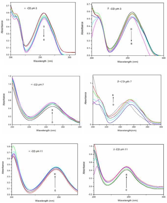
Figure 1. Absorbance spectra of 4MBA in different α-CD and β-CD concentrations (M): (1) 0, (2) 0.002, (3) 0.004, (4) 0.006, (5) 0.008 and (6) 0.01.
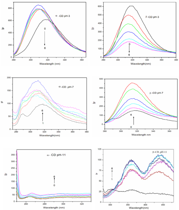
Figure 2. Fluorescence spectra of 4MBA in different α-CD and β-CD concentrations (M): (1) 0, (2) 0.002, (3) 0.004, (4) 0.006, (5) 0.008 and (6) 0.01.
At pH ≈ 3 and pH ≈ 7, the absorption maxima remain nearly unchanged at higher β-CD concentrations. The fluorescence intensities follow the order pH ≈ 3 > pH ≈ 7 > pH ≈ 11, and the progressive decrease with increasing pH arises because the carboxylate anion exhibits weaker fluorescence than the neutral species. In both CD solutions, smooth and featureless emission spectra are observed at pH ≈ 3, whereas structured emission bands appear at pH ≈ 7 and pH ≈ 11, with the most pronounced spectral structure evident in alkaline medium.
The variations in absorbance, emission intensity, and spectral maxima are attributed to the encapsulation of 4MBA within the α-CD and β-CD cavities. No significant changes are observed in the absorption spectra even after 12 h, indicating stable inclusion. The influence of α-CD and β-CD on the excited state of 4MBA is more significant than their effect on the ground state. Fluorescence enhancement is stronger at pH ≈ 3 and pH ≈ 7 than at pH ≈ 11. In α-CD, fluorescence intensity increases at both pH ≈ 3 and pH ≈ 7, whereas in β-CD it decreases relative to the CD-free solution. The distinct spectral shifts in absorption and fluorescence across different pH values clearly indicate the formation of different types of inclusion complexes.
At all three pH values, the absence of an isosbestic point and the presence of red or blue shifts in the absorption spectra confirm the formation of multiple inclusion complexes between 4MBA and CDs . Moreover, at pH ≈ 3, the absorption and emission profiles differ markedly from those at pH ≈ 7 and pH ≈ 11, implying the existence of at least two distinct inclusion species. The binding constants for the 4MBA-CD complexes were obtained from Benesi-Hildebrand plots. The negative ΔG values indicate that complex formation is spontaneous and exothermic at 303 K (Table 1).
The emission of 4MBA is strongly pH-dependent, with the Stokes shift reaching its maximum at pH ≈ 3. Such broad Stokes-shifted emission in acidic media is typical for aromatic molecules bearing electron-withdrawing substituents such as COOH . These observations may arise from several processes, including ground-state dimer formation, excimer formation, or excited-state charge-transfer interactions. The appearance of a broad band at pH ≈ 3 suggests the predominance of the monomeric form at this pH. Although the long-wavelength emission (≈ 400 nm) is weak at pH ≈ 3, dual fluorescence is observed in both CD solutions. In β-CD, the emission intensity decreases at 315 nm but increases near 400 nm. In α-CD, the well-defined emission structure at pH ≈ 7 and pH ≈ 11 indicates possible excimer formation in the excited state, likely facilitated by hydrogen bonding. Ground-state dimerization is unlikely due to ionization of the carboxyl group. The gradual increase in emission intensity around 400 nm with rising CD concentration further supports excimer formation at pH ~ 3.
In the excited state, the emission intensity of 4MBA is very weak in aqueous CD-free solutions at pH ≈ 7 and pH ≈ 11. However, upon increasing the concentration of CDs, the fluorescence intensity increases markedly and distinct vibronic bands become evident. In this molecule, the methoxy group serves as the electron donor, while the -COOH group acts as the electron acceptor. Under acidic conditions and in the presence of CDs, the carboxyl group becomes more effectively conjugated with the aromatic π-system, leading to enhanced charge separation within the molecule. The large Stokes shift observed in water indicates the involvement of dipolar resonance structures (Scheme 1).
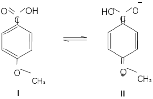
Scheme 1. Neutral and ionic form for 4MBA.
CDs preferentially encapsulate the neutral form of benzoic acid derivatives over their anionic counterparts, due to the more favorable fit of the neutral species within the hydrophobic cavity . The higher binding constant at pH ≈ 3 supports this view, suggesting that the COOH group is more effectively included inside the CD cavity than the deprotonated carboxylate form.
As noted earlier, increasing the concentration of α-CD or β-CD at pH ≈ 7 and pH ≈ 11 results in a significant enhancement of fluorescence intensity, whereas at pH ≈ 3 the emission intensity decreases. In β-CD, the red shift observed in the ground state at pH ≈ 7 suggests partial deprotonation of the carboxylate anion. These findings indicate that, in aqueous solution, structure II is predominant, while in CD solutions structure I become more favored. This shift in electronic structure accounts for the differing emission behavior observed at pH ≈ 3, pH ≈ 7, and pH ≈ 11.
The distinct spectral responses of 4MBA upon CD addition at different pH values also imply that the geometry and orientation of the guest molecule inside the CD cavity vary with pH. As is well established, hydrophobic interactions drive the encapsulation process, with the hydrophobic moiety such as the methoxy group preferentially entering the deeper nonpolar region of the cavity, while the COOH/COO⁻ group resides near the rim of the CD cavity .
3.2. Excimer Emission
The increase in long-wavelength (LW) emission intensity observed in CD solutions can be explained as follows. The appearance of a broad emission band in aqueous CD media suggests that 4MBA may form a 1: 2 inclusion complex with CDs, in which two guest molecules occupy a single CD cavity . As the CD concentration increases, the intensity of the LW band shows a slight enhancement accompanied by noticeable band broadening, while the position of the short-wavelength (SW) fluorescence maximum remains essentially unchanged. This broad LW emission is attributed to excimer fluorescence. The excimer may form either through a 1: 2 CD: (4MBA)₂ complex or through a 2: 2 (CD: 4MBA)₂ assembly generated via self-association of the 1: 1 CD-4MBA complex . The mechanism of excimer formation has been elaborated in our earlier publications .
To substantiate the excimer fluorescence in the presence of α-CD and β-CD, solvent-induced changes in the absorption and fluorescence spectra of 4MBA were examined. The spectral maxima in various solvents are listed in Table 2. The results show that the absorption and emission maxima of 4MBA in different solvents closely resemble those reported for 4-hydroxybenzoic acid (4HBA) (cyclohexane: λabs~251 nm, λflu~318 nm; acetonitrile: λabs ~258, 218 nm, λflu~328 nm; methanol: λabs ~255 nm, λflu ~338 nm; water: λabs~283, 254 nm, λflu~338 nm). In all solvents, 4MBA exhibits a single broad fluorescence band. Compared to cyclohexane, the fluorescence maxima in acetonitrile and methanol show slight red shifts. The absence of any LW emission band indicates that neither ICT nor exciplex formation occurs in these solvents.
Relative to 4-hydroxy-3-methoxy benzoic acid (HMBA) (cyclohexane: λabs ~287, 260, 216 nm, λflu~320 nm; acetonitrile: λabs~290, 258, 218 nm, λflu~330 nm; methanol: λabs ~294, 264, 220 nm, λflu ~ 340 nm; water: λabs ~283, 254, 216 nm, λflu ~340 nm) , the absorption maxima of 4MBA are blue-shifted in all solvents. This blue shift arises because HMBA undergoes intramolecular hydrogen bonding, which is not possible in 4MBA or 4HBA. Furthermore, when compared with phenol , the absorption maxima of 4MBA and 4HBA are blue-shifted in all solvents (water: λabs ≈ 272-278 nm, λflu ≈ 330 nm), indicating that conjugation between the carboxyl group and the hydroxyl/methoxy substituent is minimal.
3.3. Excited Singlet State Lifetimes
To investigate the CD-induced changes in the fluorescence behavior of 4MBA, time-resolved emission decays were recorded in water and in 0.01 M CD solutions (α-CD and β-CD) (Table 1). The fluorescence lifetime of 4MBA is strongly influenced by the presence of CDs, with the lifetimes of the inclusion complexes being markedly longer than that of the free molecule. The τ values depend on the type of CD and on the characteristics of short-lived emissive species. The lifetimes follow the order: water < α-CD < β-CD, indicating that the β-CD: 4MBA complex is more stable than the α-CD: 4MBA complex. The progressive increase in τ with increasing CD concentration reflects the encapsulation of 4MBA within the CD cavity. Longer lifetimes correspond to deeper, more rigid inclusion of the guest, whereas shorter lifetimes indicate a more weakly associated complex. The excimer decay time also increases significantly from water to CD solutions.
In CD media, the decay profile monitored at the long-wavelength (LW) emission (~400 nm) differs distinctly from that observed at the short-wavelength (SW) emission (~319 nm). This behavior can be attributed to excimer formation within the CD cavity, consistent with the LW emission band and with the steady-state fluorescence results discussed earlier. The monomer emission lifetime is very short in water and remains sensitive to CD addition. Notably, the rise time of the excimer emission which is distinct from the fast decay component of the monomer emission—increases with CD concentration, while no rise time is detected in water. These observations demonstrate that the excimer formed in the CD: 4MBA inclusion complex differs fundamentally from any excimer-like species in solution without CDs. Overall, the results confirm that excimer formation is significantly more favorable in CD media than in ordinary solvents.
3.4. Molecular Modeling
To gain deeper insight into the mode of guest encapsulation within the CD cavity, molecular docking simulations were performed for 4MBA with α-CD and β-CD using molecular modeling techniques, with the objective of constructing reliable structures of the inclusion complexes. The geometries of 4MBA and the CD ring structures were optimized using the PM3 method (Figure 3), and the corresponding energy parameters are listed in Table 2. The HOMO, LUMO, dipole moment, total energy, free energy, enthalpy, entropy, and zero-point vibrational energy values of 4MBA, α-CD, and β-CD differ markedly from those of their inclusion complexes. Mulliken charge analysis shows nearly zero charge transfer between the host and guest, confirming the absence of significant charge-transfer interactions. The thermodynamic parameters (ΔE, ΔG, and ΔH) indicate that the formation of the 4MBA-CD complexes is predominantly enthalpy-driven and energetically favorable.
The interior cavity diameters of α-CD and β-CD are approximately 4.7-5.3 Å and 6.0-6.5 Å, respectively, while their exterior diameters are 8.8 Å (α-CD) and 10.8 Å (β-CD); both CDs have an identical height of about 7.8 Å. The vertical and horizontal distances between the COOH and OCH₃ groups in 4MBA are 8.33 Å and 4.33 Å, respectively (Figure 3). The vertical dimension of 4MBA exceeds the interior cavity size of both α-CD and β-CD, whereas the horizontal dimension is smaller. Molecular docking of the inclusion complexes shows that part of the OCH₃ group protrudes outside the hydrophobic CD cavity, while the COOH group forms hydrogen bonds with the CD-OH groups. These non-bonded interactions between the carboxyl group of 4MBA and CD hydroxyl groups likely contribute to differences in structural stability among the complexes. To further validate the host-guest interactions, solid-state inclusion complexes were prepared and characterized using SEM, DSC, FT-IR, and ¹H NMR techniques. The results confirm successful encapsulation of 4MBA within the CD cavity.
Table 2. Thermodynamic parameters and HOMO-LUMO energy calculations for 4MBA and its inclusion complexes by PM3 method.

Properties

4MBA

α-CD

β-CD

4MBA: α-CD

4MBA: β-CD

EHOMO (eV)

-9.63

-10.37

-10.35

-9.12

-9.20

ELUMO (eV)

-0.74

1.26

1.23

0.54

0.58

EHOMO -ELUMO (eV)

-8.89

-11.63

-11.58

-9.66

-9.78

Dipole moment (D)

6.21

11.34

12.29

10.81

10.93

E*

-100.53

-1247.62

-1457.63

-1356.43

-1519.27

E*

-8.28

-171.12

G*

60.52

-676.37

-789.52

-649.18

-727.34

ΔG*

-33.6

-111.45

H*

91.40

-570.84

-667.55

-612.23

-685.94

ΔH

-132.79

-109.75

S**

0.103

0.353

0.409

0.462

0.495

ΔS**

0.006

-0.017

ZPE*

64.28

635.09

740.56

715.75

822.36

Mullikan charge

0.00

0.00

0.00

0.00

0.00

*kcal/mol; **kcal/mol-Kelvin; ZPE = Zero point vibration energy
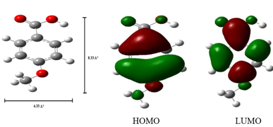
Figure 3. PM3 optimized structures of 4MBA, HOMO, LUMO of 4MBA.
3.5. Inclusion Complex Nanomaterials Studies
3.5.1. Scanning Electron Microscopy (SEM)
The powdered samples of Ag nanoparticles, 4MBA, and the nanomaterials Ag: 4MBA: α-CD and Ag: 4MBA: β-CD were examined by SEM (Figure 4). Ag nanoparticles exhibited irregularly shaped particles, while 4MBA appeared in crystalline or marble-like structures. The Ag: 4MBA: α-CD sample also showed irregular particle morphology, whereas Ag: 4MBA: β-CD displayed nanorod-like structures. SEM-EDX analysis confirmed the presence of 42.3% carbon, 48.4% oxygen, and 9.3% silver in the nanomaterials. The distinct morphological features observed for Ag nanoparticles, pure 4MBA, and the inclusion-complex nanomaterials support the successful formation of Ag: 4MBA: CD assemblies.
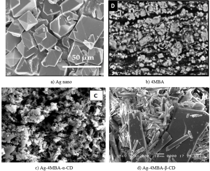
Figure 4. SEM images for a) Ag nano, b) 4MBA, c) Ag: 4MBA: α-CD and d) Ag: MBA: β-CD.
3.5.2. Differential Scanning Colorimeter
DSC thermograms of 4MBA, α-CD, β-CD, and their corresponding nanomaterials were analyzed. β-CD exhibited a broad endothermic peak at 128.6°C, while α-CD showed three endothermic transitions at 79.2°C, 109.1°C, and 137.5°C, all attributed to the release of crystal water . The DSC curve of 4MBA presented a sharp endothermic peak at 184°C, corresponding to its melting point. The broader endothermic features in α-CD, β-CD, and the nanomaterials are associated with water loss from the CDs. Notably, the nanomaterials displayed new endothermic peaks at 226°C (Ag: 4MBA: α-CD) and 249°C (Ag: 4MBA: β-CD), confirming the formation of new solid-state structures.
3.5.3. Infrared Spectral Studies
FTIR spectra of the nanomaterials were compared with those of pure 4MBA, α-CD, and β-CD. In 4MBA, the -OH stretching band appears at 3029-2984 cm⁻¹, while the COOH bending vibration occurs at 1429 cm⁻¹. Aromatic C-H and C=C stretching bands are observed at 2956 cm⁻¹ and 1518 cm-1, respectively. The O-CH₃ stretching band appears at 2845-2729 cm-1, and the C=O and COO⁻ bands occur at 1688 cm-1 and 1608 cm-1, respectively. The Ar-OCH₃ stretching band appears at 1267 cm-1, with out-of-plane ring deformation at 617-598 cm-1.
In the Ag: 4MBA: CD nanomaterials, many of these characteristic peaks either diminish in intensity or disappear. The C=O band shifts to 1605 cm-1, the aromatic ring stretching band appears at 1346 cm-1, and the Ar-OCH₃ deformation band shifts to 561 cm-1. These spectral changes indicate that 4MBA and CD surround and stabilize the Ag nanoparticles, confirming the formation of Ag: 4MBA: CD nanomaterials.
3.5.4. X RD Spectral Studies
The crystallinity of the nanoparticles was examined using their XRD patterns . Pure Ag nanoparticles exhibited distinct diffraction peaks at 38.11°, 44.30°, 64.45°, and 77.40°, characteristic of the face-centered cubic lattice of metallic silver. The XRD profile of α-CD showed crystalline reflections at approximately 11.94°, 14.11°, and 21.77°, while β-CD displayed peaks near 11.49° and 17.58°. The intensity and appearance of these peaks may vary depending on the sample preparation conditions. Several crystal structures have been reported for this compound, most of which feature O-H···O hydrogen-bonded inversion dimers. For 4MBA, characteristic peaks were observed at 13.13°, 17.26°, 19.23°, 22.74°, 25.61°, 28.82°, and 33.21°. The XRD pattern of the Ag/4MBA: β-CD nanomaterials displayed distinct reflections at 11.13°, 13.43°, 18.26°, 29.25°, 36.76°, 44.63°, 55.86°, 66.04°, and 72.75°. Variations in peak intensity and the presence of new peaks relative to the pure components confirm the formation of new nanomaterials.
3.5.5. Proton Magnetic Resonance Spectral Studies
Typically, appreciable chemical-shift changes in the guest proton signals are observed when guest molecules are encapsulated within CD cavities. The proton assignments of CDs are well established and consist of six types of protons, with H-3 and H-5 located inside the cavity. Inclusion-induced interactions with the CD interior generally influence the chemical shifts of these H-3 and H-5 protons, whereas only minor changes occur for the exterior protons (H-1, H-2, and H-4). 1H NMR spectra of isolated 4MBA and its inclusion complexes were recorded at 25°C in DMSO-d6, and the data are summarized in Table 4. Notable differences in the chemical-shift values of the nanomaterials compared with free 4MBA were observed. The aromatic protons of 4MBA exhibited upfield shifts upon complexation, indicating interactions between these aromatic protons and the CD cavity environment. The higher chemical-shift value observed for the hydroxy proton suggests hydrogen bonding between this proton and the hydroxyl groups of the CD. Additionally, the aromatic ring protons displayed a larger upfield shift than the methyl protons, implying that the aromatic ring is strongly shielded within the nanomaterial matrix and penetrates deeply into the nano-Ag-CD cavity.
Table 3. 1H-NMR chemical shift values for the 4MBA and Ag: 4MBA: CD nanomaterials.

Protons

4MBAP (δ)

Ag: 4MBA: α-CD

Ag: 4MBA: β-CD

Ha - COOH

12.70

5.71

5.74

Hb -Ortho to COOH

7.93

4.80

4.83

Hc -Meta to COOH

7.04

4.48

4.51

Hd - OCH3

3.84

2.49

2.51

4. Conclusion
The effects of α-CD and β-CD on 4-methoxybenzoic acid (4MBA) were examined at pH ~3, pH ~7, and pH ~11. Ag: 4MBA: CD nanomaterials were characterized using UV-visible and fluorescence spectroscopy, SEM-EDX, DSC, FTIR, XRD, and ^1H NMR techniques. In pH ~7 buffer, 4MBA predominantly exists as the carboxylate anion. In the ground state, no significant spectral changes were observed in α-CD or β-CD at pH ~3; however, at pH ~7, a red shift in the absorption maximum occurred in β-CD. In the excited state, blue shifts in the emission maxima were observed in both CDs at pH ~3 and pH ~7, while at pH ~11, structured multi-band emission appeared in both systems. The differing spectral responses of 4MBA upon addition of CDs at the three pH values indicate that the geometries of the inclusion complexes vary with pH. The structured emission observed at pH ~7 and pH ~11 further suggests excimer formation in the excited state. The lifetime values increased in the order: free 4MBA < α-CD < β-CD, demonstrating that the β-CD: 4MBA inclusion complex is more stable than the corresponding α-CD complex. SEM-EDX analysis confirmed the presence of silver in the nanomaterials. Results from DSC, FTIR, XRD, and NMR collectively indicate that 4MBA interacts effectively with the Ag nanoparticles.
Abbreviations

FTIR

Fourier Transform Infrared Spectroscopy

DTA

Differential Thermal Analysis

XRD

X-ray Diffraction

SEM

Scanning Electron Microscopy

HOMO

Highest Occupied Molecular Orbital

LUMO

Lowest Unoccupied Molecular Orbital

4MBA

4-methoxybenzoic Acid

Ag NPs

Silver Nanoparticles

α-CD

Alpha Cyclodextrin

β-CD

Beta Cyclodextrin

PM3

Parametric Method 3

ΔE

Iinternal Energy Change

ΔH

Enthalpy Change

ΔG

Free Energy Change

ΔS

Entropy Change

Author Contributions
Narayanasamy Rajendiran: Supervision, Resources, Methodology, Software, Writing – original draft, Writing – review & editing
Ayyadurai Mani: Formal Analysis, Investigation
Palanichamy Ramasamy: Data curation
Sengamalai Senthilmurugan: Validation
Conflicts of Interest
The authors declare no conflict of interest.
References
[1] E. Ishow, R. Guillot, G. Buntinx, O. Poizat, Photoinduced intramolecular charge-transfer dynamics of push-pull compounds: solvatochromic and time-resolved studies, J. Photochem. Photobiol. A: Chem. 234 (2012) 27-34.
[2] Y. M. Issa, H. B. Hassib, H. E. Abdelaal, I. M. Kenawi, Spectral investigation of the intramolecular charge-transfer in some aminotriazole Schiff bases, Spectrochim. Acta A 79 (2011) 1364-1371.
[3] Y. Zhu, S. Guang, X. Su, H. Xu, D. Xu, Dyes Pigm. 97 (2013) 175-181.
[4] B. K. Paul, A. Samanta, S. Kar, N. Guchhait, Evidence for excited state intramolecular charge transfer reaction in donor-acceptor molecule 5-(4-dimethylamino-phenyl)-penta-2, 4-dienoic acid methyl ester: experimental and quantum chemical approach, J. Lumin. 130 (2010) 1258-1265.
[5] M. Smoluch, H. Joshi, A. Gerssen, C. Gooijer, G. van der Zwan, J. Phys. Chem. A 109 (2005) 535-542.
[6] K. Kalyanasundaram, Photochemistry in Microheterogeneous Systems, Academic Press, New York, 1987.
[7] P. Brocos, X. Banquy, N. Díaz-Vergara, S. Pérez-Casas, M. Costas, Á. Piñeiro, Similarities and differences between cyclodextrin-sodium dodecyl sulfate host-guest complexes of different stoichiometries: Molecular dynamics simulations at several temperatures. J. Phys. Chem. B 114 (2010) 12455-12467.
[8] H. M. Ameen, S. Kunsági-Máté, L. Szente, B. Lemli, Encapsulation of sulfamethazine by native and randomly methylated β-cyclodextrins: The role of the dipole properties of guests. Spectrochim. Acta A 225 (2020) 117475.
[9] M. Jamrógiewicz, K. Milewska, Sacharides and their derivatives as pharmaceutical additives Spectrochim. Acta A 219 (2019) 346.
[10] M. A. Chouker, H. Abdallah, A. Zeiz, M. H. El-Dakdouki, Host-guest inclusion complex of quinoxaline-1, 4-dioxide derivative with 2-hydroxypropyl-β-cyclodextrin: Preparation, characterization, and antibacterial activity. J. Mol. Struct. (2021) 130273.
[11] M. Levine, B. R. Smith, Tuning fluorescence energy transfer for carcinogen detection and medical diagnostics. J. Fluoresc. 30 (2020) 1015.
[12] I. Lafifi, L. Nouar, F. Madi, A. Guendouzi, M. Cheriet, N. Boulaha, B. Houari, Computational study of inclusion complex of L-Glutamine/β-Cyclodextrin: Electronic and intermolecular interactions investigations. J. Mol. Struct. 1206 (2020) 127740.
[13] M. Akhondi, E. Jamalizadeh, A. Mohebbi, MD and DFT calculations on the structural variations of amino-cyclodextrin as a pH-sensitive carrier for smart carriage and release of Doxorubicin. J. Mol. Struct. 1230 (2021) 129855.
[14] A. Obaid, A. Khairani, M. Jamil, S. Prabu, S. M. Saharin, S. Mohamad, Spectroscopic studies for the inclusion complexation of ketoprofen enantiomers with β-cyclodextrin. Spectrochim. Acta A 225 (2020) 118674.
[15] A. Antony Muthu Prabhu, V. K. Subramanian, N. Rajendiran, Excimer formation in inclusion complexes of β-CD with salbutamol, sotalol and atenolol: Spectral and molecular modeling studies. Spectrochimica Acta, 96A (2012) 95-107.
[16] M. Jude Jenita, G. Venkatesh, V. K. Subramanian, N. Rajendiran, Excimer formation in inclusion complexes of antihypertensive drugs with HP-α- and HP-β-CDs. Indian J. Chemistry, 52A (2013) 207-216.
[17] S. Siva, R. K. Sankaranarayanan, A. Antony Muthu Prabhu, N. Rajendiran, Inclusion complexation of 3, 5-dihydroxy benzoic acid with β-CD at different pH. Indian J. Chemistry, 48A (2009)1515-1521.
[18] R. K. Sankaranarayanan S. Siva, A. Antony Muthu Prabhu, N. Rajendiran, A Study on the inclusion complexation of 3, 4, 5-trihydroxybenzoic acid with β-CD at different pH. J. Inclusion Phenomena and Macrocyclic Chemistry, 67(2010)461-470.
[19] T. Stalin, P. Vasantharani, B. Shanthi, A. Sekar, N. Rajendiran, Inclusion complex of 1, 2, 3-trihydroxybenzene with α- and β-cyclodextrins. Indian J Chemistry, 45A (2006) 1113-1120.
[20] M. Jude Jenita, A. Antony Muthu Prabhu, N. Rajendiran, Theoretical study of inclusion complexation of tricyclic antidepressant drugs with β-CD. Indian J. Chemistry A, 51A (2012) 1686-1694.
[21] N. Rajendiran, G. Venkatesh, T. Mohandoss, Fabrication of 2D nano sheet through self assembly behavior of sulfamethoxy pyridazine inclusion complex with α- and β-cyclodextrins. Spectrochim Acta A, 123A (2014) 158-166,
[22] A. Antony Muthu Prabhu, N. Rajendiran, Encapsulation of labetalol, and pseudoephedrine in β-CD cavity: Spectral and molecular modeling studies. J. Fluorescence, 22(2012)1461-1474.
[23] J. Prema Kumari, A. Antony Muthu Prabhu, G. Venkatesh, V. K. Subramanian, N. Rajendiran, Effect of solvents and pH on β-CD Inclusion complexation of 2, 4-dihydroxy azobenzene and 4-hydroxy azobenzene. J. Solution Chemistry, 40 (2011) 327-347.
[24] J. Prema Kumari, A. Antony Muthu Prabhu, G. Venkatesh, V. K. Subramanian, N. Rajendiran, Spectral characteristics of sulfadiazine, sulfisomidine: Effect of solvents, pH and β-CD. Physics and Chemistry of Liquids, 49(2011)108-132.
[25] N. Rajendiran, S. Siva, J. Saravanan, Inclusion complexation of sulfa pyridine with α- and β-CDs: Spectral and molecular modeling study. J. Molecular Structure, 1054-1055 (2013) 215-222.
[26] A. Mani, P. Ramasamy, A. Antony Muthu Prabhu, N. Rajendiran, Investigation of Ag and Ag/Co bimetallic nanoparticles with naproxen-cyclodextrin inclusion complex. J. Molecular Structure, 1284 (2023) 135301-10.
[27] A. Mani, G. Venkatesh, P. Senthilraja, N. Rajendiran, Synthesis and Characterisation of Ag-Co-Venlafaxine-Cyclodextrin Nanorods, European J Advanced Chemistry Research, 5 (2024) 9-16.
[28] A. Mani, P. Ramasamy, A. Antony Muthu Prabhu, P. Senthilraja, N. Rajendiran, Synthesis and Analysis of Ag/Olanzapine /Cyclodextrin and Ag/Co/Olanzapine /Cyclodextrin Inclusion Complex Nanorods. Physics and Chemistry of Liquids, 62 (2024) 196-209.
[29] A. Mani, P. Ramasamy, A. Antony Muthu Prabhu, P. Senthilraja, N. Rajendiran, Synthesis and Characterisation of Ag/Co/Chloroquine/Cyclodextrin Inclusion Complex Nanomaterials. J Sol-Gel Science and Technology 115 (2025) 844-856.
[30] N. Rajendiran, A. Mani, M. Venkatesan, B. Sneha, E. Nivetha, P. Senthilraja, Spectral, Microscopic, Antibacterial and Anticancer Activity of Pyrimethamine drug with Ag nano, DNA, RNA, BSA, Dendrimer, and Cyclodextrins, J Solution Chem,
[31] S. Hamai, A. Hatamiya, Excimer formation in inclusion complexes of β-cyclodextrin with 1-alkylnaphthalenes in aqueous solutions. Bull. Chem. Soc. Jpn. 69 (1996) 2469-76.
[32] S. Hamai, The excimer fluorescence of 2-methylnaphthalene in β- and γ-cyclodextrin aqueous solution. Bull. Chem. Soc. Jpn. 69 (1996) 543-549.
[33] S. Hamai, Cyclodextrin inclusion compounds. 1. Effects of β- and γ-cyclodextrins on the fluorescence of pyrene in aqueous solution. J. Phys. Chem. 93 (1989) 6527-29.
[34] T. Stalin, N. Rajendiran, A study on the spectroscopy and photophysics of 4-hydroxy-3-methoxybenzoic acid in different solvents, pH and β-cyclodextrin. J. Molecular Structure, 794 (2006) 35-45,
[35] T. Stalin, G. Sivakumar, B. Shanthi, A. Sekar, N. Rajendiran, Photophysical behaviour of 4-hydroxy-3, 5-dimethoxybenzoic acid in different solvents, pH and β-cyclodextrin. J. Photochem. Photobiol. A: Chemistry, 177 (2006) 144-155.
[36] P Ramasamy, A Mani, B Sneha, E Nivetha, M Venkatesan, N Rajendiran, Azo-hydrazo tautomerism in Sudan Red-B and Cyclodextrin/ Sudan Red-B doped ZnO nanomaterials. J Molecular Structure 1329 (2025) 141423-32.
[37] P. Ramasamy, A. Mani, B. Sneha, E. Nivetha, A. Antony Muthu Prabhu, G. Venkatesh, N. Rajendiran,* Synthesis and Characterisation of Sudan Red-G/Cyclodextrin doped ZnO Nanocrystals. American J Physical Chemistry 14 (2025) 23-32,
[38] P. Ramasamy, A. Mani, B. Sneha, E. Nivetha, A. Antony Muthu Prabhu, G. Venkatesh, P. Senthilraja, N. Rajendiran*, Synthesis and Characterisation of Cyclodextrin /Methyl Violet doped ZnO Nanocrystals. Colloid and Surface Science 9 (2025) 19-30,
[39] P. Ramasamy, A. Mani, B. Sneha, E. Nivetha, A. Antony Muthu Prabhu, G. Venkatesh, P. Senthilraja, N. Rajendiran*, Synthesis and Characterisation of Cyclodextrin/ Sudan Black-B Caped ZnO/ Nanocrystals. American J Quantum Chemistry and Molecular Spectroscopy 9 (2025) 1-11,
[40] P. Ramasamy, A. Mani, A. Antony Muthu Prabhu, G. Venkatesh, N. Rajendiran* Azo-Imino Tautomerism in Sudan Red 7B/Cyclodextrin Coated ZnO Nanocomposites: Evidence by Spectral and Microscopic Perspectives. Science Journal of Chemistry 13 (2025) 65 - 75,
[41] P. Ramasamy, A. Mani, A. Antony Muthu Prabhu, G. Venkatesh, P. Senthilraja, N. Rajendiran* PICT Effects and Anticancer Potential on Rosaniline and Spectral Characterisation of Rosaniline/Cyclodextrin Covered ZnO/ Nanocrystals. International J. Pure and Applied Chemistry 26 (2025) 107-121,
[42] P. Ramasamy, A. Mani, P. Senthilraja, N. Rajendiran Keto-Enol Tautomerism and Anticancer Potential on Sudan Blue II and Synthesis and Characterisation of Sudan Blue II/ Cyclodextrin doped ZnO Nanocrystals, J. Materials Science and Nanotechnology, 13 (2025) 1- 16,
[43] P. Ramasamy, A. Mani, P. Senthilraja, N. Rajendiran, Spectral, Microscopic and Anticancer Activity Investigation on Dimethyl Yellow/Cyclodextrin Doped ZnO Nanocomposites Journal of Chemical and Pharmaceutical Sciences (JCHPS) 18(3) (2025) 33-43.
[44] P. Ramasamy, A. Mani, P. Senthilraja, N. Rajendiran, Spectral Characteristics of ZnO/Mordent Yellow 12/ Cyclodextrin Nanomaterials, J Chemical Health Risks, (JCHR) 15(2025) 542-553
[45] P. Ramasamy, A. Mani, P. Senthilraja, S. Senthilmurugan, N. Rajendiran, Spectral, Microscopic and Anticancer Activity of 1, 8-Diaminonaphthalene Doped ZnO Nanocrystals, VVIJOURNAL 14 (2026) 135-147,
Cite This Article
  • APA Style

    Rajendiran, N., Mani, A., Ramasamy, P., Senthilmurugan, S. (2026). Inclusion Complexation of 4-methoxybenzoic Acid: Cyclodextrin at Different pH and Synthesis of Silver: 4-methoxybenzoic Acid: Cyclodextrin Nanomaterials. Advances in Materials, 15(2), 50-61. https://doi.org/10.11648/j.am.20261502.13

    Copy | Download

    ACS Style

    Rajendiran, N.; Mani, A.; Ramasamy, P.; Senthilmurugan, S. Inclusion Complexation of 4-methoxybenzoic Acid: Cyclodextrin at Different pH and Synthesis of Silver: 4-methoxybenzoic Acid: Cyclodextrin Nanomaterials. Adv. Mater. 2026, 15(2), 50-61. doi: 10.11648/j.am.20261502.13

    Copy | Download

    AMA Style

    Rajendiran N, Mani A, Ramasamy P, Senthilmurugan S. Inclusion Complexation of 4-methoxybenzoic Acid: Cyclodextrin at Different pH and Synthesis of Silver: 4-methoxybenzoic Acid: Cyclodextrin Nanomaterials. Adv Mater. 2026;15(2):50-61. doi: 10.11648/j.am.20261502.13

    Copy | Download

  • @article{10.11648/j.am.20261502.13,
      author = {Narayanasamy Rajendiran and Ayyadurai Mani and Palanichamy Ramasamy and Sengamalai Senthilmurugan},
      title = {Inclusion Complexation of 4-methoxybenzoic Acid: Cyclodextrin at Different pH and Synthesis of Silver: 
    4-methoxybenzoic Acid: Cyclodextrin Nanomaterials},
      journal = {Advances in Materials},
      volume = {15},
      number = {2},
      pages = {50-61},
      doi = {10.11648/j.am.20261502.13},
      url = {https://doi.org/10.11648/j.am.20261502.13},
      eprint = {https://article.sciencepublishinggroup.com/pdf/10.11648.j.am.20261502.13},
      abstract = {The inclusion behavior of 4-methoxybenzoic acid (4MBA) with α-cyclodextrin and β-cyclodextrin in buffer solutions of pH ~3, ~7, and ~11 was examined using UV-visible, steady-state and time-resolved fluorescence spectroscopy, along with PM3 computational analysis. Ag: 4MBA: CD nanomaterials were synthesized and characterized by SEM, DSC, FTIR, XRD, and ¹H NMR techniques. Because 4MBA predominantly exists as a carboxylate anion in pH ~7 medium, the spectra of its neutral and monoanion forms were also recorded at pH ~3 and pH ~11, respectively. In both cyclodextrin solutions, smooth emission profiles were obtained at pH ~3, while structured emission bands appeared at pH ~7 and pH ~11, with greater band structure observed in alkaline medium. The CD-induced spectral changes at different pH values indicate that the geometries of the resulting inclusion complexes vary across media. Since the carboxylate group is ionized, ground-state dimer is not formed, however, excimer formed in the excited state. Lifetime measurements reveal that the β-CD: 4MBA complex is more stable than the α-CD counterpart. The calculated HOMO-LUMO energy gap, total energy, free energy, enthalpy, entropy, dipole moment, and zero-point vibrational energy of the CD: 2AP complex differed significantly from those of the isolated 4MBA, α-CD and β-CD molecules, and both the vertical and horizontal bond lengths between the amino and hydroxy groups are smaller than the β-CD cavity size confirming the formation of an inclusion complex. SEM-EDX analysis confirms the presence of silver in the composite. FTIR, XRD, and NMR results collectively suggest strong interactions between 4MBA and the silver nanoparticles.},
     year = {2026}
    }
    

    Copy | Download

  • TY  - JOUR
    T1  - Inclusion Complexation of 4-methoxybenzoic Acid: Cyclodextrin at Different pH and Synthesis of Silver: 
    4-methoxybenzoic Acid: Cyclodextrin Nanomaterials
    AU  - Narayanasamy Rajendiran
    AU  - Ayyadurai Mani
    AU  - Palanichamy Ramasamy
    AU  - Sengamalai Senthilmurugan
    Y1  - 2026/04/10
    PY  - 2026
    N1  - https://doi.org/10.11648/j.am.20261502.13
    DO  - 10.11648/j.am.20261502.13
    T2  - Advances in Materials
    JF  - Advances in Materials
    JO  - Advances in Materials
    SP  - 50
    EP  - 61
    PB  - Science Publishing Group
    SN  - 2327-252X
    UR  - https://doi.org/10.11648/j.am.20261502.13
    AB  - The inclusion behavior of 4-methoxybenzoic acid (4MBA) with α-cyclodextrin and β-cyclodextrin in buffer solutions of pH ~3, ~7, and ~11 was examined using UV-visible, steady-state and time-resolved fluorescence spectroscopy, along with PM3 computational analysis. Ag: 4MBA: CD nanomaterials were synthesized and characterized by SEM, DSC, FTIR, XRD, and ¹H NMR techniques. Because 4MBA predominantly exists as a carboxylate anion in pH ~7 medium, the spectra of its neutral and monoanion forms were also recorded at pH ~3 and pH ~11, respectively. In both cyclodextrin solutions, smooth emission profiles were obtained at pH ~3, while structured emission bands appeared at pH ~7 and pH ~11, with greater band structure observed in alkaline medium. The CD-induced spectral changes at different pH values indicate that the geometries of the resulting inclusion complexes vary across media. Since the carboxylate group is ionized, ground-state dimer is not formed, however, excimer formed in the excited state. Lifetime measurements reveal that the β-CD: 4MBA complex is more stable than the α-CD counterpart. The calculated HOMO-LUMO energy gap, total energy, free energy, enthalpy, entropy, dipole moment, and zero-point vibrational energy of the CD: 2AP complex differed significantly from those of the isolated 4MBA, α-CD and β-CD molecules, and both the vertical and horizontal bond lengths between the amino and hydroxy groups are smaller than the β-CD cavity size confirming the formation of an inclusion complex. SEM-EDX analysis confirms the presence of silver in the composite. FTIR, XRD, and NMR results collectively suggest strong interactions between 4MBA and the silver nanoparticles.
    VL  - 15
    IS  - 2
    ER  - 

    Copy | Download

Author Information
  • Abstract
  • Keywords
  • Document Sections

    1. 1. Introduction
    2. 2. Materials and Methods
    3. 3. Result and Discussion
    4. 4. Conclusion
    Show Full Outline
  • Abbreviations
  • Author Contributions
  • Conflicts of Interest
  • References
  • Cite This Article
  • Author Information