Research Article | | Peer-Reviewed

Mineral Content, Antioxidant Properties in vitro, Reduction of Inflammation, and Liver Steatosis in vivo by Ngaoundal Propolis in Wistar Rats Fed an Atherogenic Diet

Received: 14 April 2024    Accepted: 7 May 2024    Published: 30 May 2024
Views:       Downloads:
Abstract

Several studies have reported the benefits of Propolis in the treatment of various disorders such as parasitic infections, bacterial infections, wounds, and burns. The overall aim of this study was to evaluate the preventive effects and anti-inflammatory activities of the hydroethanolic extract (EthP) and the fraction powder ≤ 125 µm of Propolis (PP) on atherogenic diet-induced non-alcoholic fatty liver disease. Dry Propolis was finely ground, a first part was macerated in a mixture (30:70 v/v water and ethanol) and a second part was fractionated by sieving with a sieve mesh (≤125 µm). The powder fraction≤ 125µm (PP) and Propolis hydroethanolic extract (EthP) obtained were used to characterize the mineral composition in vitro and in vivo antioxidant and anti-inflammatory properties. 20 male Wistar rats were divided into 5 groups EthP and PP were administered orally to the rats at the same dose (250 mg/kg bw) and fed simultaneously with an atherogenic diet for 45 days. At the end of the experiment, the lipid profile, transaminase aspartate aminotransferase (AST), and alanine aminotransferase (ALT) in serum, and antioxidants were measured at the organ level (aorta, liver, kidney, and heart). The activities of all parameters were significant (p < 0.05). The results of this study show that Propolis had a significantly (p<0.0001) lower in vitro mineral composition in Iron by 32.56%; in Zinc by 83.21%; in Calcium by 10.82% and in Manganese by 21.40% at the PP level compared to EthP. Antioxidant capacity (DPPH, TAC, and FRAP), which increased with Propolis concentration. High polyphenol content (EthP>PP). Treatment with EthP250 and PP250 significantly (p<0.05) reduced serum ALT by 34.27% and 47.36%, creatinine by 67.36% and 37.5%, TG by 63.91% and by 20.18%, IL-17 expression by 50.25% and 100% respectively. HDL-c levels were significantly increased by 47.7% (p<0.001) in serum compared with TN. NO levels increased significantly (p<0.001) by 1.38% and 1.63% in the aorta respectively. MDA levels were significantly reduced by 55.12% (p<0.0001) and 76.09% (p<0.05) in the liver respectively. This study demonstrated the efficacy of Propolis in the management of non-alcoholic hepatic steatosis and its anti-inflammatory capacity.

Published in Advances in Biochemistry (Volume 12, Issue 2)
DOI 10.11648/j.ab.20241202.13
Page(s) 60-75
Creative Commons

This is an Open Access article, distributed under the terms of the Creative Commons Attribution 4.0 International License (http://creativecommons.org/licenses/by/4.0/), which permits unrestricted use, distribution and reproduction in any medium or format, provided the original work is properly cited.

Copyright

Copyright © The Author(s), 2024. Published by Science Publishing Group

Keywords

Propolis, Non-Alcoholic Hepatic Steatosis, Hydroethanolic Extract, Powder Fraction ≤ 125 µm, Anti-inflammatory, Rat

1. Introduction
Propolis is a natural substance derived from the harvesting by specialized worker bees of plant resins from leaf buds, stems, or flowers of plants, and is used as protection in the hive .
Non-alcoholic hepatic steatosis is defined as an accumulation of fat in the liver in the form of triglycerides in more than 5% of hepatocytes in the absence of other liver disease etiologies or secondary causes of hepatic steatosis . In Africa, prevalence is estimated at 13.48% . In Cameroon, a prevalence of 48.9% has been found among patients with metabolic syndrome . According to the WHO, 80% of the population in developing countries rely on traditional medicine for basic health care . Propolis can be used in several extracted forms, namely hydroethanolic extraction or other conventional methods. However, these techniques have their limitations, notably the use of solvents, which can be harmful to the environment and can also be expensive. Faced with this situation, another new extraction technique has been developed. This is the Pulverization and Controlled Differential Screening process (CDSp). This is a dry extraction technique for extracting bioactive compounds from a body according to their granulometric size, developed by Baudelaire . However, to the best of our knowledge, no studies have been conducted on the anti-inflammatory activities of the hydroethanolic extract and the CDSp powder fraction ≤ 125 µm of Propolis on non-alcoholic hepatic steatosis. This raised the research question: what are the effects of (EthP) and (PP) on hepatic steatosis? To answer this question, our general objective is to evaluate the antioxidant and anti-inflammatory activities of the hydroethanolic extract and the CDSp powder fraction ≤ 125 µm of Propolis on atherogenic diet-induced non-alcoholic hepatic steatosis in Wistar rats. More specifically, we will determine the mineral composition, in vitro and in vivo antioxidant and anti-inflammatory activities of the hydroethanolic extract (EthP), and powder fraction ≤ 125 µm of Propolis (PP) on atherogenic diet-induced steatosis in Wistar rats. anti-inflammatory activities of the hydroethanolic extract and on atherogenic diet-induced steatosis in Wistar rats.
2. Materials and Methods
2.1. Materials
2.1.1. Plant Material
Propolis was obtained in the Ngaoundal (Adamawa Region, Cameroon) locality in December 2021. It was harvested by hand from the hives and stored at room temperature (28 ± 1°C) in the dark.
2.1.2. Experimental Animals
The animals used in this study were male rats of Wistar strain, 8-12 weeks old and weighing between 180 and 200g. They were obtained from the LABBAN animal facility (Laboratory of Biophysics, Food Biochemistry and Nutrition of ENSAI, University of Ngaoundere, Cameroon). They were kept under favorable rearing conditions, with temperature (25±3°C), humidity between 60 -70%, and a photoperiod of 12 hours during the day and 12 hours at night. The animals had free access to standard feed and water .
2.2. Methods
2.2.1. Fractionation and Preparation of Propolis Hydroethanolic Extract
i. Propolis fractionation
Propolis was harvested, dried, and cleaned of all debris. It was then crushed with a mortar to obtain a pre-powder. The pre-powder was ground in a grinder (Biobase Disintegrator MPD-102) to obtain a fine powder. Fractionation was carried out using the Controlled Differential Pulverization and Sieving (CDPS) process described by Deli et al. using an Endecotts sieve shaker (Minnor 1332-06). This method involved depositing a given mass of the powder at the top of a column of sieves with decreasing mesh size, to which a vertical vibratory motion would be applied for a given time. In our study, 370 g of Propolis powder, divided into batches of 30 g, were deposited at the top of a column of sieves with decreasing mesh sizes (250 µm, 200 µm, and 125 µm), placed on a platform directly connected to the sieve shaker motor shaft. The sieve shaker was directly subjected to a continuous vertical vibratory motion for 15 minutes. After passing through the mesh, the powders are retained according to their particle size. The powder fraction used for our study was from the sieve with a mesh diameter of less than 125 µm. Of this fraction, the mass obtained after weighing on a balance (Biobase Biodustry Shandong) was 16.23g. The yield was 7.77%, according to the following formula (1):
R=MM0x100(1)
R: Yield; M: Mass in grams of the fraction obtained; M0: Mass in grams of the Propolis powder used.
ii. Preparation of hydroethanolic extract of Propolis (EthP)
EthP was prepared as described by Djongra et al. . After clearing the crude Propolis of debris, 298 g of crude Propolis were cut into small pieces. In a clean container, these pieces were mixed with 1400 mL of a hydroethanolic mixture (30: 70 v/v water and ethanol), then stirred 3 times a day for 72 hours at 37°C. The macerate was filtered through Whatman N°4 filter paper. After filtration, we obtained 900 mL, which was evaporated under study (40°C), yielding 19.91g of concentrated Propolis hydroethanolic extract in the form of a brown oily paste, stored at +4°C. The extract yield was 6.6%.
2.2.2. Total Phenolics, Flavonoids, and Condensed Alkaloids Contents
The spectrophotometric technique used as the Folin-Ciocalteu reagent described by Wafa et al. was used for the determination of total polyphenols. The results were expressed in mg gallic acid equivalent per gram of dry weight (GAE/g DW).
Total flavonoids were evaluated by the colorimetric method described by Dewanto et al. . The results were expressed in mg quercetin equivalent per gram of dry weight (mg QE/g DW).
The condensed alkaloid content of each particle size class was assessed according to the method described by Sun et al. with slight modifications. The condensed alkaloid content was expressed as gram catechin equivalent per 100 grams of dry weight (g CE/100 g DW).
2.2.3. Minerals-Elements Content
Minerals elements (Manganese, Iron, Selenium, Zinc, Copper, and Vitamin C) content of hydroethanolic extract and fraction powder ≤ 125µm of Propolis powder were determined by employing the AOAC .
2.2.4. In-vitro Antioxidant Activity of Hydroethanolic Extract and Fraction Powder ≤ 125µm of Propolis
i. DPPH radical-scavenging activity assay
The DPPH radical scavenging activity of the hydroethanolic extract and the powder fraction ≤ 125 µm of Propolis have been measured according to the method of Zhang and Hamauzu with slight modifications. IC50 values were obtained by plotting the DPPH scavenging effect (%) as a function of concentration followed by extrapolation (2) and (3).
Y(EthP) = 29.502 ln(x) + 28.268; R2= 0.9662(2)
Y(PP) = 25.963 ln(x) – 7.6658; R2= 0.803(3)
ii. Total antioxidant capacity (TAC)
The total antioxidant capacity of hydroethanolic extract and the powder fraction ≤ 125 µm of Propolis of different samples was evaluated by the phosphomolybdenum method of Prieto et al. . Total antioxidant capacity was expressed as micrograms equivalents of ascorbic acid per gram of dry weight (µgAA/g DW).
iii. Ferric Reducing Ability (FRAP) Test
The FRAP assay was carried out according to Benzie and Strain with some modifications . The FRAP assay was expressed as micrograms equivalents of ascorbic acid per gram of dry weight (µgAA/g DW).
2.2.5. Induction of Non-Alcoholic Fatty Liver Disease and Treatment of Animals
After one week of acclimatization, twenty (20) male rats were divided into 5 groups of 4 rats, and the normal diet and atherogenic diet were formulated according to Tsague et al. for 45 days as follows:
1. Normal control group: received distilled water (1 mL/100 g bw/day, per os) and fed a normal diet;
2. Negative control group: received distilled water (1 mL/100 g bw/day, per os) and was fed an atherogenic diet;
3. Positive control group: Atorvastatin (10 mg/kg bw/day, per os) and was fed an atherogenic diet;
4. Test group 1: received a hydroethanolic extract of Propolis (EthP) (250mg/kg bw/day, per os) and was fed an atherogenic diet;
5. Test group 2: received Propolis fraction ≤ 125 µm (PP) (250 mg/kg bw/day, per os) and was fed an atherogenic diet.
During 45 days of experimentation, the body weight of each rat was recorded every 5 days, and the food intake was recorded weekly, or even according to the formula (4).
consumptionwaterfood=Average consumptionwaterfoodAverage lot weight x 100(4)
At the end of the treatment, the animals were fasted for 12 hours and finally sacrificed under anesthesia with Ketamine/Valium (0.2 mL/0.1 mL per 100g bw in IP). Arterial blood was collected in dry tubes, by sectioning the jugular artery, then centrifuged at 2500 rpm for 15 min. Serum was collected, aliquoted, and stored at -20°C for analysis of biochemical parameters of interest. The organs (aorta, heart, liver, and kidneys) were carefully isolated, then washed in 0.9% NaCl solution, wrung out, and weighed. A 0.6 g fraction of each organ was removed and triturated in 2 mL Phosphate buffer (0.1M, pH 7.2) for the liver and kidneys, and in 2 mL Mac Ewen buffer (pH 7.4) for the aorta and heart. The remaining liver was placed in 10% formalin for histological sections. Homogenates were centrifuged at 3500 rpm for 15 minutes. The supernatant was collected and stored at -20°C for analysis (Tsague et al. ).
Table 1. Composition of the normal diet and the atherogenic diet was modified .

Composition

Normal diet (%)

Atherogenic diet (%)

Protein: fish powder

12

10

Carbohydrates: 50% pulped corn flour + 50% wheat flour

71

61

Sugar: table sugar

05

05

Fatty acid: lard

05

16

Salts: cooking salts 3%+calcium 1%

04

04

Fiber: cellulose

02

01

Cholesterol: cooked egg yolk

-

01

vitamins

01

02

Total weight (g)

100

100

2.2.6. Evaluation of Biochemical Parameters
Biochemical analyses were performed using an APPEL PD-303S spectrophotometer.
2.2.7. Alanine Aminotransferase (ALT) and Aspartate Aminotransferase (AST), and Creatinine Assay
i. Determination of alanine aminotransferase (ALT) and aspartate aminotransferase (AST)
The determination of alanine aminotransferase (ALT) and aspartate aminotransferase (AST) was performed using SBio reagents, Biosciences PTELtd. Malaga, Spain, Ref.90770075.
The activity of ALT and AST was described by Wallnofer et al. . Activity values are expressed in IU/L.
ii. Creatinine Assay
Creatinine was determined by the enzymatic colorimetric method using the kit: MonlabTest, Ref: MO-165082, and the optical density reading was taken at a wavelength of 492 nm. Results are expressed in mg/dL .
2.2.8. Assessment of Lipid Profile Parameters, Inflammation Marker Assay: Interleukin-17, Measurement of Oxidative Stress Marker Activity in the Aorta, Heart, Liver, and Kidney
i. Total cholesterol assay technique
Total cholesterol was determined by the enzymatic colorimetric method using the Human kit Ref: 10017 and read at a wavelength of 593nm. Results are expressed in mg/dL .
ii. Triglyceride assay technique
Triglycerides were determined by the enzymatic colorimetric method using the SBio kit, Biosciences Ref: 90810075. Optical density was read at a wavelength of 546 nm. Results are expressed in mg/dL .
iii. HDL-c assay technique
HDL-cholesterol was determined by the enzymatic colorimetric method using the Human kit Ref:10084. Optical density was read at a wavelength of 593 nm. Results are expressed in mg/dL .
iv. Determination of LDL-cholesterol (LDL-c)
LDL-cholesterol was determined using Friedwald's formula.
[LDL-c] (mg/dL) = [Total Cholesterol] - [HDL-c] -[TG]/5 .
v. Inflammation Marker Assay: Interleukin-17
The IL-17 assay was performed using a RayBiotech ELISA kit, and optical densities (O. D.) were read using the HumanReader HS automated system .
vi. Measurement of tissue-reduced glutathione, superoxide dismutase (SOD); malondialdehyde (MDA), and nitric oxide (NO) A activities
Reduced glutathione activity was measured by the method of Ellman . Absorbance is read at a wavelength of 412 nm. Tissue glutathione concentration in µmol/g of the organ.
Superoxide dismutase activity was measured by the method of Misra and Fridovish . Absorbance is read at a wavelength of 480 nm. SOD activity is determined in units of SOD/g of the organ.
Malondialdehyde activity was measured using the method of Wilbur et al. . Absorbance is read at a wavelength of 530 nm. Malondialdehyde concentration in tissue in µmol/g of the organ.
Nitric oxide activity was measured using the method of Grand et al. . Absorbance is read at a wavelength of 570 nm. Tissue nitric oxide concentration in mmol/g of organ.
2.2.9. Histological Section of Liver
Stained liver tissue sections were carefully examined under an Olympus CH2 photonic microscope in the histology laboratory of the Yaounde University Hospital. Tissue sections from all groups were examined for fatty deposits compared with the normal control group. Photomicrographs of selected slides from the different groups were taken under 10× and 40× magnification using an Olympus 101 integrated digital camera.
2.2.10. Statistical Analysis
Results were expressed as mean ± standard error of the mean. Analysis of variance (ANOVA) and Tukey's multiple comparison tests for significant differences were performed using GraphPad Prism 8.0.1.244 software. Graphs were plotted using Microsoft Excel 2016 software. Statistical significance is defined for p<0.05.
3. Results
The study aimed to evaluate the powerful antioxidant and anti-inflammatory of Propolis.
3.1. Minerals-Elements Content
Vitamin C, Iron, Manganese, Zinc, Copper, Selenium, and Calcium were determined in the EthP and PP, as demonstrated in Table 2.
Table 2. Composition in some minerals of hydroethanolic extract of Propolis (EthP) and powder fraction ≤ 125µm (PP).

Constituents (g/100g DW)

Hydroethanolic extract

Powder fraction

EthP

PP

Dry matter content

80.44±0.42

92.62±5.36****

Ash content

6.79±0.33

7.87±0.92***

Moisture

19.56±0.42

7.37±5.36****

Vitamin C

79.53±2.86

56.93±4.13****

Copper

4.36±0.43

4.66±1.1

Zinc

10.56±0.26

12.69±0.25***

Selenium

0.023±0.023

0.026±0.002

Manganese

2.53±0.33

11.85±0.04****

Iron

12.11±0.41

37.19±0.41****

Calcium

213.4±12.56

253.10±3.3****

Values are expressed as means±SEM; n=3; *p<0.05; **p<0.01; ***p<0.001; ****p<0.0001 significant difference from EthP.
EthP: Hydroethanolic extract of Propolis; PP: Powder fraction ≤125µm of Propolis; ≤125µm: Powdery fractions with a diameter inferior at 125µm
Ash content was significantly (P˂0.0001) higher in PP (86.27%) than in EthP. Moisture content was significantly (P=0.001) decreased in PP (37.67%) compared to EthP. Zinc, Manganese, Iron, and Calcium were significantly (P˂0.05) increased in PP 83.21%, 21.35%, 32.56%, and 84.31% respectively compared to EthP. While Vitamin C was significantly decreased (P=0.0001) in PP (71.58%) compared to EthP.
3.2. In-vitro Antioxidant Activity of Hydroethanolic Extract and Fraction Powder ≤ 125µm of Propolis
Figure 1 shows the bioactive compounds: polyphenols, flavonoids, and alkaloids (A), and antioxidant activities: DPPH scavenging activity (B), Total Antioxidant Capacity (C), and Ferric Reducing activity Potential (D) of EthP and PP.
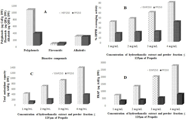
Figure 1. Bioactive compounds (A) and antioxidant activities: Percentage of DPPH scavenging activity (B), Total antioxidant capacity (C), and Ferric Reducing activity Potential (D) of hydroethanolic extract and powder fraction ≤125µm of Propolis at different concentrations.
µg: Microgramme; AAE: Ascorbic Acid Equivalents; CaE: Catechin Equivalent; g: Gramme; DW: Dry Weight; EQ: Quercetin Equivalent; QiE: Quinine Equivalent; EthP: Hydroethanolic extract of Propolis; PP: Propolis powder fraction ≤ 125µm; ≤125µm: Powdery fractions with a diameter inferior at 125 µm
Figure 1A showed that polyphenol content was significantly (p<0.05) high in EthP (1037.80±16.99 µg CaE/ g DW) compared to PP (354±5.08 (μg CaE/g DW)); flavonoid content was significantly (p<0. 05) higher in PP (62.25±3.61 (μg QE/g DW)) than in EthP (51.20±2.68 (μg QE/g DW)). However, the alkaloid content in EthP (267.3±13.43 µg QiE/ g DW) was significantly (p<0.05) lower than in PP (257.05±4.23 µg QiE/ g DW).
The DPPH radical scavenging activity of the EthP and PP is shown in Figure 1B. After observation, it was noted that the different samples trapped this synthetic radical in a concentration-dependent manner (activity increases in parallel with increasing extract or powder concentrations). The PP with a CI50 of 93.90mg/mL exhibited the best activity.
Y(EthP) = 29.502 ln(x) + 28.268; R2= 0.9662 IC50= 143.68 mg/mL(5)
Y(PP) = 25.963 ln(x) – 7.6658; R2= 0.803 IC50= 93.90 mg/mL(6)
3.3. Effect of Hydroethanolic Extract and Fraction Powder ≤ 125 µm of Propolis on Weight Change
The hydroethanolic extract and the fraction power ≤ 125 µm of Propolis were administered to the rats during the 45 days of experimentation at the end of which the weight change was recorded and is mentioned in Figure 1. The normal diet group of rats increased body weight up to the 45th day of experimentation. The negative control group, which was fed the atherogenic diet, saw weight increase significantly (p<0.01) up to day 10, followed by a significant (p<0.001) decrease up to day 45. On day 5, the EthP250 and PP250 groups significantly (p<0.05) increased weight by 60.97% and 61.24% respectively compared with the normal control group.
However, from days 15 and 20 onwards, there was a significant (p<0.001) decrease in body weight for EthP250 and PP250, followed by a significant (p<0.001) increase in weight on day 25, before a significant (p<0.0001) decrease in weight on day 45 respectively compared with the normal control. At day 45, the EthP250 and PP250 groups showed a small significant decrease (p<0.001) compared with the negative control group.
Table 3. Effect of hydroethanolic extract and powder fraction ≤ 125 µm of Propolis on transaminase activities, creatinine level, and the lipidic profile.

ND

AD

AD

Atorvastatin (10mg/kg)

EthP250

PP250

ALT (U/L)

34.15±2,59

64.48±0.14***

38.98±6.55##

22.10±2.52####

30.54±2.33####

AST (U/L)

34.54±0.11

85.75±0.21****

62.24±4.53**#

46.3±1.26###

68.14±5.51***α

AST/ALT

1.01

1.32

1.59

2.09

2.23

Creatinine (mg/dL)

1.28±0.1

1.9±0.04*

1.68±0.16

0.6±0.07**###

1.6±0.14ααα

TC (mg/dL)

115.3±1.8

161.2±4.5***

143.05***##

119.6±2.6###

118.8±2.5###

TG (mg/dL)

91.5±1.4

98.1±0.9*

85.2±1.7*###

62.7±1.34***###

19.8±0.67***###ααα

LDL-C (mg/dL)

42.7±5.9

104.8±9.4*

36.96±9.15#

29.47±11.46##

27.3±17.62##

HDL-C (mg/dL)

53.23±1.19

39.42±1.3**

84.65±4.16***###

82.63±1.36****####

95.56±3.01****####αα

CT/HDL-C

2.16

4.08

1.68

1.44

1.24

(%) Protection

-

-

58.82

64.70

69.60

ND: Normal diet; AD: Atherogenic diet; AST: Aspartate aminotransferase; ALT: Alanine aminotransferase; TC: Total cholesterol; TG: Triglycerides; LDL: Low-density lipoprotein; HDL: High-density lipoprotein; EthP250: Hydroethanolic extract of Propolis at the dose 250mg/kg bw; PP250: Propolis powder fraction ≤ 125µm at the dose 250mg/kg bw; ≤12µm: Powdery fractions with a diameter inferior at 125 µm.
Values are expressed as means ± SEM; n=3; *p<0.05; **p<0.01; ***p<0.001; ****p<0.0001 significant difference from the control group; p<0.05; ≠≠p<0.01; ≠≠≠p<0.001; ≠≠≠≠p<0.0001 significant difference from the negative control group; αp<0.05; ααp<0.01; αααp<0.001; ααααp<0.0001 significant difference from the EthP group.
3.4. Effect of Propolis Hydroethanolic Extract and Fraction Powder ≤ 125 µm on the Liver and the Kidney Function Markers
Effect on transaminase (ALT, and AST) activity and creatinine levels
Table 3 shows the effect of treatment with EthP250 and PP250 on ALT, AST, and creatinine levels in rats after 45 days of experimentation. It was found that feeding the atherogenic diet induced a significant increase in ALT, AST, and creatinine activities of 52.96% (P˂0.001), 40.27% (P˂0.0001), and 67.36% (P˂0.05) respectively in the TN group compared with the normal control group. Treatment of animals with EthP250 resulted in a significant decrease in ALT, AST, and creatinine activities of 34.27% (P˂0.0001), 53.99% (P˂0.001), and 31.57% (P˂0.05) respectively compared to the TN group. Whereas PP250 only resulted in a 47.33% (P˂0.0001) decrease in AST activity compared with the TN group. However, EthP250 significantly decreased serum AST and creatinine levels by 67.94% (P˂0.05) and 37.5% (P˂0.001) versus the PP250 group.
3.5. Effect of Hydroethanolic Extract and Propolis Fraction Powder ≤ 125 µm on Lipid Profile
Feeding the rats an atherogenic diet produced the results on the lipid profile of the animals shown in Table 3.
This table shows that the serum total cholesterol concentration of animals in the TN group increased significantly (p<0.001) by 71.52% compared with the normal control. On the other hand, treatment with EthP250, and PP250 significantly (p<0.001) lowered cholesterol levels by 74.19% and 73.69% respectively, compared with the TN group.
After 45 days of experimentation, rats on the atherogenic diet (TN) showed a significant (p<0.05) 93.27% increase in TG levels compared with the normal control group. The administration of EthP250, and PP250 as preventive treatment resulted in a significant (p<0.001) decrease in serum triglyceride levels of 63.91% and 20.18% respectively compared to the TN group. However, the PP250 significantly (p<0.001) reduced triglyceride levels by 31.57% compared with the group receiving the EthP250 at the same dose.
At the end of the experiment, LDL-c levels increased significantly (p<0.05) by 40.74% in the TN group compared with the normal control. Treatment with EthP250 and PP250 significantly (p<0.01) reduced LDL-c concentration by 28.12% and 26.04% respectively, compared with the TN group.
The HDL-c results presented in Table 4 show a significant (p<0.01) decrease in HDL-c levels of 74.05% in the TN group compared with the normal control. There was a significant increase (p<0.0001) of 47.7% and 41.25% when EthP250 and PP250 were administered compared with the TN group. However, treatment with PP250 showed a significant (p<0.01) 86.46% increase in HDL-c concentration, compared with treatment with EthP250, Table 3 showed that rats fed an atherogenic diet for 45 days were at risk of developing atherosclerosis and consequently cardiovascular disease. Propolis reduced this risk by 64.70% and 69.60% respectively with EthP250 and PP250.
Table 4. Effect of hydroethanolic extract and powder fraction ≤ 125 µm of Propolis on some Biomarkers.

AD

ND

AD

Atorvastatin (10mg/kg)

EthP250

PP250

NO (mmol/g of organ)

Liver

1.21±0.24

0.44±0.12

1.51±0.15#

1.21±0.13

1.16±0.14

Kidney

1.05±0.15

0.7±0.19

0.55±0.08

1.31±0.13

1.59±0.28

Heart

0.33±0.03

0.22±0.02

0.37±0.03

0.67±0.19

0.33±0.08

Aorta

0.42±0.008

0.01±0.002**

0.58±0.12####

0.72±0.075####

0.61±0.03*####

Glutathione (µmol/g of organ)

Liver

34.87±2.25

63.07±5.87*

32.48±2.91#

52.57±8.88

35.85±3.45

Kidney

39.37±3.5

52.61±6.1

33.46±0.94##

56.83±1.37**

58.85±2.14**

Heart

98.5±0.5

138.1±4.7**

101.3±8.5##

97.71±5.43##

126.6±7.74β

Aorta

31.5±3.6

15.69±0.49

45.28±2.51

46.81±0.28

31±0.14

SOD (UI/g of organ)

Liver

56.3±8.7

139.1±11.35***

86.19±8.7#

96.22±3.3

88.77±5.79

Kidney

46.4±3.3

209.1±41.47***

66.32±3.3

109.1±5.6

129.2±1.04

Heart

69.65±17.23

182.5±14.4

63.02±3.31***

86.02±3.21##

82.92±11.96##

Aorta

74.63±14.36

238.6±5.85****

99.38±0.07####

69.65±11.49####

34.33±3.16####

MDA (µmol/g of organ)

Liver

1.38±0.13

2.05±0.001

1.49±0.12##

1.13±0.06####

1.56±0.08

Kidney

1.004±0.08

2.009±0.02****

1.15±0.02####

1.15±0.07####

1.28±0.07####

Heart

1.02±0.09

1.85±0.06****

1.06±0.001

1.21±0.03###

1.23±0.04###

Aorta

1.53±0.04

3.07±0.81

1.47±0.01#

1.43±0.03#

1.49±0.001#

ND: Normal diet; AD: Atherogenic diet; NO: Nitrite oxide; SOD: Superoxide dismutase; MDA: Malondialdehyde; EthP250: Hydroethanolic extract of Propolis at the dose 250mg/kg bw; PP250: Propolis powder fraction ≤ 125µm at the dose 250mg/kg bw; ≤12µm: Powdery fractions with a diameter inferior at 125 µm.
Values are expressed as means ± SEM; n=3; *p<0.05; **p<0.01; ***p<0.001; ****p<0.0001 significant difference from the control group; p<0.05; ≠≠p<0.01; ≠≠≠p<0.001; ≠≠≠≠p<0.0001 significant difference from the negative control group; αp<0.05; ααp<0.01; αααp<0.001; ααααp<0.0001 significant difference from the EthP group.
3.6. Effect of Hydroethanolic Extract and Fraction Powder ≤ 125µm of Propolis on Interleukin-17
IL-17 expression levels are shown in Figure 2. It can be seen that the TN group showed a significant increase (P˂0.0001) in IL-17 expression levels of 35.5% compared with the normal control. Treatment with EthP250 and PP250 significantly (p<0.001) reduced IL-17 expression levels by 50.25% and 100% respectively compared with the TN group. Results showed that serum IL-17 expression was significantly reduced (p<0.001) in the PP250 group by 100% compared with the EthP250 group.
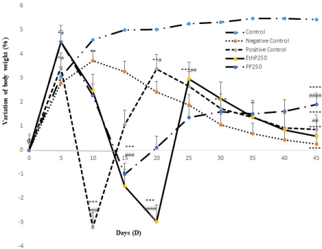
Figure 2. Variation of body weight.
EthP250: Hydroethanolic extract of Propolis at the dose of 250mg/kg bw; PP250: Propolis powder fraction ≤ 125µm at the dose of 250mg/kg bw; ≤12µm: Powdery fractions with a diameter inferior at 125 µm.
Values are expressed as means ± SEM; n=3; *p<0.05; **p<0.01; ***p<0.001; ****p<0.0001 significant difference from the control group; p<0.05; ≠≠p<0.01; ≠≠≠p<0.001; ≠≠≠≠p<0.0001 significant difference from the negative control group.
Effects of EthP250 and PP250 on markers of oxidative stress in preventive treatment of cardiovascular disease. Propolis reduced this risk by 64.70% and 69.60% respectively with EthP250 and PP250.
3.7. Effect of Hydroethanolic Extract and Fraction Powder ≤ 125µm of Propolis on Some Biomarkers
At the end of the experiment, the administration of the atherogenic diet had an impact on markers of oxidative stress. The data received are shown in Table 4.
Table 3 shows that aortic nitrite levels in the TN group decreased significantly (p<0.01) by 2.38% compared with the normal control group. On the other hand, when EthP250 and PP250 were administered, nitrite levels in the aorta increased significantly (p<0.0001) by 1.38% and 1.63% respectively, compared with the TN group.
Tissue glutathione levels rose significantly (p<0.05) by 55.28% in the liver and 71.32% in the heart (p<0.01) respectively, compared with the normal control group (Table 4). Only EthP250 caused a significant (p˂0.01) 70.75% drop in glutathione levels compared with the negative control. Propolis powder, however, significantly increased (p˂0.05) tissue glutathione levels by 77.18% compared with Propolis hydroethanolic extract.
According to Table 4, SOD activity increased significantly (p<0.001) by 40.47% in the liver, 22.19% in the kidney, and 31.27% (p<0.0001) in the aorta, respectively in TN compared with the normal control. Rats given the EthP250 and PP250 of Propolis showed a significant (p<0.01) decrease of 47.13% and 45.43% respectively in the heart; 29.19% and 14.38% (p<0.0001) respectively in the aorta compared with TN.
Malondialdehyde levels assessed in experimental rats showed a significant increase (p<0.0001) in TN of 49.97% and 55.13% respectively in the kidney and heart compared with the normal control (Table 4). Compared with NT, rats treated with EthP250 and PP250 showed a significant decrease in MDA levels in the liver by 55.12% (p<0.0001) and 76.09% (p<0.05) respectively; in the kidney by 57.24% (p<0.0001) and 63.71% (p<0.0001) respectively. In the heart, these values were 65.40% (p<0.001) and 66.48% (p<0.001) respectively; in the aorta, 46.57% (p<0.05) and 48.53% (p<0.05).
However, the administration of PP250 significantly (p<0.05) increased MDA levels by 72.43% compared with EthP250.
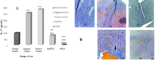
Figure 3. Effect of hydroethanolic extract and powder fraction ≤ 125µm of Propolis on the Interleukin-17, determined by ELISA assays (A), Histological section of the liver, and effects of Propolis on lipid accumulation (B).
Values are expressed as means ± SEM; n=4;***p<0,001; ****p<0.0001 significant difference from the control group; ≠≠≠≠p<0.0001 significant difference from the negative control group; ααααp<0.0001 significant difference from the EthP group. ####p<0.00: significant differences from negative group; ααααp<0.001 significant differences from EthP treatment group EthP250: Hydroethanolic extract of Propolis at the dose of 250mg/kg bw; PP250: Propolis powder fraction ≤ 125µm at the dose of 250mg/kg bw; IL-17: Interleukin 17; Negative Control: Moderate steatosis x 10; Positive Control: mild steatosis and mild inflammation x10; EthP250: mild steatosis x 40; PP250: mild steatosis x 10.
Presence of hepatic steatosis
Presence of inflammation
4. Discussion
This study evaluated the content of certain mineral elements (Vitamin C, Iron, Manganese, Zinc, Copper, Selenium, and Calcium), determined antioxidant activity in vitro (DPPH, TAC, and FRAP), demonstrated the reduction in inflammation and hepatic steatosis in vivo of Propolis hydroethanolic extract (EthP) and powder fraction ≤ 125 µm (PP) in Wistar rats fed an atherogenic diet.
4.1. Minerals-Elements Content
Fractionation and sieving of Propolis to a powder fraction≤125µm resulted in a significant increase in dry matter, center content, Zinc, Magnesium, Iron, and Calcium. This may be explained by the fact that sieving favors the concentration of certain bioactive compounds. The increase in ash content with the reduction in particle size could be explained by the fact that during sieving the ash, which was less heavy, was found in the smaller particle size classes .
4.2. In-vitro Antioxidant Activity of Hydroethanolic Extract and Fraction Powder ≤ 125µm of Propolis
EthP showed better activity than PP, which could be explained by the fact that the polyphenols present in EthP would have a high polarity, as the water-ethanol mixture would favor polar compounds. Similar results were reported by Tsague et al. .
The DPPH radical scavenging activities of the EthP and PP are shown in Figure 1B. After observation, it was noted that the different samples trapped this synthetic radical in a concentration-dependent manner (activity increases in parallel with increasing extract or powder concentrations). The PP with a CI50 of 93.90mg/mL exhibited the best activity.
Taking a look at Figure 1C for data on the total antioxidant capacity of the PP and EthP, these results confirm previous trends observed through DPPH radical scavenging and iron reduction activities, which showed that the antioxidant potential of our extracts was concentration-dependent. However, the best activity here was shown by the PP, in contrast to the result observed for FRAP potential.
Figure 1D shows the ferric iron reduction capacity of the EthP and PP. These data show that, as in the case of DPPH radical scavenging, the iron reduction potential increased positively with increasing hydroethanolic extract and powder concentration. The EthP exhibited the best activity.
4.3. Effect of Hydroethanolic Extract and Fraction Powder ≤ 125 µm of Propolis on Weight Change
In this study, rats subjected to the atherogenic diet lost weight throughout the experiment. This observed weight loss is thought to be due to the secretion of leptin . Indeed, leptin is a hormone secreted mainly by adipose tissue and acts at the level of the hypothalamus to stimulate the sensation of satiety and stop food intake. This result is in line with those of Tsague et al. , who found that this atherogenic diet reduced the weight of the animals simultaneously with food intake. Feeding with EthP250 and PP250 significantly improved this weight loss compared with the TN group. These results suggest that EthP250 and PP250 may have an appetite-stimulating effect on weight gain. At the end of the experiment, PP250 showed greater activity than Eth250, proving that Pulverization and Controlled Differential Screening process concentrate bioactive compounds in addition to polyphenols. Which stimulated appetite in rats.
4.4. Effect of Propolis Hydroethanolic Extract and Fraction Powder ≤ 125 µm on the Liver and the Kidney Function Markers
Effect on transaminase (ALT, and AST) activity and creatinine levels
Transaminases are enzymes synthesized by hepatocytes, ensuring the synthesis of amino acids by transferring an amino group from an amino acid to ketoglutarate . Excessive fat accumulation in the liver would have caused oxidative stress and liver inflammation, which would have damaged liver cells and led to their lysis, hence the observed increase . They are found in the cytosol (ALT and AST) and mitochondria (AST) of liver cells. They are the most sensitive biomarkers implicated in liver dysfunction and tissue damage . The enzymatic activity of transaminases is increased in the bloodstream due to increased lysis of liver parenchyma cells . When this decreases, there is restoration of plasma membrane stability as well as protection and regeneration of damaged liver cells . The hepatoprotective effect of Propolis is due to its richness in bioactive compounds such as polyphenols and the presence of certain mineral compounds such as Zinc, Cu, Mn, Iron, Ca, Se, and Vit C. These results are in agreement with work presented by Tsague et al. who demonstrated that the presence of these minerals in powder fraction ≤ 125 µm of Eribroma oblongum promoted a decrease in anti-inflammatory expression in Wistar strain rats. For this practical reason, the use of Propolis will be essential in solving the problem of non-alcoholic fatty liver disease , 40, 41] showed that polyphenols in aqueous extracts of Scolymus hispanicus and Salvia plebeia reduced liver damage in non-alcoholic fatty liver disease by improving transaminase levels.
The kidney is the site of the excretion of toxic wastes released by the liver . When these wastes exceed the kidney's filtration capacity, we observe a decrease in glomerular filtration in the kidney and a consequent increase in certain substances such as creatinine in the blood . Creatinine is a metabolite of creatine, which in skeletal muscle is phosphorylated to creatine phosphate, a compound rich in free energy . It is a marker of renal function.
4.5. Effect of Hydroethanolic Extract and Propolis Fraction Powder ≤ 125µm on Lipid Profile
The liver is the main site of lipid metabolism in the body . Following a meal, chylomicrons transport triglycerides and dietary cholesterol to store some in tissue reserves and some in the liver . In the liver, these TGs are incorporated into VLDLs, which are responsible for supplying fatty acids to user tissues or depositing them in adipose tissue. The circulating VLDLs, as they supply free fatty acid, become IDLs, which are enriched with cholesterol, forming LDLs responsible for supplying esterified cholesterol to user tissues .
An atherogenic diet rich in saturated fatty acids and cholesterol can therefore be the source of TG overload in the liver, with an increase in LDL in the circulation followed by a drop in HDL . Propolis may therefore have acted by reducing the secretion of chylomicrons into the lymph and inhibiting the secretion of pancreatic lipase, a key enzyme in the intestinal absorption of dietary lipids. It could also act by increasing the expression of carnitine palmitoyl transferase receptors, which will increase the beta-oxidation of fatty acids to produce energy and thus reduce fat accumulation in the liver . These results are in line with those of Afsharinasab et al. who showed that the hydroalcoholic extract of Berberis integerrima improved the lipid profile. Propolis increased HDL-c levels in rats. HDL-c is responsible for capturing cholesterol and returning it to the liver for excretion. Non-alcoholic fatty liver is particularly prone to death from cardiovascular or liver-related causes, as is the inflammation seen in NAFLD .
The lipid profile enables direct confirmation of the diagnosis associated with cardiovascular disease. It can be combined with the calculation of the atherogenic index to determine cardiovascular risk, more specifically intermediate risk . The atherogenic index, also known as the Castelli index, is a lipid ratio used to assess the risk of atherosclerosis and cardiovascular disease . Administration of PP250 reduced the risk of atherosclerosis by 93.05%. These results are similar to those of Tsague et al. , who showed that administration of Eribroma oblongum (100mg/kg bw) in rats reduced cardiovascular risk by 89.09%. Propolis (250mg/kg bw) therefore has an anti-steatosis and anti-atherosclerosis effect.
4.6. Effect of Hydroethanolic Extract and Fraction Powder ≤ 125µm of Propolis on Interleukin-17
IL-17 is a pro-inflammatory cytokine that is ubiquitously expressed in liver cells, which explains its involvement in liver damage . Atherogenic feeding of rats in the TN group resulted in a significant 35.5% increase in IL-17 expression compared with rats in the normal control group. This result is similar to that of Che et al. , who showed that adult male C57BL/6 mice fed a high-fat diet strongly expressed IL-17 levels. In the same study, administration of the 200 mg/Pc aqueous extract of Cirsium japonicum lowered IL-17 expression levels in mice. Duan et al. demonstrated the efficacy of Dahuang Zhechong pills in reducing IL-17-secreting Th17 cells in NAFLD patients. Rats on the atherogenic diet had elevated levels of IL-17 due to hepatocyte damage. Fat accumulation in the liver can lead to the release by macrophages, of certain cytokines such as IL-6 and TNF alpha . When fat accumulates in the liver, IL-17 stimulates kupffer cells to secrete cytokines such as IL-6, which in turn, when associated with TGF-β, stimulates the differentiation and activation of Th17 cells, which in turn produces IL-17 to trigger the inflammatory process via neutrophil infiltration . Treatment with EthP250 and PP250 significantly reduced IL-17 expression by 50.25% and 100% respectively, compared with the TN group. This could be due to a decrease in IL-17 receptors on Th17 cells, or to inhibition of IL-17 secretion by macrophages. Also, progression from simple steatosis to non-alcoholic hepatic steatosis is characterized by increased accumulation of Th17 cells 57]. Results showed that serum IL-17 expression was significantly reduced in the PP250 group by 100% compared to the EthP250 group. Administration of Propolis to rats on the atherogenic diet lowered IL-17 expression levels and thus reduced or ameliorated inflammation.
The atherogenic diet significantly reduced nitrite levels by 2.38% in the TN group compared with the normal control group. In contrast, treatment with EthP250 and PP250 significantly increased aortic nitrite levels by 1.38% and 1.63% respectively, compared with the TN group.
Treatment with EthP250 and PP250 significantly reduced IL-17 expression by 50.25% and 100% respectively, compared with the TN group. This could be due to a decrease in IL-17 receptors on Th17 cells, or to inhibition of IL-17 secretion by macrophages. Also, progression from simple steatosis to non-alcoholic hepatic steatosis is characterized by increased accumulation of Th17 cells . Results showed that serum IL-17 expression was significantly reduced in the PP250 group by 100% compared to the EthP250 group. Administration of Propolis to rats on the atherogenic diet lowered IL-17 expression levels and thus reduced or ameliorated inflammation. Sokeng et al. demonstrated that the effect of Arachic Acid Ethyl Ester Isolated from Propolis has Anti-Inflammatory and Analgesic.
4.7. Effect of Hydroethanolic Extract and Fraction Powder ≤ 125µm of Propolis on Some Biomarkers
MDA is the end product of the ROS peroxidation reaction on lipids. It is therefore used as an indicator of free radical damage to cells . Excess fat in hepatocytes can increase the beta-oxidation capacity of mitochondria and consequently incomplete lipid oxidation and increased free radical production .
The TN group showed a significant increase in MDA of 49.97% and 55.13% respectively in the kidney and heart in the TN group compared with the normal control. This increase is thought to be due to ROS-mediated peroxidation of cell membrane lipids .
Rats given EthP250 and PP250 showed a significant decrease in MDA levels in the liver by 55.12% and 76.09% respectively; in the kidney by 57.24% and 63.71% in the heart, these values were 65.40% and 66.48% respectively; in the aorta by 46.57% and 48.53%. Propolis administration improved MDA levels in treated rats, opposing lipid peroxidation and preventing free radical damage .
This result proves that Propolis, with its wealth of bioactive compounds such as polyphenols, reduced the production of free radicals in the liver by neutralizing them, thus preventing them from destroying membrane lipids. Our results are similar to those of Amirinejad et al. who showed that the hydroalcoholic extract of spinach ameliorated oxidative stress in NADFL by reducing MDA levels.
However, administration of PP250 significantly increased MDA levels by 72.43% compared with EthP250, due to the high concentration of bioactive compounds present in Propolis powder.
Oxidative stress is the imbalance between pro-oxidants and antioxidants in favor of pro-oxidants . Antioxidants refer to all substances that, in low concentrations relative to the oxidizable substrate, delay or inhibit oxidation of the substrate . Feeding rats an atherogenic diet for 45 days increased organ levels of reduced glutathione (GSH) and SOD. This increase is thought to be because when the liver is attacked by lipid accumulation, free radicals are produced. In response to this aggression, the body is predisposed to a cascade of antioxidant secretions to neutralize the free radicals generated by the atherogenic diet. The role of reduced glutathione (GSH) is to trap superoxide radicals and protect protein thiol groups from oxidation .
5. Conclusion and Perspectives
This study aimed to evaluate the preventive effects and anti-inflammatory activity of Propolis on atherogenic diet-induced non-alcoholic fatty liver disease in Wistar rats.
The in vitro antioxidant activity demonstrated that Propolis was very rich in phenolic compounds, which increased with Propolis concentration. A higher concentration of Zinc, Iron, Calcium, Manganese, and Vitamin C in the hydroethanolic extract compared to the powder fraction ≤ 125 µm of Propolis. Administration of the hydroethanolic extract and the Propolis powder fraction ≤ 125 µm in rats on an atherogenic diet limited liver damage by lowering transaminase levels. Propolis reduced fat accumulation in the liver by lowering total cholesterol, TG, and LDL-c levels and increasing HDL-c levels in rat serum. Anti-inflammatory activity was reduced by decreased IL-17 expression levels after administration of the hydroethanolic extract and the powder fraction ≤ 125 µm of Propolis. However, the most striking effects were obtained with the Propolis powder fraction ≤ 125 µm. Propolis was shown to protect rats against non-alcoholic hepatic steatosis and to have anti-inflammatory activity. These results suggest that Propolis could be a promising adjunct in the management of non-alcoholic fatty liver, confirming its use in traditional medicine. However, further studies are needed to better understand Propolis' mechanisms of action and assess its short- and long-term deleterious effects.
Abbreviations

CDSp

Pulverization and Controlled Differential Screening Process

PP

The Powder Fraction≤ 125µm

EthP

Propolis Hydroethanolic Extract

ND

Normal Diet

AD

Atherogenic Diet

AST

Aspartate Aminotransferase

ALT

Alanine Aminotransferase

TC

Total Cholesterol

TG

Triglycerides

LDL

Low-Density Lipoprotein

HDL

High-Density Lipoprotein

VLDL

Very Low-Density Lipoprotein

TAC

Total Antioxidant Capacity

DPPH

-2,2-Diphenyl-1-Picrylhydrazyl

FRAP

Ferric Reducing Antioxidant Power

ROS

Reactive Oxygen Species

NO

Nitric Oxide

GSH

Reduced Glutathione

MDA

Malondialdehyde

SOD

Superoxide Dismutase

NADFLD

Nonalcoholic Fatty Liver Disease

Il-17

Interleukin 17

Th17

T-helper 17

TNF

Tumor Necrosis Factor

TGF-β

Transforming Growth Factor β

Acknowledgments
The author would like to thank the team at the Laboratory of Biophysics, Food Biochemistry and Nutrition of ENSAI, University of Ngaoundere, Cameroon for permission to use their research laboratory.
Data Availability Statement
All data are available on request from the corresponding author.
Conflicts of Interest
The authors declare no conflict of interest.
References
[1] Ghedira K, Goetz P, le Jeune R. Propolis. Phytothérapie. 2009; 7(2): 100–105.
[2] Dezmirean DS, Paşca C, Moise AR, Bobiş O. Plant Sources Responsible for the Chemical Composition and Main Bioactive Properties of Poplar-Type Propolis. Plants (Basel). 2020; 10(1): 22.
[3] Tagkou NM, Goossens N. (2023). Stéatose hépatique non alcoolique: diagnostic et traitement en 2022. Schweizer Gastroenterologie, 4(1), 27–37.
[4] Fang Z, Shen G, Wang Y, Hong F, Tang X, Zeng Y, Zhang T, Liu H, Li Y, Wang J, Zhang J, Gao A, Qi W, Yang X, Zhou T, Gao G. Elevated Kallistatin promotes the occurrence and progression of non-alcoholic fatty liver disease. Signal Transduct Target Ther. 2024; 9(1): 66.
[5] Nga WTB, Etoa MC, Marina BG, Bagnaka SAE, Ndam AWN, Malongue A, Namme LH. (2023). Prevalence and Factors Associated with Hepatic Steatosis in Patients with Metabolic Syndrome in Cameroon: Cases of 4 Reference Hospitals. Open Journal of Gastroenterology, 13(3), 99-110.
[6] Okaiyeto K, Oguntibeju OO. African Herbal Medicines: Adverse Effects and Cytotoxic Potentials with Different Therapeutic Applications. Int J Environ Res Public Health. 2021; 18(11): 5988.
[7] Njintang YN, Tatsadjieu NL, Ngakou A, Danra D, Tchuenguem-Fohouo FN. (2012). Antiradical activity and polyphenol content of ethanolic extracts of Propolis. International Journal of Biosciences (IJB), 2(4), 56-63.
[8] Zingue S, Nde CBM, Michel T, Ndinteh DT, Tchatchou J, Adamou M, Fernandez X, Fohouo FT, Clyne C, Njamen D. (2017). Ethanol-extracted Cameroonian Propolis exerts estrogenic effects and alleviates hot flushes in ovariectomized Wistar rats. BMC complementary and alternative medicine, 17(1), 65.
[9] Zingue S, Maxeiner S, Rutz J, Ndinteh DT, Chun FK, Fohouo FT, Njamen D, Blaheta RA. (2020). Ethanol-extracted Cameroonian propolis: Antiproliferative effects and potential mechanism of action in prostate cancer. Andrologia, 52(9), e13698.
[10] Baudelaire E. Comminution and controlled differential screening method for the dry extraction of natural active principles 2013; Brevet: PCT/FR2011/0005616.
[11] Mai-Mbe Baiva C, Ngatchic Metsagang TJ, Njintang Yanou N. Antihyperlipidemic Properties of Powder Fractions and Extracts of Diospyros Mespiliformis HOCHST Fruits. South Asian Research Journal of Biology and Applied Biosciences. 2019; 1(3): 55-61.
[12] Deli M, Ndjantou EB, Ngatchic Metsagang JT, Petit J, Njintang Yanou N, Scher J. (2019). Successive grinding and sieving as a new tool to fractionate polyphenols and antioxidants of plants powders: Application to Boscia senegalensis seeds, Dichrostachys glomerata fruits, and Hibiscus sabdariffa calyx powders. Food science and nutrition, 7(5), 1795–1806.
[13] Djongra M, Mando NB, Djafsia B, Tchuenguem FN, Ndjonka D. (2022). In vitro evaluation of the Anthelmintic Activity of PROMAXC and propolis extract on the Nematode Parasite Onchocerca ochengi Bwangamoi, 1969 (Spirurida: Onchocercidae). American Journal of PharmTech Research, 12(03), 36-54.
[14] Wafa G, Amadou D, Larbi KM. (2014). Activité larvicide, composition phytochimique et propriétés antioxydantes de différentes parties de cinq populations de Ricinus communis L. Cultures et produits industriel, 56, 43-51.
[15] Dewanto V, Wu X, Adom KK, Liu RH. (2002). Thermal processing enhances the nutritional value of tomatoes by increasing total antioxidant activity. Journal of Agricultural and Food Chemistry, 50: 3010-3014.
[16] Sun S, Lee S, Wu A, Chen C. (1998). Détermination des alcaloïdes bisbenzylisoquinoléines par chromatographie liquide haute performance. Journal de Chromatographie A, 799(1-2), 337-342.
[17] AOAC. (Association of Official Analytical Chemists) Official method of analysis 1990; (13th ed.). Washington, DC: Association of Official Analytical Chemists.
[18] Zhang D, Hamauzu Y. (2004). Phenolics, ascorbic acid, carotenoids and antioxidant activity of broccoli and their changes during conventional and microwave cooking. Food chemistry. 88(4): 503-509.
[19] Prieto P, Pineda M, Aguilar M. (1999). Spectrophotometric quantitation of antioxidant capacity through the formation of a phosphomolybdenum complex: specific application to the determination of vitamin E. Analytical biochemistry. 269(2): 337-341.
[20] Benzie IFF, Strain JJ: The ferric reducing ability of plasma (FRAP) as a measure of “antioxidant power”: the FRAP assay. Anal Biochem 1996, 239: 70–76.
[21] Tsague MV, Njiki Bikoï J, Sokamte Tegang A, Edoun Ebouel FL, Nodem Sohanang FS, Ahamat Abakar, Mahoumo Fodop AO, M’bann Nsonngan S, Ze Minkande J. In Vitro Antioxidant Properties and Interleukin-17 Expression Levels of Eribroma oblongum (Malvaceae) in Wistar Rats with Atherogenic Diet-Induced Steatotic Liver. Advances in Biochemistry. 2024; 12(1): 10-19.
[22] Tsague MV, Fokunang Ntungwen C, Ngameni B, Tembe-fokunang EA, Guedje NM, Ngo Lemba Tom E, Atogho-Tiedeu B, Zintchem RF, Mecthi Dongmo M, Ngoupayo J, Sokeng Dongmo S, Dzeufiet Djomeni, Oben Eyong J, Dimo T, Ze Minkande J and Ngadjui Tchaleu B. Pre-clinical evaluation of the hypotensive and antiatherogenic activity of hydroethanolic extract of Eribroma oblongum (Malvaceae) stem bark on wistar rats models. British Journal of Pharmaceutical research. 2015; 5(1): 1-14.
[23] Wallnofer HE, Schmidt FW. Schmidt eds (1974). Synopsis Der Leberkrannkheiten. Georg Dtsh. Med. Wschr. 99, 343.
[24] Koller A, Kaplan A. (1984). The CV Mosby Co St. Louis toronto princeton. Clin Chem, 418, 1316-24.
[25] Trinder P. (1969). Standard methods of clinical chemistry. Ann. Clin. Biochem, 6, 24.
[26] Fossati P, Prencipe L. (1982). Serum triglycerides determined colorimetrically with an enzyme that produces hydrogen peroxide. Clinical chemistry, 28(10), 2077–2080.
[27] Friedewald W, Levy R, Fredrickson D. (1972). Estimation of the concentration of Low Density Lipoprotein Cholesterol in plasma, without use of preparative of ultracentrifuge. Clinical Chemistry. 18(6): 499-502.
[28] Ellman G. (1959). Tissue sulfhydryl groups. Archives of Biochemistry and Biophysics. 82(1): 70-7.
[29] Misra HP, Fridovich I. (1972). The role of superoxide anion in the autoxidation of epinephrine and a simple assay for superoxide dismutase. The Journal of biological chemistry. 247(10); 3170–3175.
[30] Wilbur KM, Bernheim F, Shapiro OW. (1949). The thiobarbituric acid reagent as a test for the oxidation of unsaturated fatty acids by various agents. Archives of biochemistry, 24(2); 305–313.
[31] Grand F, Guitton J, Goudable J. (2001). Optimisation des paramètres du dosage des nitrites et nitrates sériques par la technique de Griess. In Annales de biologie clinique (Paris). 59(5); 559-565.
[32] Ahmed J, Al-Attar H, Arfat YA. (2016). Effect of particle size on compositional, functional, pasting and rheological properties of commercial water chestnut flour. Food Hydrocoll. 52: 888–895.
[33] Paz-Filho G, Mastronardi CA, Licinio J. (2015). Leptin treatment: facts and expectations. Metabolism: clinical and experimental. 64(1): 146–156.
[34] Vroon DH, Israili Z. Aminotransferases (1990). In: Walker HK, Hall WD, Hurst JW, editors. Clinical Methods: The History, Physical, and Laboratory Examinations. 3rd edition. Boston: Butterworths; Chapter 99.
[35] Berdja S, Boudarene L, Smail L, Neggazi S, Boumaza S, Sahraoui A, Haffaf EM, Kacimi G, Aouichat Bouguerra S. (2021). Scolymus hispanicus (Golden Thistle) Ameliorates Hepatic Steatosis and Metabolic Syndrome by Reducing Lipid Accumulation, Oxidative Stress, and Inflammation in Rats under Hyperfatty Diet. Evidence-based complementary and alternative medicine: eCAM, 5588382.
[36] McGill MR. The past and present of serum aminotransferases and the future of liver injury biomarkers. EXCLI J. 2016; 15: 817-828.
[37] Gnanadesigan M, Ravikumar S, Anand M. (2017). Hepatoprotective activity of Ceriops decandra (Griff.) Ding Hou mangrove plant against CCl4 induced liver damage. Journal of Taibah University for Science. 11(3): 450-457.
[38] Jodynis-Liebert J, Nowicki M, Murias M, Adamska T, Ewertowska M, Kujawska M, Piotrowska H, Konwerska A, Ostalska-Nowicka D, Pernak J. (2010). Cytotoxicity, acute and subchronic toxicity of ionic liquid, didecyldimethylammonium saccharinate, in rats. Regulatory toxicology and pharmacology: RTP. 57(2-3); 266–273.
[39] Touzani S, Imtara H, Katekhaye S, Mechchate H, Ouassou H, Alqahtani AS, Noman OM, Nasr FA, Fearnley H, Fearnley J, Paradkar A, ElArabi I, et Lyoussi B. (2021). Determination of Phenolic Compounds in Various Propolis Samples Collected from an African and an Asian Region and Their Impact on Antioxidant and Antibacterial Activities. Molecules (Basel, Switzerland). 26(15); 4589.
[40] Tutunchi H, Arefhosseini S, et Ebrahimi-Mameghani M. (2023). Clinical effectiveness of α-lipoic acid, myo-inositol and propolis supplementation on metabolic profiles and liver function in obese patients with NAFLD: A randomized controlled clinical trial. Clinical nutrition ESPEN. 54: 412–420.
[41] Bae S, Lee YH, Lee J, Park J, et Jun W. (2022). Salvia plebeia R. Br. Water Extract Ameliorates Hepatic Steatosis in a Non-Alcoholic Fatty Liver Disease Model by Regulating the AMPK Pathway. Nutrients. 14(24): 5379.
[42] Pizzorno J. The Kidney Dysfunction Epidemic, Part 1: Causes. Integr Med (Encinitas). 2015; 14(6): 8-13. PMID: 26807064.
[43] Sejpal KN, S PP, Ponnusamy M, Mattewada NK, Parameswaran S, Kashiv P, Dubey S. Renal Functional Reserve in Acute Kidney Injury Patients Requiring Dialysis. Cureus. 2024; 16(1): e52901.
[44] Bonilla DA, Kreider RB, Stout JR, Forero DA, Kerksick CM, Roberts MD, Rawson ES. Metabolic Basis of Creatine in Health and Disease: A Bioinformatics-Assisted Review. Nutrients. 2021; 13(4): 1238.
[45] Alves-Bezerra M, Cohen DE. Triglyceride Metabolism in the Liver. Compr Physiol. 2017; 8(1): 1-8.
[46] Fon Tacer K, Rozman D. Nonalcoholic Fatty liver disease: focus on lipoprotein and lipid deregulation. J Lipids. 2011; 2011: 783976.
[47] Braakhuis A. Evidence on the Health Benefits of Supplemental Propolis. Nutrients. 2019; 11(11): 2705.
[48] Afsharinasab, M., Mohammad-Sadeghipour, M., Reza Hajizadeh, M., Khoshdel, A., Mirzaiey, V., et Mahmoodi, M. (2020). The effect of hydroalcoholic Berberis integerrima fruits extract on the lipid profile, antioxidant parameters and liver and kidney function tests in patients with nonalcoholic fatty liver disease. Saudi journal of biological sciences. 27(8): 2031–2037.
[49] Bebawi E, Takla M, Leonard J. Nonalcoholic fatty liver disease. CMAJ. 2023; 195(40): E1388-E1389.
[50] Abid H, Abid Z, et Abid S. (2021). Atherogenic indices in clinical practice and biomedical research: a short review. Baghdad Journal of Biochemistry and Applied Biological Sciences. 2(02): 60-70.
[51] Meng T, Li X, Ao X, Zhong Y, Tang R, Peng W, Yang J, Zou M, et Zhou Q. (2014). Hemolytic Streptococcus may exacerbate kidney damage in IgA nephropathy through CCL20 response to the effect of Th17 cells. PLoS One. 9(9): 108723.
[52] Che DN, Shin JY, Kang HJ, Cho BO, Park JH, Wang F, Hao S, Sim JS, Sim DJ, et Jang SI. (2021). Ameliorative effects of Cirsium japonicum extract and main component cirsimaritin in mice model of high-fat diet-induced metabolic dysfunction-associated fatty liver disease. Food science and nutrition. 9(11): 6060–6068.
[53] Duan X, Lv J, Jiang H, Zheng K, et Chen Y. (2022). Inflammatory Cytokines, Adipocytokines, and Th17/Treg Balance in Patients with Nonalcoholic Fatty Liver Disease following Administration of Dahuang Zhechong Pills. Genetics research. 8560831.
[54] Wong VW, Wong GL, Yeung DK, Lau TK, Chan CK, Chim AM, Abrigo JM, Chan RS, Woo J, Tse YK, Chu WC, et Chan HL. (2015). Incidence of non-alcoholic fatty liver disease in Hong Kong: a population study with paired proton-magnetic resonance spectroscopy. Journal of hepatology. 62(1): 182–189.
[55] Giles DA, Moreno-Fernandez ME, et Divanovic S. (2015). IL-17 Axis Driven Inflammation in Non-Alcoholic Fatty Liver Disease Progression. Current drug targets. 16(12): 1315–1323.
[56] Tang Y, Bian Z, Zhao L, Liu Y, Liang S, Wang Q, Han X, Peng Y, Chen X, Shen L, Qiu D, Li Z, et Ma X. (2011). Interleukin-17 exacerbates hepatic steatosis and inflammation in non-alcoholic fatty liver disease. Clinical and Experimental Immunology. 166(2): 281-90.
[57] Beringer, A., et Miossec, P. (2018). IL-17 and IL-17-producing cells and liver diseases, with focus on autoimmune liver diseases. Autoimmunity reviews, 17(12), 1176–1185.
[58] Sokeng SD, Talla E, Sakava P, Fokam Tagne MA, Henoumont C, Sophie L, Mbafor JT, Tchuenguem Fohouo FN. Anti-Inflammatory and Analgesic Effect of Arachic Acid Ethyl Ester Isolated from Propolis. Biomed Res Int. 2020 May 3; 2020: 8797284.
[59] Sies H. (1991). Role of reactive oxygen species in biological processes. Klinische Wochenschrift, 69(21-23), 965–968.
[60] Halliwell B, et Gutteridge JM. (1990). The antioxidants of human extracellular fluids. Archives of biochemistry and biophysics, 280(1), 1–8.
[61] Pasavei AG, Mohebbati R, Boroumand N, Ghorbani A, Hosseini A, Jamshidi ST, et Soukhtanloo M. (2020). Anti-Hypolipidemic and Anti-Oxidative Effects of Hydroalcoholic Extract of Origanum majorana on the Hepatosteatosis Induced with High-Fat Diet in Rats. The Malaysian journal of medical sciences: MJMS, 27(1), 57–69.
[62] Amirinejad A, Totmaj AS, Mardali F, Hekmatdoost A, Emamat H, Safa M, et Shidfar F. (2021). Administration of hydro-alcoholic extract of spinach improves oxidative stress and inflammation in high-fat diet-induced NAFLD rats. BMC complementary medicine and therapies, 21(1), 221.
[63] Hopps E, Noto D, Caimi G, et Averna MR. (2010). A novel component of the metabolic syndrome: the oxidative stress. Nutrition, metabolism, and cardiovascular diseases: NMCD, 20(1), 72–77.
[64] Gusdon AM, Song KX, et Qu, S. (2014). Nonalcoholic Fatty liver disease: pathogenesis and therapeutics from a mitochondria-centric perspective. Oxidative medicine and cellular longevity, 637027.
[65] El-Guendouz S, Al-Waili N, Aazza S, Elamine Y, Zizi S, Al-Waili T, Al-Waili A, et Lyoussi B. (2017). Antioxidant and diuretic activity of co-administration of Capparis spinosa honey and propolis in comparison to furosemide. Asian Pacific journal of tropical medicine, 10(10), 974–980.
Cite This Article
  • APA Style

    Valentine, T. M., Flaure, M. D. M., Steve, N. S. F., Mireille, K. T. B., Larissa, O. E. N. M., et al. (2024). Mineral Content, Antioxidant Properties in vitro, Reduction of Inflammation, and Liver Steatosis in vivo by Ngaoundal Propolis in Wistar Rats Fed an Atherogenic Diet. Advances in Biochemistry, 12(2), 60-75. https://doi.org/10.11648/j.ab.20241202.13

    Copy | Download

    ACS Style

    Valentine, T. M.; Flaure, M. D. M.; Steve, N. S. F.; Mireille, K. T. B.; Larissa, O. E. N. M., et al. Mineral Content, Antioxidant Properties in vitro, Reduction of Inflammation, and Liver Steatosis in vivo by Ngaoundal Propolis in Wistar Rats Fed an Atherogenic Diet. Adv. Biochem. 2024, 12(2), 60-75. doi: 10.11648/j.ab.20241202.13

    Copy | Download

    AMA Style

    Valentine TM, Flaure MDM, Steve NSF, Mireille KTB, Larissa OENM, et al. Mineral Content, Antioxidant Properties in vitro, Reduction of Inflammation, and Liver Steatosis in vivo by Ngaoundal Propolis in Wistar Rats Fed an Atherogenic Diet. Adv Biochem. 2024;12(2):60-75. doi: 10.11648/j.ab.20241202.13

    Copy | Download

  • @article{10.11648/j.ab.20241202.13,
      author = {Tsague Marthe Valentine and Metchi Donfack Mireille Flaure and Nodem Sohanang Francky Steve and Kingha Tekombo Bernice Mireille and Ondo Eyi Ned Merlyne Larissa and Hassimi Moussa and Dang’ne Madoue Denis and M’bann Nsonngan Salomon and Ahamat Abakar and Tchuenguem-Fohouo Fernand Nestor and Ze Minkande Jacqueline},
      title = {Mineral Content, Antioxidant Properties in vitro, Reduction of Inflammation, and Liver Steatosis in vivo by Ngaoundal Propolis in Wistar Rats Fed an Atherogenic Diet
    },
      journal = {Advances in Biochemistry},
      volume = {12},
      number = {2},
      pages = {60-75},
      doi = {10.11648/j.ab.20241202.13},
      url = {https://doi.org/10.11648/j.ab.20241202.13},
      eprint = {https://article.sciencepublishinggroup.com/pdf/10.11648.j.ab.20241202.13},
      abstract = {Several studies have reported the benefits of Propolis in the treatment of various disorders such as parasitic infections, bacterial infections, wounds, and burns. The overall aim of this study was to evaluate the preventive effects and anti-inflammatory activities of the hydroethanolic extract (EthP) and the fraction powder ≤ 125 µm of Propolis (PP) on atherogenic diet-induced non-alcoholic fatty liver disease. Dry Propolis was finely ground, a first part was macerated in a mixture (30:70 v/v water and ethanol) and a second part was fractionated by sieving with a sieve mesh (≤125 µm). The powder fraction≤ 125µm (PP) and Propolis hydroethanolic extract (EthP) obtained were used to characterize the mineral composition in vitro and in vivo antioxidant and anti-inflammatory properties. 20 male Wistar rats were divided into 5 groups EthP and PP were administered orally to the rats at the same dose (250 mg/kg bw) and fed simultaneously with an atherogenic diet for 45 days. At the end of the experiment, the lipid profile, transaminase aspartate aminotransferase (AST), and alanine aminotransferase (ALT) in serum, and antioxidants were measured at the organ level (aorta, liver, kidney, and heart). The activities of all parameters were significant (p p) lower in vitro mineral composition in Iron by 32.56%; in Zinc by 83.21%; in Calcium by 10.82% and in Manganese by 21.40% at the PP level compared to EthP. Antioxidant capacity (DPPH, TAC, and FRAP), which increased with Propolis concentration. High polyphenol content (EthP>PP). Treatment with EthP250 and PP250 significantly (p) reduced serum ALT by 34.27% and 47.36%, creatinine by 67.36% and 37.5%, TG by 63.91% and by 20.18%, IL-17 expression by 50.25% and 100% respectively. HDL-c levels were significantly increased by 47.7% (p) in serum compared with TN. NO levels increased significantly (p) by 1.38% and 1.63% in the aorta respectively. MDA levels were significantly reduced by 55.12% (p) and 76.09% (p) in the liver respectively. This study demonstrated the efficacy of Propolis in the management of non-alcoholic hepatic steatosis and its anti-inflammatory capacity.
    },
     year = {2024}
    }
    

    Copy | Download

  • TY  - JOUR
    T1  - Mineral Content, Antioxidant Properties in vitro, Reduction of Inflammation, and Liver Steatosis in vivo by Ngaoundal Propolis in Wistar Rats Fed an Atherogenic Diet
    
    AU  - Tsague Marthe Valentine
    AU  - Metchi Donfack Mireille Flaure
    AU  - Nodem Sohanang Francky Steve
    AU  - Kingha Tekombo Bernice Mireille
    AU  - Ondo Eyi Ned Merlyne Larissa
    AU  - Hassimi Moussa
    AU  - Dang’ne Madoue Denis
    AU  - M’bann Nsonngan Salomon
    AU  - Ahamat Abakar
    AU  - Tchuenguem-Fohouo Fernand Nestor
    AU  - Ze Minkande Jacqueline
    Y1  - 2024/05/30
    PY  - 2024
    N1  - https://doi.org/10.11648/j.ab.20241202.13
    DO  - 10.11648/j.ab.20241202.13
    T2  - Advances in Biochemistry
    JF  - Advances in Biochemistry
    JO  - Advances in Biochemistry
    SP  - 60
    EP  - 75
    PB  - Science Publishing Group
    SN  - 2329-0862
    UR  - https://doi.org/10.11648/j.ab.20241202.13
    AB  - Several studies have reported the benefits of Propolis in the treatment of various disorders such as parasitic infections, bacterial infections, wounds, and burns. The overall aim of this study was to evaluate the preventive effects and anti-inflammatory activities of the hydroethanolic extract (EthP) and the fraction powder ≤ 125 µm of Propolis (PP) on atherogenic diet-induced non-alcoholic fatty liver disease. Dry Propolis was finely ground, a first part was macerated in a mixture (30:70 v/v water and ethanol) and a second part was fractionated by sieving with a sieve mesh (≤125 µm). The powder fraction≤ 125µm (PP) and Propolis hydroethanolic extract (EthP) obtained were used to characterize the mineral composition in vitro and in vivo antioxidant and anti-inflammatory properties. 20 male Wistar rats were divided into 5 groups EthP and PP were administered orally to the rats at the same dose (250 mg/kg bw) and fed simultaneously with an atherogenic diet for 45 days. At the end of the experiment, the lipid profile, transaminase aspartate aminotransferase (AST), and alanine aminotransferase (ALT) in serum, and antioxidants were measured at the organ level (aorta, liver, kidney, and heart). The activities of all parameters were significant (p p) lower in vitro mineral composition in Iron by 32.56%; in Zinc by 83.21%; in Calcium by 10.82% and in Manganese by 21.40% at the PP level compared to EthP. Antioxidant capacity (DPPH, TAC, and FRAP), which increased with Propolis concentration. High polyphenol content (EthP>PP). Treatment with EthP250 and PP250 significantly (p) reduced serum ALT by 34.27% and 47.36%, creatinine by 67.36% and 37.5%, TG by 63.91% and by 20.18%, IL-17 expression by 50.25% and 100% respectively. HDL-c levels were significantly increased by 47.7% (p) in serum compared with TN. NO levels increased significantly (p) by 1.38% and 1.63% in the aorta respectively. MDA levels were significantly reduced by 55.12% (p) and 76.09% (p) in the liver respectively. This study demonstrated the efficacy of Propolis in the management of non-alcoholic hepatic steatosis and its anti-inflammatory capacity.
    
    VL  - 12
    IS  - 2
    ER  - 

    Copy | Download

Author Information
  • Abstract
  • Keywords
  • Document Sections

    1. 1. Introduction
    2. 2. Materials and Methods
    3. 3. Results
    4. 4. Discussion
    5. 5. Conclusion and Perspectives
    Show Full Outline
  • Abbreviations
  • Acknowledgments
  • Data Availability Statement
  • Conflicts of Interest
  • References
  • Cite This Article
  • Author Information