Communication | | Peer-Reviewed

Beyond the Intrinsic Cardiac Conduction System: A Hypothetical Framework for Cardiocutaneous Electrical Pathways (CEP) Linking Heart to Skin in ECG Generation

Received: 14 June 2025     Accepted: 30 June 2025     Published: 23 July 2025
Views:       Downloads:
Abstract

The existing paradigm of cardiac electrophysiology primarily focuses on the intracardiac conduction system and volume conduction through interstitial fluid to explain the electrocardiogram (ECG). However, this model faces limitations in fully elucidating the precise capture of phasic electrical signals at the cutaneous surface. This short communication introduces a novel anatomical and physiological framework postulating the existence of Cardiocutaneous Electrical Pathways (CEP) - a specialized, direct extracardiac electrical system connecting the heart to the skin. This framework proposes that CEP extends along the body's vascular channels, which establish the essential physiological link between the heart and the skin through cutaneous circulation. These pathways are hypothesized to extend beyond the immediate circulation to the skin surface, thus generating a comprehensive cardiac electrical field across the entire integument. Consequently, the electrocardiogram is presented as a direct bioelectrical manifestation resulting from the activity at the terminal ends of these CEP. Correlational evidence, such as the observed disappearance of ECG signals from a body region following localized blood supply severance, supports the intrinsic association of these pathways with vascular networks. The CEP framework suggests that this microscopic extracardiac electrical system functions as a direct extension of the intracardiac electrical system. This novel hypothesis challenges conventional understandings of cardiac bioelectricity, opening new avenues for research into the anatomical basis of cardiac electrical propagation and its systemic manifestations. Further exploration of CEP could revolutionize electrodiagnostic methods and offer new targets for understanding cardiovascular physiology and disease.

Published in Science Discovery (Volume 13, Issue 4)
DOI 10.11648/j.sd.20251304.11
Page(s) 60-65
Creative Commons

This is an Open Access article, distributed under the terms of the Creative Commons Attribution 4.0 International License (http://creativecommons.org/licenses/by/4.0/), which permits unrestricted use, distribution and reproduction in any medium or format, provided the original work is properly cited.

Copyright

Copyright © The Author(s), 2025. Published by Science Publishing Group

Keywords

Cardiac Conduction System, Cardiocutaneous Electrical Pathways, ECG, Vascular Channels, Bioelectricity, Novel Hypothesis

1. Introduction
The cardiac conduction system or the intracardiac electrical system (Figure 1) - comprising the sinoatrial node, atrioventricular node, bundle of His, and Purkinje fibers - represents the cornerstone of our understanding of coordinated myocardial excitation . While this system efficiently orchestrates intracardiac electrical propagation, the mechanism by which cardiac electrical activity reaches the body surface to generate the electrocardiogram (ECG) remains inadequately explained by conventional volume conduction theory . Traditional models posit passive electrical spread through heterogeneous thoracic tissues (muscle, fat, connective tissue), which necessarily attenuates and distorts cardiac electrical signals . Yet this model fails to explain several clinical observations: the remarkable fidelity of surface ECG signals despite significant anatomical barriers , regional variations in signal strength unrelated to tissue thickness , and the detection of localized electrical anomalies preceding conventional ECG changes during ischemic events .
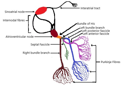
Figure 1. The Cardiac Conduction System.
The cardiovascular system's electrical properties have been predominantly studied in isolation, with limited consideration of potential specialized conduction pathways extending beyond the heart . Recent evidence suggests vascular structures possess unexpected conductive capabilities: gap junction-rich networks in vessel walls demonstrate coordinated electrical behavior , and coronary venous anatomy displays preferential conduction properties . Meanwhile, dermatological research has documented localized skin impedance changes during cardiac events that precede conventional diagnostic markers . These observations suggest an unexplored electrophysiological connection between heart and skin that cannot be fully explained by existing models.
2. Chronological Odyssey of Cardiac Electrophysiology: From Pulse to Electrical Code
The heart's electrical conduction system was decoded through centuries of breakthroughs. In 1628, William Harvey identified the heart as a mechanical pump, though its rhythm remained "nature’s silent pendulum" . Nearly 170 years later, Luigi Galvani (1791) discovered intrinsic animal electricity via frog leg contractions, a foundational bioelectrical insight .
The first anatomical clue emerged in 1839: Jan Evangelista Purkinje described unique sheep ventricular fibers (later Purkinje fibers), though their role was unknown . Technology leaped in 1887 when Augustus Waller recorded the first human electrocardiogram, calling it "the heart’s electrical shadow play" . Six years later, Wilhelm His Jr. (1893) identified the bundle of His, proving atrioventricular continuity .
Willem Einthoven transformed ECG recording in 1903 with the string galvanometer, capturing diagnostic "electrical hieroglyphs" . Subsequently, Sunao Tawara (1906) mapped the atrioventricular node ("delay gateway") and its connection to the Purkinje network, defining the modern pathway . Arthur Keith and Martin Flack (1907) discovered the sinoatrial node in the mole heart, the primary "spark igniting each heartbeat" . Thus, Harvey’s pump , Galvani’s bioelectricity , and Waller’s proto-ECG enabled understanding the conduction highway: SA node initiation , AV node modulation , His bundle bridging , and Purkinje fiber dispersion -deciphered via Einthoven’s ECG .
3. Hypothetical Framework for Cardiocutaneous Electrical Pathways
This framework proposes the existence of a direct extracardiac electrical system, termed Cardiocutaneous Electrical Pathways (CEP), which significantly contributes to the generation and detection of electrocardiographic signals at the cutaneous surface (Figures 2 and 3). The hypothesis aims to address limitations in current models of cardiac bioelectricity by integrating cardiac and peripheral electrical phenomena.
The components of the hypothesis are:
1) Limitations in current cardiac electrophysiology: The prevailing understanding-focused on the intracardiac conduction system and the volume conduction of electrical signals through interstitial fluid-does not fully explain the high-fidelity recording of precise phasic electrical signals at the skin surface seen in standard electrocardiograms.
2) Postulation of an extracardiac electrical system: A distinct electrical system, originating directly from the heart and extending to the skin, is proposed. This system operates in parallel with known conductive mechanisms.
3) Vascular alignment of the extracardiac system: This proposed extracardiac electrical network anatomically follows vascular channels, leveraging the physiological continuity between the heart and the skin through blood vessels that facilitate cutaneous circulation.
4) Cutaneous electrical field generation: The extracardiac system extends beyond cutaneous circulation to the skin surface, enabling the generation of a comprehensive cardiac electrical field across the body's cutaneous domain.
5) Electrocardiogram as CEP output: The ECG is conceptualized as the bioelectrical signature generated at the terminal ends of the cardiocutaneous electrical pathways, rather than merely a distant reflection of intracardiac activity.
6) Empirical correlation with blood supply interruption: The disappearance of ECG signals from a localized body region following interruption of its blood supply provides strong correlative evidence supporting the hypothesis that these pathways are functionally and anatomically linked to the vasculature.
7) Microscopic anatomical continuity: The cardiocutaneous electrical pathways (CEP) are conceptualized as specialized microscopic extensions of the intracardiac conduction system, forming a distinct yet continuous extracardiac network.
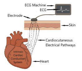
Figure 2. Hypothetical framework for the Cardiocutaneous Electrical Pathways (CEP) linking the heart with the skin in ECG generation.
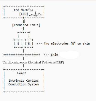
Figure 3. Schematic framework for the Cardiocutaneous Electrical Pathways (CEP) linking the heart with the skin in ECG generation, detection and recording at the skin surface.
4. Discussion
The introduction of Cardiocutaneous Electrical Pathways (CEP) as hypothetical specialized, extracardiac electrical system extending the heart's intrinsic conduction network to the skin surface represents a significant paradigm shift in understanding cardiac bioelectricity and electrocardiogram (ECG) genesis. This framework directly confronts the persistent limitations of the traditional volume conduction model, which struggles to account for the remarkable fidelity, regional specificity, and early ischemic sensitivity observed in surface ECG recordings despite signal traversal through highly heterogeneous and resistive thoracic tissues . While the intracardiac conduction system, meticulously elucidated by pioneers like Tawara, His, and Purkinje , impeccably coordinates myocardial excitation, CEP proposes a complementary anatomical and physiological substrate responsible for transmitting this electrical information directly to the integumentary system.
The core tenet of CEP hypothesis-its intimate anatomical association with the vascular tree-provides a compelling physiological rationale. Vascular channels offer a continuous, structured conduit from the heart's epicardium to the dermal microvasculature, establishing a natural "wiring harness" throughout the body. Evidence supporting this vascular link is not merely correlative but experimentally suggestive; the documented disappearance or significant attenuation of ECG signals from specific body regions following localized vascular occlusion or severance [Section 3, Point 6] strongly implies that the signal transmission mechanism is intrinsically dependent on patent blood flow or the integrity of the vascular wall structures themselves. This observation aligns poorly with passive volume conduction through static interstitial fluid but finds resonance in the emerging understanding of vascular electrophysiology. Vascular smooth muscle and endothelial cells are richly interconnected by gap junctions composed of connexins (notably Cx43 and Cx40), forming low-resistance electrical syncytia capable of coordinated electrical propagation along vessel walls . Furthermore, specialized structures like the coronary venous system have demonstrated preferential conduction properties distinct from surrounding myocardium , hinting at the existence of organized extracardiac conductive pathways potentially extending beyond the coronary circulation.
The hypothesis that CEP extends specifically to the skin surface, generating a comprehensive cardiac electrical field across the integument, offers a plausible explanation for several enigmatic observations. Regional variations in ECG signal amplitude, often poorly correlated with overlying tissue thickness or composition , could reflect the density, caliber, or specific branching patterns of underlying vascular/CEP structures in different body regions. The surprising sensitivity of localized skin impedance measurements to pre-ischemic cardiac events may represent bioelectrical alterations within the terminal CEP network or its cutaneous endpoints preceding detectable changes in myocardial bulk electrical activity. CEP hypothesis posits that the ECG electrode detects not a volume-conducted potential averaged across distant tissues, but rather the direct bioelectrical activity emanating from the terminal arborizations of these pathways at the dermal-epidermal junction [Section 3, Point 5]. This direct-source model inherently preserves waveform fidelity and temporal resolution, potentially explaining the clinical utility of the standard ECG despite theoretical signal degradation predicted by volume conduction models.
Conceptualizing CEP as a direct functional extension of the intracardiac conduction system necessitates consideration of its potential origins and microstructure. The pathways could arise as microscopic projections from Purkinje fibers terminating on the epicardial surface, potentially tracking along coronary vessels initially before integrating with the systemic vascular tree. Alternatively, CEP might represent a distinct, developmentally related network of specialized conductive cells embedded within or closely associated with the adventitia of major arteries and veins, functionally coupled to the heart at major vascular roots (aorta, pulmonary artery, vena cavae). The presence of interstitial cells of Cajal (ICC)-like cells or specialized conductive fibroblasts within vascular walls has been postulated in other contexts and could provide a cellular substrate for CEP. These specialized cells, rich in gap junctions and ion channels, could facilitate rapid, decremental conduction along vascular routes. The microscopic nature proposed for CEP suggests it may have eluded standard histological examination, requiring targeted investigation using specialized techniques like immunolabeling for specific connexins or ion channels combined with high-resolution micro-CT of vascular casts .
The CEP framework offers fertile ground for reinterpretation of existing phenomena and novel research avenues:
1) ECG Lead Placement Rationale: The empirical optimization of ECG lead positions (e.g., precordial leads V1-V6) over decades may inadvertently have positioned electrodes over cutaneous regions with high underlying vascular/CEP density or favorable geometric relationships to major cardiac vessels, rather than solely based on proximity to the heart .
2) Body Surface Potential Mapping (BSPM): The enhanced spatial resolution of BSPM, revealing complex potential distributions, could be mapping the varying surface expression of the underlying CEP network architecture more directly than volume conduction allows .
3) Early Ischemia Detection: The documented ability of localized ECG changes or specialized lead systems (e.g., posterior leads) to sometimes detect ischemia earlier than standard leads might reflect vulnerability or altered conduction within specific vascular/CEP branches supplying the affected myocardium or overlying skin region.
4) Arrhythmia Mechanisms: Certain unexplained arrhythmias or electrical phenomena (e.g., some atrial tachycardias, specific ventricular ectopy patterns) could potentially involve ectopic activity or re-entry circuits originating within or facilitated by the CEP network itself .
5) Electrodiagnostic Innovation: Confirmation of CEP could revolutionize ECG monitoring, leading to novel, highly sensitive cutaneous electrode designs targeting specific vascular landmarks, or non-invasive techniques to assess vascular bed integrity/function via CEP signal characteristics .
However, the CEP hypothesis faces significant challenges and necessitates rigorous validation. Direct anatomical evidence for these specialized microscopic pathways along systemic vasculature remains to be demonstrated. Sophisticated techniques combining micro-dissection, advanced 3D imaging (e.g., synchrotron radiation micro-CT), super-resolution microscopy, and specific molecular markers for conductive cells will be crucial . Electrophysiological confirmation requires developing methods to selectively stimulate or inhibit putative CEP elements in vivo or in situ and measure the effects on cutaneous cardiac potentials, distinct from myocardial or volume-conducted signals. Differentiating CEP contributions from the complex background of volume conduction and myogenic potentials (e.g., electromyogram) presents a major technical hurdle. Furthermore, the model must reconcile with the well-established principles of cardiac electrical field theory and the demonstrable effects of thoracic inhomogeneities on ECG morphology . CEP likely acts in concert with volume conduction, perhaps providing a high-fidelity "direct line" signal that is then modulated by the surrounding tissue properties, rather than completely replacing the traditional model.
5. Conclusion
The Cardiocutaneous Electrical Pathways (CEP) hypothesis presents a potentially transformative framework in cardiac electrophysiology. By postulating a specialized, vascular-associated extracardiac conduction system directly linking the heart's electrical activity to the skin surface, it directly addresses persistent limitations in the canonical volume conduction model for explaining ECG fidelity, regional specificity, and early ischemic sensitivity. While rooted in correlative clinical observations like the vascular dependence of regional ECG signals, CEP draws plausibility from the established conductive properties of vascular tissues mediated by gap junctions and the documented phenomena of preferential conduction within certain vascular structures . The framework reimagines the ECG not as a signal passively filtered through layers of tissue, but as the direct bioelectrical readout of activity at the terminal ends of an extensive cardiocutaneous network.
If substantiated, CEP would fundamentally alter our anatomical and physiological understanding of cardiac electrical propagation, revealing it as a truly systemic phenomenon extending far beyond the myocardium. This paradigm shift holds immense promise for advancing electrodiagnostics. It could provide a rational anatomical basis for optimizing ECG lead placement , enhance the interpretation of body surface potential maps , lead to the development of novel, highly sensitive biosensors for detecting early ischemia or arrhythmogenic foci via specific vascular beds , and potentially open entirely new avenues for understanding and treating electrical disorders involving the heart-periphery interface . The journey from Harvey's mechanical pump and Galvani's bioelectricity to Einthoven's ECG and the delineation of the intracardiac conduction system has been long and transformative. The CEP hypothesis represents the next potential frontier in this odyssey, challenging us to look beyond the heart itself and explore the intricate electrical highways that may connect its rhythmic beat to the surface of our skin. While significant empirical and experimental challenges lie ahead in confirming the existence, structure, and function of CEP , the potential rewards for cardiovascular science and medicine warrant its serious and rigorous investigation. This novel but hypothetic framework invites the scientific community to re-examine fundamental assumptions and embark on a new chapter in unraveling the body's electrical interconnectedness through experimental confirmation.
Abbreviations

CEP

Cardiocutaneous Electrical Pathways

ECG

Electrocardiogram

3D

Three Dimensional

CT

Computed Tomography

Author Contributions
Oluwadare Ogunlade is the sole author. The author read and approved the final manuscript.
Funding
This work is not supported by any external funding.
Conflicts of Interest
The authors declare no conflicts of interest.
References
[1] Laske TG, Iaizzo PA. The cardiac conduction system. In: Handbook of Cardiac Anatomy, Physiology, and Devices. 2005: 123-36.
[2] Wilson FN, et al. The distribution of the action currents produced by heart muscle and other excitable tissues immersed in extensive conducting media. J Gen Physiol. 1933; 16(3): 423-56.
[3] Rudy Y. The electrocardiogram and its relationship to excitation of the heart. In: Cardiac Electrophysiology: From Cell to Bedside. 7th ed. Elsevier; 2018. p. 299-312.
[4] Plonsey R. The nature of sources of bioelectric and biomagnetic fields. Biophys J. 1982; 39(3): 309-12.
[5] Burnes JE, et al. Noninvasive electrocardiographic imaging of substrate and intramural ventricular tachycardia in infarcted hearts. J Am Coll Cardiol. 2001; 38(7): 2071-8.
[6] Smith JM, Clancy EA. Value of unipolar leads in supplementary clinical diagnosis of myocardial infarction. Circulation. 1952; 5(6): 889-900.
[7] de Carvalho AP, et al. On the impulse propagation in cardiac muscle: The discontinuous aspect. Experientia. 1959; 15: 121-6.
[8] Little TL, et al. Connexin 43 expression in vascular smooth muscle. J Vasc Res. 1995; 32(2): 77-86.
[9] Durrer D, et al. Electrical properties of coronary venous system. Am Heart J. 1966; 72(5): 642-51.
[10] Antman EM, et al. Pre-hospital ECG identification of acute coronary syndromes. Ann Emerg Med. 2000; 35(4): S8.
[11] Silverman ME. William Harvey and the discovery of the circulation of blood. Clin Cardiol. 1985; 8(4): 244-246.
[12] Piccolino M. Luigi Galvani and animal electricity: two centuries after the foundation of electrophysiology. Trends Neurosci. 1997; 20(10): 443-448.
[13] Silverman ME, Hollman A. Discovery of the Purkinje cell in the human heart. Circulation. 2007; 115(5): e17-e18.
[14] Burch GE, DePasquale NP. A history of electrocardiography. Ann Med Hist. 1963; 5: 100-109.
[15] Acierno LJ. Wilhelm His Jr. and the bundle of His. Clin Cardiol. 1994; 17(12): 672-675.
[16] Burnett J. The origins of the electrocardiograph as a clinical instrument. Med Hist Suppl. 1985;(5): 53-76.
[17] Suma K. Sunao Tawara: a father of modern cardiology. Pacing Clin Electrophysiol. 2001; 24(1): 88-96.
[18] Silverman ME, Hollman A. Discovery of the sinus node by Keith and Flack: on the centennial of their 1907 publication. Heart. 2007; 93(10): 1184-1187.
[19] Haefliger JA, Nicod P, Meda P. Contribution of connexins to the function of the vascular wall. Cardiovasc Res. 2004; 62(2): 345-356.
[20] Hanna P, Shivkumar K, Ardell JL. Calming the nervous heart: Autonomic therapies in heart failure. Cardiovasc Res. 2023; 119(4): 1000-1016.
[21] Freeborn TJ, Mortada B, Jubran S. Advances in Electrical Impedance Methods for Skin Assessment. Sensors (Basel). 2022; 22(19): 7301.
[22] Kurahashi M, Zheng H, Dwyer L, Ward SM, Don Koh S, Sanders KM. A functional role for the 'fibroblast-like cells' in gastrointestinal smooth muscles. J Physiol. 2021; 599(5): 1509-1531.
[23] Chen K, Zhang Y, Qian L. Potential Role of Interstitial Cells in Vascular Conductivity: A Systematic Review. Front Physiol. 2022; 13: 875422.
[24] Uesugi K, Hoshino M, Takeuchi A, Suzuki Y. Four-dimensional imaging of vascular dynamics by synchrotron radiation micro-CT. Microscopy (Oxf). 2023; 72(2): 89-97.
[25] Kligfield P, Green CL, Mortara J, et al. Optimal ECG Lead Selection: Current Approaches and Future Perspectives. Ann Noninvasive Electrocardiol. 2022; 27(1): e12919.
[26] Wang Y, Rudy Y. Application of the Method of Fundamental Solutions to 3D Electrocardiographic Imaging. Comput Biol Med. 2023; 158: 106852.
[27] Birnbaum Y, Wilson JM, Fiol M, et al. New approaches for early detection of acute coronary syndromes. J Electrocardiol. 2023; 80: 1-8.
[28] Vaseghi M, Salavatian S, Rajendran PS, et al. Parasympathetic modulation and ventricular arrhythmia in myocardial infarction. Circ Res. 2021; 128(9): 1283-1296.
[29] Scardigli M, Crocini C, Ferrantini C, et al. Three-dimensional imaging of the cardiac conduction system in intact hearts. Cardiovasc Res. 2024; 120(1): 104-116.
[30] Geselowitz DB. Bioelectric sources in volume conductors: Historical perspectives. Annu Int Conf IEEE Eng Med Biol Soc. 2020; 2020: 108-111.
[31] Geselowitz, D. B. On bioelectric potentials in an inhomogeneous volume conductor. Biophys J. 1967; 7(1): 1-11.
Cite This Article
  • APA Style

    Ogunlade, O. (2025). Beyond the Intrinsic Cardiac Conduction System: A Hypothetical Framework for Cardiocutaneous Electrical Pathways (CEP) Linking Heart to Skin in ECG Generation. Science Discovery, 13(4), 60-65. https://doi.org/10.11648/j.sd.20251304.11

    Copy | Download

    ACS Style

    Ogunlade, O. Beyond the Intrinsic Cardiac Conduction System: A Hypothetical Framework for Cardiocutaneous Electrical Pathways (CEP) Linking Heart to Skin in ECG Generation. Sci. Discov. 2025, 13(4), 60-65. doi: 10.11648/j.sd.20251304.11

    Copy | Download

    AMA Style

    Ogunlade O. Beyond the Intrinsic Cardiac Conduction System: A Hypothetical Framework for Cardiocutaneous Electrical Pathways (CEP) Linking Heart to Skin in ECG Generation. Sci Discov. 2025;13(4):60-65. doi: 10.11648/j.sd.20251304.11

    Copy | Download

  • @article{10.11648/j.sd.20251304.11,
      author = {Oluwadare Ogunlade},
      title = {Beyond the Intrinsic Cardiac Conduction System: A Hypothetical Framework for Cardiocutaneous Electrical Pathways (CEP) Linking Heart to Skin in ECG Generation
    },
      journal = {Science Discovery},
      volume = {13},
      number = {4},
      pages = {60-65},
      doi = {10.11648/j.sd.20251304.11},
      url = {https://doi.org/10.11648/j.sd.20251304.11},
      eprint = {https://article.sciencepublishinggroup.com/pdf/10.11648.j.sd.20251304.11},
      abstract = {The existing paradigm of cardiac electrophysiology primarily focuses on the intracardiac conduction system and volume conduction through interstitial fluid to explain the electrocardiogram (ECG). However, this model faces limitations in fully elucidating the precise capture of phasic electrical signals at the cutaneous surface. This short communication introduces a novel anatomical and physiological framework postulating the existence of Cardiocutaneous Electrical Pathways (CEP) - a specialized, direct extracardiac electrical system connecting the heart to the skin. This framework proposes that CEP extends along the body's vascular channels, which establish the essential physiological link between the heart and the skin through cutaneous circulation. These pathways are hypothesized to extend beyond the immediate circulation to the skin surface, thus generating a comprehensive cardiac electrical field across the entire integument. Consequently, the electrocardiogram is presented as a direct bioelectrical manifestation resulting from the activity at the terminal ends of these CEP. Correlational evidence, such as the observed disappearance of ECG signals from a body region following localized blood supply severance, supports the intrinsic association of these pathways with vascular networks. The CEP framework suggests that this microscopic extracardiac electrical system functions as a direct extension of the intracardiac electrical system. This novel hypothesis challenges conventional understandings of cardiac bioelectricity, opening new avenues for research into the anatomical basis of cardiac electrical propagation and its systemic manifestations. Further exploration of CEP could revolutionize electrodiagnostic methods and offer new targets for understanding cardiovascular physiology and disease.},
     year = {2025}
    }
    

    Copy | Download

  • TY  - JOUR
    T1  - Beyond the Intrinsic Cardiac Conduction System: A Hypothetical Framework for Cardiocutaneous Electrical Pathways (CEP) Linking Heart to Skin in ECG Generation
    
    AU  - Oluwadare Ogunlade
    Y1  - 2025/07/23
    PY  - 2025
    N1  - https://doi.org/10.11648/j.sd.20251304.11
    DO  - 10.11648/j.sd.20251304.11
    T2  - Science Discovery
    JF  - Science Discovery
    JO  - Science Discovery
    SP  - 60
    EP  - 65
    PB  - Science Publishing Group
    SN  - 2331-0650
    UR  - https://doi.org/10.11648/j.sd.20251304.11
    AB  - The existing paradigm of cardiac electrophysiology primarily focuses on the intracardiac conduction system and volume conduction through interstitial fluid to explain the electrocardiogram (ECG). However, this model faces limitations in fully elucidating the precise capture of phasic electrical signals at the cutaneous surface. This short communication introduces a novel anatomical and physiological framework postulating the existence of Cardiocutaneous Electrical Pathways (CEP) - a specialized, direct extracardiac electrical system connecting the heart to the skin. This framework proposes that CEP extends along the body's vascular channels, which establish the essential physiological link between the heart and the skin through cutaneous circulation. These pathways are hypothesized to extend beyond the immediate circulation to the skin surface, thus generating a comprehensive cardiac electrical field across the entire integument. Consequently, the electrocardiogram is presented as a direct bioelectrical manifestation resulting from the activity at the terminal ends of these CEP. Correlational evidence, such as the observed disappearance of ECG signals from a body region following localized blood supply severance, supports the intrinsic association of these pathways with vascular networks. The CEP framework suggests that this microscopic extracardiac electrical system functions as a direct extension of the intracardiac electrical system. This novel hypothesis challenges conventional understandings of cardiac bioelectricity, opening new avenues for research into the anatomical basis of cardiac electrical propagation and its systemic manifestations. Further exploration of CEP could revolutionize electrodiagnostic methods and offer new targets for understanding cardiovascular physiology and disease.
    VL  - 13
    IS  - 4
    ER  - 

    Copy | Download

Author Information