1. Introduction
Infectious diseases in humans are illnesses brought on by bacteria, viruses, fungi, or parasites. Numerous microbes inhabit our bodies. Usually, they are neither harmful nor even beneficial. However, some microbes have the potential to cause illness in specific situations. Certain infectious diseases can spread from person to person. While some infections that are not life-threatening may be treated at home with rest and home treatments,
| [25] | Ranjbar, R., Bagheri, H., Ghasemi, F., Guest, P. C., & Sahebkar, A. (2021). Effects of curcumin and Its analogs on infectious diseases. Studies on Biomarkers and new targets in aging research in Iran: Focus on Turmeric and Curcumin. 75-101. https://doi.org/10.1007/978-3-030-56153-6_5 |
[25]
. Hospitals are the source of infection for many infectious disorders, including urinary tract infections caused by bacteria. Additionally, frequent and thorough hand washing helps shield us from the majority of infectious diseases
| [15] | Jump, R. L., Crnich, C. J., Mody, L., Bradley, S. F., Nicolle, L. E., & Yoshikawa, T. T. (2018). Infectious diseases in older adults of long‐term care facilities: update on approach to diagnosis and management. Journal of the American Geriatrics Society. 66(4), 789-803. https://doi.org/10.1111/jgs.15248 |
[15]
.
A little fragment of DNA contains instructions that are read by single-celled bacteria. We are surrounded by bacteria, which are also found on our skin and in our bodies
. While many bacteria are beneficial or even innocuous, certain bacteria can cause illness by releasing toxins.
Escherichia coli is a bacterium; a unicellular organism too small to be seen with the naked eye.
E. coli got its name, Escherichia, from the German pediatrician Theodor Escherich, who discovered the bacterium in 1885. Many animals, including humans, have gastrointestinal tracts or intestines where the majority of
E. coli reside and grow innocuously
.
Unfortunately, the
E. coli strains that people are most familiar with are the ones that cause disease.
Escherichia coli is a gram-negative, motile, facultatively anaerobic, rod-shaped bacterium, non-spore-forming, flagellated.
Escherichia coli continues to be a major concern in clinical microbiology research offices because of the heavy workload related to UTIs. Such bacterial contamination is urinary tract infections (UTI), including the nearness of microbes within the urinary tract which is sterile
| [20] | Nas, F. S., Ali, M., Abdallah, M. S., & Zage, A. U. (2019). Prevalence and antibiotic susceptibility pattern of Escherichia coli isolated from urine samples of urinary tract infection patients. ARC Journal of Urology. 4(1), 14-20. https://doi.org/10.20431/2456‑060X.0401004 |
[20]
. A urinary tract contamination may be a condition in which one or more parts of the urinary framework (the kidneys, ureters, bladder, and urethra) get sullied
.
Virulence factors linked to the pathogenicity of uropathogenic
E. coli are diverse and possess various functions, ranging from facilitating bacterial colonization to enhancing virulence. These factors are typically encoded on pathogenicity islands (PAIs), plasmids, and other mobile genetic elements. These are the molecules that bacteria make that allow them to enter a host, get past the host's defenses, and spread illness. These factors are secretory, membrane-associated, or cytosolic in nature strains of UPEC.
E. coli virulence factors can be broadly categorized into two main types: surface virulence factors and export factors, as identified by Bien
et al.
| [4] | Bien, J., Sokolova, O., & Bozko, P. (2012). Role of uropathogenic Escherichia coli virulence factors in the development of urinary tract infection and kidney damage. International journal of nephrology. 1-11. https://doi.org/10.1155/2012/681473 |
[4]
. The primary surface virulence elements of UPEC comprise lipopolysaccharide, capsule, type 1 fimbriae, and P fimbriae, crucial for initiating colonization within the urinary tract. Additionally, surface-displayed virulence factors such as hemolysin, cytotoxic necrotizing factor 1, cytolethal distending toxin, secreted autotransporter toxin, and siderophore facilitate nutrient acquisition from the host cells
| [22] | Pakbin, B., Brück, W. M., & Rossen, J. W. (2021). Virulence factors of enteric pathogenic Escherichia coli: A review. International journal of molecular sciences. 22(18), 9922. https://doi.org/10.3390/ijms22189922 |
[22]
.
Fimbriae adhesins such as
PapG and
CsgA are hurtful components that energize the association of
E. coli | [6] | Hrovat, K., Molan, K., Seme, K. (2024). Molecular characterization of extended-spectrum β-lactamase-producing Escherichia coli isolated from lower respiratory tract samples between 2002 and 2019 in the Central Slovenia region. Ann Clin Microbiol Antimicrob 23, 6. https://doi.org/10.1186/s12941-023-00664-1 |
[6]
. Close to these mechanisms, UPEC can undermine the host's immune system through various means, including damage and evasion of host defense systems, collectively referred to as released damaging tendency components. The era of these damaging tendency factors by UPEC may cause a provocative response which makes a conceivable pathway for UTI signs. In this review, various critical virulence factors of UPEC are discussed, particularly those encoded within pathogenicity islands. These factors encompass adherence mechanisms, toxins, and evasion strategies, collectively constituting significant risk factors for UPEC pathogenicity. This review aims to provide a comprehensive analysis of the major virulence factors associated with UPEC and to evaluate their specific roles in the pathogenesis of UTIs.
2. Review of the Literature
2.1. Surface Virulence Factors
The urine of an uninfected person is sterile due to the urinary stream and antimicrobial development of uric destruction. A typical stream of urine does not allow the microorganism to colonize the insides of the urinary tract. However, the adherence of E. coli to uroepithelial cells enables them to resist the impact of urinary flow. Surface damaging tendency components of UPEC incorporate several unmistakable sorts of cement organelles (fimbriae), which progress bacterial association to have tissues interior the urinary tract
| [4] | Bien, J., Sokolova, O., & Bozko, P. (2012). Role of uropathogenic Escherichia coli virulence factors in the development of urinary tract infection and kidney damage. International journal of nephrology. 1-11. https://doi.org/10.1155/2012/681473 |
[4]
. The presence of cement molecules (adhesins) by UPEC is the first basic determinant of pathogenicity. UPEC adhesins contribute to harm in various ways: (i) by activating host and bacterial signaling pathways, (ii) by facilitating the delivery of bacterial components to host tissues, and (iii) by promoting bacterial invasion.
Numerous pathogenic microorganisms are considered as the essential step inside the colonization plans and both the have and
E. coli work in this handle. UPEC's ability to colonize relies on the expression of specific fimbrial adhesins. For a viable adherence to the have cell surface, UPEC communicates various adherence components that are imperative for association and in this way regarded as harmfulness components. Bacterial pathogens must adhere to host cells to colonize the body, a process known as tissue tropism. This involves specific interactions with target receptors and tissue surfaces. Various surface structures play a significant role in facilitating specific adhesion
| [29] | Sarowska, J., Futoma-Koloch, B., Jama-Kmiecik, A., Frej-Madrzak, M., Ksiazczyk, M., Bugla-Ploskonska, G., & Choroszy-Krol, I. (2019). Virulence factors, prevalence and potential transmission of extraintestinal pathogenic Escherichia coli isolated from different sources: recent reports. Gut pathogens. 11, 1-16. https://doi.org/10.1186/s13099‑019‑0305‑2 |
[29]
.
2.1.1. Fimbriae
Various bacterial adhesions are organized in a lean filamentous structure called fimbriae or pili showing disdain toward the reality that there are affirmations of the closeness of adhesins inside the cell surface of organisms. Adhesins of fimbrial nature are crucial amid association handling
| [7] | Gahlot, D. K., Taheri, N., & MacIntyre, S. (2022). Diversity in the genetic regulation of bacterial fimbriae assembled by the chaperone usher pathway. International Journal of Molecular Sciences. 24(1), 161. https://doi.org/10.3390/ijms24010161 |
[7]
. Fimbriae are long hair-like structures contained inside the cell
. Microbial pros and inclining components surface of organisms that recognize specific compounds commonly carbohydrates of the target have cells. Fimbriae contain oligomeric pilin proteins. These slim, helical, circular, and hollow proteinaceous structures, shorter than flagella, are expressed in uropathogenic strains of E. coli and are regarded as harmful components due to their role in specific adhesion. Most of the receptor’s fimbriae are carbohydrates. They consolidate type 1 fimbriae, P fimbriae, and incline aggregative fimbriae. Various bacterial pathogens can make a cluster of these adhesins, and the regular obstacle of a single grip may fetch adequate for a bacterium to lose its danger. In gram-negative tiny living beings like UPEC, adhesins are uncovered by a chaperone-usher-assisted pathway. This pathway includes two proteins, one may be a periplasmic chaperone, and the other may be a protein called usher. The usher serves as the foundation of the structure, while the chaperone's role involves folding and recruiting the subunits
. Usher makes a differentiation to make the fimbriae and its transportation through the exterior guaranteeing cleverness of the exterior layer. In uropathogenic
E. coli strains, chaperone-usher family fimbriae are more abundant.
Type 1 fimbriae: Type 1 fimbriae are common among
E. coli strains from all clinical categories of UTI
| [7] | Gahlot, D. K., Taheri, N., & MacIntyre, S. (2022). Diversity in the genetic regulation of bacterial fimbriae assembled by the chaperone usher pathway. International Journal of Molecular Sciences. 24(1), 161. https://doi.org/10.3390/ijms24010161 |
[7]
. The adherence of type 1-fimbriated strains to have cells within the urinary tract may advance the advancement of cystitis, their adherence to and incitement of hPMNLs may advance bacterial murdering but may moreover contribute to renal scarring. Several E. coli strains, qualities to encode type 1 fimbriae are shown
| [33] | Vigil, Patrick D., Christopher J. Alteri, and Harry LT Mobley., (2011). Identification of in vivo-induced antigens including an RTX family exoprotein required for uropathogenic Escherichia coli virulence. Infection and Immunity. 6, 35-44. https://doi.org/10.1128/IAI.00110-11 |
[33]
, and amid urinary tract contaminations, they hurt urinary tract cells by mediating an extended bothering.
In organize to enter into the cells of the urinary tract; Type 1 fimbriae play an extraordinary portion. Type 1 fimbriae are shockingly adaptable damaging tendency factors of UPEC that can stabilize the association of the organisms to different types of cells all through the urinary tract. Strong enjoying of Type 1 fimbriae was found in proximal tubules and vessel dividers. Inside the bladder, they tie unequivocally to solid layers and decently to vessel dividers. Receptors for type 1 fimbriae were additionally found inside the distal tubules and collecting channels. They can start their official to the surface of macrophages
. Type 1 fimbriae recognize uroplakin from bladder epithelial cells as well as mannose-containing proteins. Colonization by type 1 fimbriae may be restored by expressing plasmids containing appropriate subunits.
FimH in affiliation with LPS can stimulate toll-like receptor 4 (TLR4) to begin a particular signaling cascade that will order the humoral safe response. Various considerations approximately revealed that the expression of type 1 fimbriae comes almost in damaging tendency and incident of expression
. In any case; type 1 fimbriae-mediated association might be a significant arrange of cystitis. The cement tip of these fimbriae,
FimH, ties to α-D-mannosylated proteins such as UPs, which are communicated by the separated urothelium and in this way relates this adhesin to the lower urinary tract
. Due to its authoritative liking,
FimH-mediated attachment or aggregation of eukaryotic cells (red blood cells) is hindered by mannose. Authoritative of FimH to its receptors intervenes in attachment and invasion.
P fimbriae: P fimbriae are vital within the pathogenesis of UTI since they intercede alpha-
DGalp-(1-4)-beta-D-Galp adherence to epithelial cells inside the human urinary tract, subsequently allowing bacterial colonization and fortifying inflammation
. In compromised the prerequisite for P fimbriae in starting genuine UTI is diminished, proposing that P fimbriae are essential for
E. coli to overcome certain components of the types have a defense framework. Anti-P-fimbriae resistance secures humans against renal contamination with homologous P-fimbriated strains, but the serological differences of P fimbriae and the restricted effect of anti-P-fimbrial antibodies on adherence complicate endeavors to create anti-P-fimbrial antibodies for human utilize. Inside the kidney, they tie unequivocally to Bowman's capsule, glomerulus, and endothelial cells of vessel dividers. P fimbriae communicated
E. coli enters the urinary tract; they construct up bacteriuria and offer help to cross the epithelial boundary to enter the circulatory framework and can cause hemagglutination of erythrocytes
.
There are at smallest nine qualities inside the pap quality cluster with two restriction regions at two closes. The regulatory portion starts the taking after Eco R1 comprising of papI and papa. At that point
papA, papH, papC, papD, papE, papF, and
papG are organized. Around 1000 subunits outline a P fimbria, being joined together in a helical way. The protein
PapC, which is the greatest one with 80 KD of mass, makes a difference in the strategy by transporting the subunits outside the portion of the cell. The minor subunits at the tip of the fimbria choose the specificity of the receptor.
PapH closes the get-together of the fimbriae and joins in this way portion of
PapG was found in several varieties of P fimbriae which means they can begin subunit polymerization
| [7] | Gahlot, D. K., Taheri, N., & MacIntyre, S. (2022). Diversity in the genetic regulation of bacterial fimbriae assembled by the chaperone usher pathway. International Journal of Molecular Sciences. 24(1), 161. https://doi.org/10.3390/ijms24010161 |
[7]
.
Various tests show that expression of these fimbriae isn't important to urinary tract defilement, though more present-day other tests have concluded around their portion in pathogenesis. It shows disdain toward the reality that P fimbriae can begin incendiary responses by actuating TLR4
. It secures UPEC from human polymorph nucleus leukocytes (hPMNLs). In the dynamic environment of the urinary tract, conditions influence the expression of P fimbriae.
Thin aggregative fimbriae (Curli): Curli strands are extracellular amyloid fibrils that are dynamically communicated by
E. coli. Characteristic of amyloids, curli strands are exceedingly steady, insoluble, tall molecular-weight protein complexes ruled by a beta-sheet auxiliary structure
| [11] | Hung, C., Marschall, J., Burnham, C. A. D., Byun, A. S., & Henderson, J. P. (2014). The bacterial amyloid curli is associated with urinary source bloodstream infection. PloS one. 9(1), 86-89. https://doi.org/10.1371/journal.pone.0086009 |
[11]
. Curli is the as it is known amyloid filament encoded by
E. coli and other Enterobacteriaceae.
Curli filaments are intentionally gathered by committed bacterial apparatus. Curli's advanced adherence to epithelial cells and resistance against the human antimicrobial peptide, additionally cause acceptance of the proinflammatory cytokine IL-8. They display a select part in advancing UPEC biofilms and speak to one of the major biofilm components
| [5] | Foroogh, N., Rezvan, M., Ahmad, K., & Mahmood, S. (2021). Structural and functional characterization of the FimH adhesin of uropathogenic Escherichia coli and its novel applications. Microbial Pathogenesis, 161, 105288. https://doi.org/10.1016/j.micpath.2021.105288 |
[5]
. Curli is delivered at the restriction of supplements and salts, at decreased oxygen pressure, and at a temperature below 30°C. In any case, it is accepted that numerous pathogenic microbes and commensal strains can moreover express curli at 37°C amid disease in people.
F1C/ S fimbriae: F1C may be a destructiveness figure capable of urinary tract diseases, which is encoded by an operon of seven qualities, i.e., focAICDFGH, where FocA is the major subunit and FocH is the tip adhesin
| [3] | Arafi, V., Hasani, A., Sadeghi, J., Varshochi, M., Poortahmasebi, V., Hasani, A., & Hasani, R. (2023). Uropathogenic E. coli endeavors: An insight into the characteristic features, resistance mechanism, and treatment choice. Archives of Microbiology. 205(6), 226. https://doi.org/10.1007/s00203-023-03553-5 |
[3]
. F1C receptors are shown in the bladder endothelium and solid layer. They tie to glomeruli, distal tubules, collecting channels, and vascular endothelial cells. Thinks about appears that F1C fimbriae and pyelonephritis are correlated though there's a small contrast within the predominance of type 1 fimbriae in UTI strains. The predominance of F1C fimbriae is 16% in UTI strains
| [3] | Arafi, V., Hasani, A., Sadeghi, J., Varshochi, M., Poortahmasebi, V., Hasani, A., & Hasani, R. (2023). Uropathogenic E. coli endeavors: An insight into the characteristic features, resistance mechanism, and treatment choice. Archives of Microbiology. 205(6), 226. https://doi.org/10.1007/s00203-023-03553-5 |
[3]
. S fimbriae are hereditarily indistinguishable from F1C fimbriae and contrast as it were by the tip adhesin SfaS. Criteria that are required to be recognized as harmfulness calculates were decided by distinctive ponders concerning S fimbriae.
2.1.2. Lipopolysaccharide
Lipopolysaccharide (LPS) is an irreplaceably component of the cell divider and comprises the exceedingly protected lipid and repeating O-antigen subunits that differentiate massively between strains based on the sugar build-ups and their linkage plans interior the rehashing subunits
| [28] | Sarkar, S., Ulett, G. C., Totsika, M., Phan, M. D., & Schembri, M. A. (2014). Role of capsule and O antigen in the virulence of uropathogenic Escherichia coli. PloS one. 9(4), 94786. https://doi.org/10.1371/journal.pone.0094786 |
[28]
. LPS is especially well known to enact a response and to incite nitric oxide and cytokine (IL-1, TNF-α) era which progresses the incendiary response. It additionally activates the amalgamation of specific antibodies to the considerable antigen and applies an immune-adjuvant effect that propels the humoral safe response to other antigens of the pathogen. Be that it may, certain antigenic sorts of LPS are also included in the resistance of the pathogen to the killing effect of the commonplace human serum. In animal studies, severe renal failure due to LPS depends on systemic response rather than kidney TLR4 expression. However, LPS's role in UTI-related renal failure in patients is still unclear.
2.1.3. Flagellum
Flagella is an organelle that's capable of bacterial motility and plays a part in the beginning attachment stage of biofilm arrangement. A recent study showed that motility is included in the relocation of the disease from the bladder to the kidneys
| [3] | Arafi, V., Hasani, A., Sadeghi, J., Varshochi, M., Poortahmasebi, V., Hasani, A., & Hasani, R. (2023). Uropathogenic E. coli endeavors: An insight into the characteristic features, resistance mechanism, and treatment choice. Archives of Microbiology. 205(6), 226. https://doi.org/10.1007/s00203-023-03553-5 |
[3]
. Approximately 70–90% of all urinary tract contaminations are caused by flagellated UPEC, and pathogenesis includes contact between the microbes and epithelial cell surface of the urinary tract. Be that as it may, flagella motility upgrades the capacity of
E. coli by versatile reactions to appealing or repellent natural
| [35] | Zegadło, K., Gieroń, M., Żarnowiec, P., Durlik-Popińska, K., Kręcisz, B., Kaca, W., & Czerwonka, G. (2023). Bacterial motility and its role in skin and wound infections. International Journal of Molecular Sciences. 24(2), 1707. https://doi.org/10.3390/ijms24021707 |
[35]
.
2.1.4. Capsule
The primary function of the capsule is to envelop the outer surface of the cell wall, shielding bacteria from phagocytosis and the bactericidal effects of the complement system. Its main purpose is to provide a protective layer, safeguarding the bacterium from various adverse conditions. This protective framework primarily consists of polysaccharides
| [4] | Bien, J., Sokolova, O., & Bozko, P. (2012). Role of uropathogenic Escherichia coli virulence factors in the development of urinary tract infection and kidney damage. International journal of nephrology. 1-11. https://doi.org/10.1155/2012/681473 |
[4]
. The capsule protects against engulfment, complement-mediated bactericidal effects, antimicrobial resistance, and antiserum action in the host
| [13] | Jahandeh, N., Ranjbar, R., Behzadi, P., & Behzadi, E. (2015). Uropathogenic Escherichia coli virulence genes: invaluable approaches for designing DNA microarray probes. Central European Journal of Urology. 68(4), 452. https://doi.org/10.5173/ceju.2015.625 |
[13]
. Certain capsular, such as K1 and K5, avoid an appropriate humoral safe reaction of the contaminated by appearing as an atomic mimicry to tissue components. The K1 polysaccharide, connected to a sialic corrosive homopolymer, includes an exceptionally vital part in advancement as well as within the numerous stages of UTI pathogenesis.
2.2. Exported Virulence Factors
2.2.1. Toxins
Poisons offer assistance to the pathogen spreading into more profound tissues after disturbing cell judgment; to pick up get to supplements interior they have cell; or to crush safe effector cells and in this way sidestep their potential antibacterial action
. A few poisonous substances or proteins emitted by uropathogenic strains of
E. coli play a considerable part as harmful components in UTIs. Be that as it may, poisons can modify the cell signaling cascade and balance fiery reactions.
A few in vitro and in vivo studies appeared that poisons too contribute to the incitement of the cell passing and discharging of fundamental supplements, which give the capacity to get to more profound tissues inside the urinary tract
. In 1987, CDT poison (Cyclomodulins) was, to begin with, detailed as harmful poison in UPEC
| [31] | Soltani, S., Emamie, A. D., Dastranj, M., Farahani, A., Davoodabadi, A., & Mohajeri, P. (2018). Role of toxins of uropathogenic Escherichia coli in the development of urinary tract infection. British Journal of Pharmaceutical Research. 21(1). https://doi.org/10.9734/BJPR/2018/42340 |
[31]
, which opened an unused entryway within the think of the pathogenesis of UTIs.
2.2.2. Α-hemolysin
Hemolysin production is associated with human pathogenic strains of
E. coli, especially those causing more clinically severe forms of UTI. The pro-virulence activity of hemolysin is multifactorial, including disruption of phagocyte function, and direct toxicity to host tissues. Antihemolysin immunity protects against infection with hemolytic strains and should be explored for human use. Among toxins, α-hemolysin (HlyA) is noteworthy, being a lipoprotein and part of the RTX (repeats in toxin) toxin family
| [2] | Al-Dulaimi, T. H., Bunyan, I. A., & Banimuslem, T. A. (2022). Genomic analysis study of virulence potential α-hemolysin from uropathogenic E. coli. Journal of Complementary Medicine Research. 13(4), 164-164. https://doi.org/10.5455/jcmr.2022.13.04.28 |
[2]
. It acts as a pore-forming toxin, leading to cell membrane damage and apoptosis mediated by inducible nitric oxide synthase (iNOS). HlyA breaks erythrocytes and nucleated cells, potentially compromising host immune cells, and aiding UPEC's access to host nutrients and iron stores
| [4] | Bien, J., Sokolova, O., & Bozko, P. (2012). Role of uropathogenic Escherichia coli virulence factors in the development of urinary tract infection and kidney damage. International journal of nephrology. 1-11. https://doi.org/10.1155/2012/681473 |
[4]
. But when the concentration is low, HlyA can actuate the apoptosis of target cells and advance the peeling of bladder epithelial cells
| [3] | Arafi, V., Hasani, A., Sadeghi, J., Varshochi, M., Poortahmasebi, V., Hasani, A., & Hasani, R. (2023). Uropathogenic E. coli endeavors: An insight into the characteristic features, resistance mechanism, and treatment choice. Archives of Microbiology. 205(6), 226. https://doi.org/10.1007/s00203-023-03553-5 |
[3]
.
A later ponder appeared that HlyA controls the dephosphorylation, which may be a multifunctional signaling regulator and mindful for controlling provocative reactions within the have, as well as the cell cycle control. HlyA has the part within the expanded generation of IL-6 and IL-8 by actuating Ca2+ motions in renal epithelial cells.
2.2.3. Cytotoxic Necrotizing Factor 1
Cytotoxic necrotizing factor 1 (CNF1) is a virulence factor produced by
E. coli, is plays a role in UTIs by promoting actin stress fiber formation and facilitating E. coli entry into cells
| [16] | Loubet, P., Ranfaing, J., Dinh, A., Dunyach-Remy, C., Bernard, L., Bruyère, F., & Sotto, A. (2020). Alternative therapeutic options to antibiotics for the treatment of urinary tract infections. Frontiers in microbiology. 11, 1509. https://doi.org/10.3389/fmicb.2020.01509 |
[16]
. This toxin has a significant effect on the actin cytoskeleton, leading to the formation of large vacuoles within cells
| [31] | Soltani, S., Emamie, A. D., Dastranj, M., Farahani, A., Davoodabadi, A., & Mohajeri, P. (2018). Role of toxins of uropathogenic Escherichia coli in the development of urinary tract infection. British Journal of Pharmaceutical Research. 21(1). https://doi.org/10.9734/BJPR/2018/42340 |
[31]
. However, a few in vitro and in vivo consider appeared that this protein interferes with polymorphonuclear phagocytosis and inspires apoptotic passing of bladder epithelial cells. In expansion, there's moreover a plausibility of the association of CNF1 with the hemolysin within the harmfulness component, which is useful for the microbes
| [35] | Zegadło, K., Gieroń, M., Żarnowiec, P., Durlik-Popińska, K., Kręcisz, B., Kaca, W., & Czerwonka, G. (2023). Bacterial motility and its role in skin and wound infections. International Journal of Molecular Sciences. 24(2), 1707. https://doi.org/10.3390/ijms24021707 |
[35]
.
2.2.4. Secreted Autotransporter Toxin (SAT)
Secreted autotransporter toxin (SAT) could be significant as a virulence factor in the development of UTIs due to its toxin activity against cell lines originating from the bladder or kidney. SAT is a serine protease autotransporter belonging to a subgroup of autotransporters recently categorized as the SPATE (serine protease autotransporters of
Enterobacteriaceae) family, often found in pyelonephritic
E. coli strains
| [9] | Habouria, H., Pokharel, P., Maris, S., Garénaux, A., Bessaiah, H., Houle, S., & Dozois, C. M. (2019). Three new serine-protease autotransporters of Enterobacteriaceae (SPATEs) from extra-intestinal pathogenic Escherichia coli and combined role of SPATEs for cytotoxicity and colonization of the mouse kidney. Virulence. 10(1), 568-587. https://doi.org/10.1080/21505594.2019.1624102 |
[9]
. SAT may exert cytopathic effects, damaging host tissue and potentially enhancing UPEC proliferation. Additionally, this toxin might facilitate the entry of pyelonephritogenic strains into the bloodstream through specific damage to the glomeruli and proximal tubules
| [35] | Zegadło, K., Gieroń, M., Żarnowiec, P., Durlik-Popińska, K., Kręcisz, B., Kaca, W., & Czerwonka, G. (2023). Bacterial motility and its role in skin and wound infections. International Journal of Molecular Sciences. 24(2), 1707. https://doi.org/10.3390/ijms24021707 |
[35]
.
2.2.5. Cytolethal Distending Toxin (CDT)
The cytolethal-distending toxin, notable for its ability to harm the DNA of target cells, was initially discovered in pathogenic
E. coli in 1987
| [31] | Soltani, S., Emamie, A. D., Dastranj, M., Farahani, A., Davoodabadi, A., & Mohajeri, P. (2018). Role of toxins of uropathogenic Escherichia coli in the development of urinary tract infection. British Journal of Pharmaceutical Research. 21(1). https://doi.org/10.9734/BJPR/2018/42340 |
[31]
. This toxin can disrupt the cell cycle and contribute to the development of UTIs. CDT originates from an operon containing three proteins: CdtA, CdtB, and CdtC, which are encoded by the cdtA, cdtB, and cdtC genes, respectively
| [23] | Parvez, S. A., & Rahman, D. (2018). Virulence factors of uropathogenic E. coli. Microbiology of Urinary Tract Infections-Microbial Agents and Predisposing Factors. 7-21. https://doi.org/10.5772/intechopen.79557 |
[23]
. CDT has DNase I-like enzymatic movement and assaults DNA, whereas the other bacterial poisons assault the cell layer or diverse targets inside the cytoplasm. This special property of assaulting DNA harms the target cell DNA which comes about in dynamic cell distending driving to cell passing
| [31] | Soltani, S., Emamie, A. D., Dastranj, M., Farahani, A., Davoodabadi, A., & Mohajeri, P. (2018). Role of toxins of uropathogenic Escherichia coli in the development of urinary tract infection. British Journal of Pharmaceutical Research. 21(1). https://doi.org/10.9734/BJPR/2018/42340 |
[31]
.
2.3. Iron Acquisition Systems
Siderophores are little natural atoms created by microorganisms beneath iron-limiting conditions which enhance the uptake of iron to the microorganisms
| [26] | Saha, M., Sarkar, S., Sarkar, B., Sharma, B. K., Bhattacharjee, S., & Tribedi, P. (2016). Microbial siderophores and their potential applications: a review. Environmental Science and Pollution Research. 23, 39-45. https://doi.org/10.1007/s11356-015-4294-0 |
[26]
. Iron is crucial for various biological processes, including oxygen transport, DNA synthesis, electron transport, and peroxide metabolism in E. coli. However, iron availability decreases in the host urinary tract during UTIs
| [32] | Sora, V. M., Meroni, G., Martino, P. A., Soggiu, A., Bonizzi, L., & Zecconi, A. (2021). Extraintestinal pathogenic Escherichia coli: Virulence factors and antibiotic resistance. Pathogens. 10(11), 1355. https://doi.org/10.3390/pathogens10111355 |
[32]
. In reaction to this,
E. coli has a few numerous practically repetitive frameworks that intercede press take-up by emitting low-molecular-weight Fe
3+ chelating atoms.
Iron utilization, interceded by these siderophores, is basic for the colonization of the urinary tract by UPEC.
E. coli possesses four siderophore systems: yersiniabactin, aerobactin, enterobactin, and salmochelin
| [23] | Parvez, S. A., & Rahman, D. (2018). Virulence factors of uropathogenic E. coli. Microbiology of Urinary Tract Infections-Microbial Agents and Predisposing Factors. 7-21. https://doi.org/10.5772/intechopen.79557 |
[23]
. These systems are activated in low-iron environments and are controlled by ferrous iron and the ferric uptake regulator Fur (
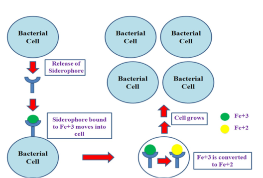
Figure 1).
Under iron-limited conditions, the bacterial cell releases a siderophore. The siderophore forms a complex with insoluble ferric ions (Fe
3+), which then bind to the surface of the bacterial cell. This complex is transported into the cell, where the insoluble ferric iron (Fe
3+) is transformed into the soluble ferrous form (Fe
2+). Siderophores are either degraded within the bacterial cell or released into the extracellular environment in their free form. The bacterial cell then utilizes this ferrous iron for its growth, leading to an increase in its population
| [23] | Parvez, S. A., & Rahman, D. (2018). Virulence factors of uropathogenic E. coli. Microbiology of Urinary Tract Infections-Microbial Agents and Predisposing Factors. 7-21. https://doi.org/10.5772/intechopen.79557 |
[23]
.
2.3.1. Aerobactin
Aerobactin could be a little particle shaped from the condensation of two lysine particles and one citrate taken after discharge by
E. coli cells; aerobactin extricates (separate) Fe3+ from iron-binding proteins and is taken up through external film receptor protein
. Strains with the aerobactin framework have a development advantage in low-iron conditions. The chromosomal aerobactin framework is related to other uropathogenic harmfulness component determinants, though the plasmid aerobactin framework is often carried by plasmids encoding different antimicrobial specialist resistance
.
2.3.2. Enterobactin
Enterobactin, another specialized and widely prevalent catecholate siderophore, is less soluble and stable than aerobactin. However, it exhibits a higher affinity for iron and can bind transferrin more rapidly than aerobactin in a liquid environment. In any case, the press is discharged from enterobactin through the hydrolysis of this siderophore
| [30] | Sheldon, J. R., Laakso, H. A., & Heinrichs, D. E. (2016). Iron acquisition strategies of bacterial pathogens. Virulence Mechanisms of Bacterial Pathogens. 43-85. https://doi.org/10.1128/9781555818265.ch3 |
[30]
. Apart from these, enterobactin enables UPEC to thrive in iron-limited environments like the urinary tract. However, this siderophore has a drawback as it can be deactivated by proteins like serum albumin and siderocalin, which are components of mammalian defense mechanisms functioning as antimicrobial agents
| [30] | Sheldon, J. R., Laakso, H. A., & Heinrichs, D. E. (2016). Iron acquisition strategies of bacterial pathogens. Virulence Mechanisms of Bacterial Pathogens. 43-85. https://doi.org/10.1128/9781555818265.ch3 |
[30]
.
2.3.3. Yersiniabactin
A phenolate siderophore is prevalent in Enterobacteriaceae, including
E. coli, and is encoded on the high-pathogenicity island
. Yersiniabactin includes a tall iron partiality and delivers yersiniabactin-Fe
3+ complex official to the press atom which recognizes the particular bacterial external film TonB-dependent receptor and Fyu (Psn). The press molecule is discharged from yersiniabactin inside the cytosol with the help of membrane-embedded proteins
. In expansion, this siderophore increments resistance to copper push by chelating Cu
2+.
2.3.4. Salmochelin
Enterobacteriaceae, including
E. coli, produce a glucosylated form of enterobactin, termed salmochelin, to evade host immune responses like siderocalin recognition. Salmochelin is synthesized by glucosylation via glucosyltransferase, preventing its binding by siderocalin. However, recent studies indicate salmochelin may facilitate urothelial cell invasion, implicating iron in urinary tract infections
| [27] | Sargun, A., Johnstone, T. C., Zhi, H., Raffatellu, M., & Nolan, E. M. (2021). Enterobactin-and salmochelin-β-lactam conjugates induce cell morphologies consistent with inhibition of penicillin-binding proteins in uropathogenic Escherichia coli CFT073. Chemical science. 12(11), 4041-4056. https://doi.org/10.1039/D0SC04337K |
[27]
. Apart from enterobactin, the hemin uptake system, involving ChuA and Hma, is crucial for iron acquisition during UTIs and biofilm formation. ChuA expression is regulated by RfaH, while Hma operates independently, both aiding in hemin utilization essential for efficient kidney colonization
| [8] | Garcia, E. C., Brumbaugh, A. R., & Mobley, H. L. (2011). Redundancy and specificity of Escherichia coli iron acquisition systems during urinary tract infection. Infection and immunity. 79(3), 25-35. https://doi.org/10.1128/IAI.01222-10 |
[8]
.