
 ,
Friday Obinwa Uhegbu,
Peter Anani Dabesor,
Amaka Omekara Maduabuchi,
Christiana Ezekiel Atat
,
Friday Obinwa Uhegbu,
Peter Anani Dabesor,
Amaka Omekara Maduabuchi,
Christiana Ezekiel Atat
In addition to the highly valued sweet and aromatic fragrance of Tetrapleura tetraptera fruit, it is used in ethnomedicine to treat and manage a wide range of diseased conditions. In this study, the methanol extract of the fruits of Tetrapleura tetraptera was analyzed by Gas Chromatography - Mass Spectrometry (GC-MS). Twenty-six different phytochemical compounds and their percentages have been characterized including: 3,4-Dihydroxyphenyl(2,3-trans-Catechin) (3.10%), 9-Octadecenal (3.96%), hexadecane (3.16%), pinene (4.80%), 1-methyl-4-cyclohexene (7.45%), humulene (3.60%), carbonic acid (3.34%), ferulic acid (4.03%), 5,6-dihydroxy-7-Oglucuronide flavone (22.46%), 2,4-Di-tert-butylphenol (8.08%) and oleic acid (10.99%). Molecular docking was done using the Maestro Schrodinger suite and glide tool which uses a genetic algorithm to optimize the placement of ligands within a receptor binding site, and employs an empirical scoring function to evaluate and rank the binding affinity between a ligand and a receptor. Molecular docking showed that bioactive compounds in the fruit extract; Naringenin and Hesperidin exhibit strong binding affinities to receptors of interest; AMP-activated protein kinase (AMPK) and endothelial nitric oxide synthase (eNOS), which are diabetes and hypertension-related target proteins; facilitating insightful structural assessments and predictive analyses. The interaction network of Naringenin and Metformin with AMPK residues showed that Naringenin exhibits greater interaction stability, stronger binding affinity and lower binding energy compared to Metformin, suggesting a potentially higher interaction with the protein target. The interaction network of Hesperidin and Nitroglycerine with eNOS amino acid residues also showed that Hesperidin forms a greater number of stabilizing interactions with more diverse interaction network compared to Nitroglycerine, indicating a stronger and more stable interaction which may contribute to its stronger binding affinity. These findings present T. tetraptera as a promising reservoir of active pharmaceutical ingredients warranting further exploration for novel therapeutic avenues and gives credence to the use of T. tetraptera in herbal medicine for the treatment of various diseases.
| Published in | Journal of Diseases and Medicinal Plants (Volume 12, Issue 2) |
| DOI | 10.11648/j.jdmp.20261202.12 |
| Page(s) | 88-102 |
| Creative Commons |
This is an Open Access article, distributed under the terms of the Creative Commons Attribution 4.0 International License (http://creativecommons.org/licenses/by/4.0/), which permits unrestricted use, distribution and reproduction in any medium or format, provided the original work is properly cited. |
| Copyright |
Copyright © The Author(s), 2026. Published by Science Publishing Group |
Phytocompounds, Gas Chromatography-mass Spectrometry, Molecular Docking, T. tetraptera, Diabetes, Hypertension
Chromatogram peak | Compound name | Molecular Formula | Molecular Weight (g/mol) | Retention Time (mins) | Peak Area (%) | Nature of Compound |
|---|---|---|---|---|---|---|
1 | Butyl 9-octadecenoate | C22H42O2 | 338.58 | 5.25 | 1.48 | fatty acid ester |
2 | 3,4-Dihydroxyphenyl (Epicatechin) | C15H14O6 | 290.27 | 5.74 | 3.10 | Polyphenolic flavonoid |
3 | 9-Octadecenal | C18H34O | 266.46 | 6.89 | 3.96 | Fatty Aldehyde |
4 | Hexadecenoic acid | C16H32O2 | 256.43 | 7.14 | 1.45 | Fatty acid |
5 | Benzene | C6H6 | 78.11 | 7.78 | 1.78 | Aromatic compound |
6 | 4,5,6,7,8-Polymethoxy flavone | C20H20O7 | 372.37 | 8.34 | 2.42 | Flavonoid |
7 | 2-Ethyl-1-dedecanol | C14H30O | 214.38 | 8.52 | 1.89 | Aliphatic alcohol |
8 | 1-octylsulfinyloctane | C16H34OS | 274.51 | 8.61 | 2.45 | Organosulfur compound |
9 | 4,5,7-Trihydroxyflavonone | C15H12O5 | 272.26 | 8.63 | 2.48 | Polyphenol |
10 | Hexadecane | C16H34 | 226.45 | 8.89 | 3.16 | Alkane hydrocarbon |
11 | Pinene | C10H16 | 136.24 | 9.10 | 4.80 | Terpene |
12 | Hesperitin,7-O-rutinoside | C28H34O15 | 610.56 | 9.12 | 1.34 | Flavonoid |
13 | 1-methyl-4-cyclohexene (Limonene) | C10H16 | 136.24 | 9.43 | 7.45 | Monoterpene |
14 | Humulene | C15H24 | 204.36 | 9.74 | 3.60 | Terpene |
15 | Carbonic acid | H2CO3 | 62.03 | 10.16 | 3.34 | Weak acid |
16 | Ferulic acid | C10H10O4 | 194.18 | 10.27 | 4.03 | Phenolic compound |
17 | 3,5,4-Trihydroxy-trans-Stilbene (Resveratrol) | C14H12O3 | 228.24 | 12.38 | 1.57 | Polyphenol |
18 | 1-4-Methanonaphthalene | C11H10O2 | 174.20 | 15.87 | 0.84 | Aromatic compound |
19 | 5,6-dihydroxy-7-O-glucuronide (baicalin) | C21H18O11 | 446.36 | 16.53 | 22.46 | Flavone glycoside |
20 | Terpinene | C10H16 | 136.24 | 18.28 | 1.31 | Monoterpene |
21 | 3,7-Dimethyloct-en-1-ol (citronellol) | C10H20O | 156.27 | 19.36 | 0.67 | Monoterpenoid |
22 | Naphthalene | C10H8 | 128.17 | 19.42 | 0.68 | Aromatic compound |
23 | 2,4-Di-tert-butylphenol | C14H22O | 206.33 | 22.13 | 8.08 | Phenol |
24 | Cyclopentadecanone | C15H28O | 224.38 | 25.96 | 0.26 | Ketone |
25 | 4-Hydroxy-3-Methoxy | C8H8O4 | 168.14 | 30.70 | 2.05 | Methoxy-benzoic acid |
26 | Oleic acid | C18H34O2 | 282.46 | 31.03 | 10.99 | Fatty acid |
Compound Names (CID) | Binding Affinity (kcal/mol) | Hydrogen Bond Score | Binding Free Energy (kcal/mol) |
|---|---|---|---|
AMP-Protein Kinase (6BX6) | |||
NARINGENIN (439246) | -6.192 | -1.442 | -32.43 |
METFORMIN (4091) | -2.15 | -1.072 | -11.15 |
Compound Names (CID) | Binding Affinity (kcal/mol) | Hydrogen Bond Score | Binding Free Energy (kcal/mol) |
|---|---|---|---|
Endothelial Nitric Oxide Synthase (8UFU) | |||
NITROGLYCERIN (4510) | -2.299 | -0.83 | -17.26 |
HESPERIDIN (10621) | -13.876 | -6.342 | -30.83 |
GC-MS | Gas Chromatography-Mass Spectrometry |
| [1] | Elekwa, I. (2015). “Plants. Man’s Friend Indeed” 19th Inaugural Lecture. Abia State University, Uturu, Abia State, Nigeria. pp. 12; 27. |
| [2] | Rahman, S. Z. and Singhal, K. C. (2002). Problems in pharmacovigilance of medicinal products of herbal origin and means to minimize them. Uppsalla: Uppsalla Reports., 17: 1-4. |
| [3] | Mazzari A. L. D. A and Prieto J. M. (2014). Herbal medicines in Brazil: Pharmacokinetic profile and potential herb drug interactions. Pharmacol., 5: 162 |
| [4] | Tagboto, S. and Townson, S. (2001). Antiparasitic properties of medicinal plants and other naturally occurring products. Adv. Parasitol., 50: 199-295. |
| [5] | Hudaib, M., Mohammad, M., Bustanji, Y., Tayyem, R., Yousef, M., Aburjaie, M. and Aburjai, T. (2008). Ethnopharmacological survey of Medicinal plants in Jordan, Mujib nature reserve and surrounding area. J. Ethnopharmacol., 120: 63-71. |
| [6] | Uhegbu, F. O., Nwaogwugwu, C. J., Onwuegbuchulam, C. H., Oriaku, C. E., Atasie, O. C. and Akatobi, K. U. (2017). Renal Protective Properties of Aqueous Extract of Bryophyllum pinnatum (Lam.) Oken Leaf against Petrol Vapour-Induced Toxicity on Male Albino Rats. European Journal of Medicinal Plants. 19(3): 1-8. |
| [7] | Shills, M. Y. G. and Young, V. R. (1992). Modern Nutrition in Health and Disease In: Nutrition, Nieman, D. C. D. E. Butter Worth and C. N. Nieman (Eds). WMC Brown Publishers, Dubugue, USA Pg. 276-282. |
| [8] | Edmonds, J. M. and Chweya, J. A. (1997). Promoting the conservation and use of underutilized and neglected crops in: Black Nightshade (Solanum nigrum L.) and related species. Edmonds J. M. and J. A Chweya (Eds.) International Plant Genetic Resources Institute, Rome Italy, Pg. 73-87. |
| [9] | Okorondu, S. I., Braide, W., Ogbuile, J. N. and Akujobi, C. O. (2006). Antimicrobial and Phytochemical properties of some traditional Spices. Nig. J. Microbiol., 20: 1361-1308. |
| [10] | Suresh, k., Saravanna, B. S. and Harisranry, R. (2008). Studies on in vitro antimicrobialactivity of ethanol extract of Rauvofia tetraphylla. Ethnobotanical leaflets., 12: 586 – 590. |
| [11] | Agwu, C. N., Nkwe, C. V., Inya-Agha, S. I. and Okonta, J. M. (2010). Antidiabetic effect of Picralima nitida aqueous extracts in experimental rabbit model. J. Nat. Remedies 1: 35-139. |
| [12] | Ogunnowo, A. A., Ezekiek, C. N., Anokwuru, C. P., Ogunshola, Y. A. and Kulyop, O. O. (2010). Anticandidai and antistaphylococcal activity of soap fortified with Ocimum gratissmum extract. J. of Applied Sci., 10: 2121-2126. |
| [13] | Mishra, B. B. and Tiwari, V. K. (2011). Natural products: an evolving role in future drug discovery. Eur. J. Pharmacol. Med. Chem., 46: 4769-4807. |
| [14] | Hughes, J. P., Rees, S., Kalindjian, S. B. and Philpott, k. L. (2011). Principles of early drug discovery. British Journal of Pharmacology. 162(6): 1239-1249. |
| [15] | Uhegbu, F. O., Imo, C. and Onwuegbuchulam, C. H. (2016). Lipid lowering, hypoglycemic and antioxidant activities of Chromolaena odorata (L) and Ageratum conyzoides (L) ethanolic leaf extracts in albino rats. Journal of Medicinal Plants Studies. 4(2): 155-159. |
| [16] | Adimoeljia, A. (2000). Phytochemicals and the breakthrough of traditional herbs in the management of sexual dysfunction. Int. J. Androl., 23: 82-84. |
| [17] | Akubugwo, I. E. Obasi, A. N. and Ginika, S. C. (2007). Nutritional Potential of the leaves and seeds of black nightshade, Solanum, nigrum L. Varvirginicum from Afikpo Nigeria. Pak. J. Nutr., 6: 323-326. |
| [18] | Aja, P. M., Okaka, A. N. C., Onu, P. N., Ibiam, U. and Urako, A. J. (2010). Phytochemical Composition of Talinum triangulare (water leaf) leaves. Pak. J. Nutr., 9: 527-530. |
| [19] | Thomas, M. J. and Carolyn, F. S. (1996). “Ethnopharmacology: The conjunction of Medical Ethnography and the Biology of Therapeutic Action” Medical Anthropology: Contemporary Theory and Method. Wesport Connecticut: Praeger Publisher: Pg. 132- 133, 151. |
| [20] | Farnsworth, N. R., Akerele, O., Bingel, A. S., Soejarto, D. D. and Guo, Z. (1985). Medicinal Plants in therapy. Bull. World Heath Organiz., 1983, 63. |
| [21] | Sofowora, A. (1993). Medicinal Plants and Traditional Medicine in Africa. Spectrum Books Ltd. Ibadan, Nigeria Pg. 191-289. |
| [22] | Edeoga, H. O., Okwu, D. E. and Mbaebie, B. O. (2005). Phytochemical constituents of some Nigerian Medicinal plants. Afr. J. Biotechnol., 4(7): 685-688. |
| [23] | Krishaiah, D., Devi, T., Bono, A. and Sarbatly, R. (2009). Studies on Phytochemical Constituents of Six Malaysian medicinal plants. J. Med. Plants. Res., 3(2): 062-072. |
| [24] | Okigbo, R. N., Anuagasi, C. I. and Amadi, J. E. (2009). Advances in selected medicinal and aromatic plants indigenous to Africa. J. Med. Plants Res., 3(2): 086-095. |
| [25] | Pickhardt, M., Gazova, Z., Von Bergen, M., Khlistunva, I., Wong, Y., Hascher, A., Mandelkow, E. M., Biernat, J. and Mandelkow, E. (2005). “Anthraquinones inhibit Tau Aggregation and Dissolve Alzheimer’s paired, Helical Filaments in vitro and in cells” The Journal of biological chemistry., 280(5): 3628-3635. |
| [26] | Gaynes, R. (2017). The Discovery of Penicillin – New Insights After More Than 75 Years of Clinical Use. Emerging Infectious Diseases. 23(5): 849-853. |
| [27] | Fleming, A. (1999). “Penicillin: Nobel Lecture, December 11, 1945” In Samuelsson, Bengt; Sohlman, Michael; Lindsten, Jan E. (eds). Nobel Lectures, Physiology of Medicine, 1942-1962. |
| [28] | Kemigisha, E., Owusu, E. O., Elusiyan, C. A., Omujal, F., Tweheyo, M. and Bosu, P. P. (2018). Tetrapleura tetraptera in Ghana, Nigeria, and Uganda: households uses and local market. Forests, Trees and Livelihoods., 27(1): 1-14. |
| [29] | Adetunji, J. A. (2006). Tetrapleura tetraptera: Molluscicidal Activity and Chemical Constituents. African Journal of Tradit. Complement. Altern. Med. 4(1): 23-36. |
| [30] | Lengauer, T. and Rarey, M. (1996). “Computational methods for biomolecular docking.” Current Opinion in Structural Biology., 6(3): 402-406. |
| [31] | Mostashari-Rad, T., Arian, R., Mehridehnavi, A., Fassibi, A. and Ghasemi, F. (2019).” Study of CXCR4 Chemokine receptor inhibitors using QSPR and molecular docking methodologies.” Journal of Theoretical and Computational Chemistry., 18(4): 1950018. |
| [32] | Kitchen, D. B., Decornez, H., Furr, J. R. and Bajorath, J. (2004). Docking and Scorring in virtual screening for drug discovery: “methods and applications.” Reviews. Drug Discovery., 3(11): 935-949. |
| [33] | Wei, B. G., Weaver, L. H. Ferrari, A. M., Matthews, B. W. and Shoichet, B. K. (2004). “Testing a flexible-receptor docking algorithm in a model binding site.” Journal of Molecular Biology., 337(5): 1161-1182. |
| [34] | Igwe, O. U. and Okwu, D. E. (2013). GC-MS Evaluation of Bioactive Compounds and Antibacterial Activity of the Oil Fraction from the Stem Bark of Brachystegia eurycoma Harms. International Journal of Chemical Sciences., 11(1): 357-371. |
| [35] | Igwe, O. U. and Okwu, D. E. (2013). GC-MS Evaluation of Bioactive Compounds and Antibacterial Activity of the Oil Fraction from the Seeds of Brachystegia eurycoma Harms. Asian Journal of Plant Science and Research., 3(2): 45-47. |
| [36] | Madhavi, S. G., Adzhigirey, M., Day, T., Annabhimoju, R. and Sherman, W. (2013). Protein and ligand preparation: parameters, protocols and influence on virtual screening enrichments. J. Comput. Aided Mol. Des. 27: 221-234. |
| [37] | Olugbogi, E., Arobadade, O., Balogun, S., Akingbola, I., Adelakun, O., Popoola, T. and Arietarhire, I. (2023). Application of in-silico methologies in exploring the antagonistic potential of trigonella foenum-graecum on cyclooxygenase-z-(Cox-2) in cancer treatment. IPS Journal of Molecular Docking Simulations., 2(1): 26-36. |
| [38] | Borkotoky, S., Meena, C. K. and Murali, A. (2016). Interaction analysis of T7 RNA Polymerase with heparin and its low molecular weight derivatives-an in silico-approach. Bioinf Biol Insights., 10: BBI-S40427. |
| [39] | Juang, Y. P. and Liang, P. H. (2020). Biological and Pharmacological Effects of Synthetic Saponins. Molecules. 25(21): 4974. |
| [40] | Sapna, D., Desasi, D. G. and Harmeet, K. (2009). Saponins and their biological activities. Pharma Times. 4(3): 13-16. |
| [41] | Hostettmann, K. and Marston, A. (1995). Saponins. Cambridge: Cambridge University Press. p. 3ff. |
| [42] | Sonawane, P. D., Poller, J., Panda, S. and Szvmanski, J. (2016). Plant Cholesterol biosynthetic pathway overlaps with phytosterol metabolism. Nature Plants. 3(1): 1-13. |
| [43] | Shen, M., Yuan, L., Zhang, J., Wang, X., Zhang, M., Li, H., Jing, Y., Zeng, F. and Xie, J. (2024). Phytosterols: Physiological Functions and Potential Applications. Foods. 13(11): 1754. |
| [44] | Bradford, P. G. and Awad, A. B. (2010). Modulation of Signal Transduction in Cancer Cells by Phytosterols. Biofactors. 36: 241-247. |
| [45] | Bradford, P. G. and Awad, A. B. (2007). Phytosterols as Anticancer Compounds. Mol. Nutr. Foods Res. 51: 161-170. |
| [46] | Antonella, S., Davide, B., Ersilla, B. and Domenico, T. (2016). Proanthocyanindins and hydrolysable tannins: occurrence, dietary intake and pharmacological effects. British Journal of Pharmacology. 174(11): 1244-1262. |
| [47] | Rahman, M. M., Saidur. M., Rahman, R. I., Firoza, M., Faria, M. M., Taha, A., Mohannad, A. A., Samia, Q. A., Abdullah, S. A., Hossain, S. Ahmed, M., Rajib, D., Talha, B. E. and Uddin, M. S. (2021). Role of Phenolic Compounds in Human Disease: Current Knowledge and Future Prospects. Molecules. 27(1): 233. |
| [48] | Yin, P., Wang, Y., Yang, L., Sui, J. and Liu, Y. (2018). Hypoglycemic Effects in Alloxan-Induced Diabetic Rats of Phenolic Extract from Mongolian Oak Cups Enriched in Ellagic Acid, kaemferol and their Derivatives. Molecules. 23(5): 1046. |
| [49] | Asad, U., Munir, S., Seyed, L. B., Noreen, K., Ghani, I., Benjamin, G. P., AbdulHamid, E. and Jaremko, M. (2020). Important Flavonoids and Their Role as a Therapeutic Agent. Molecules. 25(22): 5243. |
| [50] | Brito-Arias, M. (2007). Synthesis and Characterization of Glycosides. Spriger. ISBN 978-0-387-26251-2. |
| [51] | Robert, H. G. S., Henning, B., Jochen, M. E. and Keld, K. (2003). The Na, K-ATPase in the failing human heart. Cardiovascular Research. 57(4): 913-920. |
| [52] | Ved, P. (2017). Terpenoids as source of anti-inflammatory compounds. Asian Journal of Pharmaceutical and Clinical Research. 10(3): 68-76. |
| [53] | Winder, W. W. and Hardle, D. G. (1999). “AMP-activated protein kinase, a metabolic master switch: possible roles in type 2 diabetes” The American Journal of Physiology., 227(1): E1-10. |
| [54] | Sang-Min, J. (2016). Regulation and function of AMPK in physiology and diseases. Exp. Mol. Med. 48(7): e245. |
| [55] | Koo, S. H., Flechner, L., Qi, L., Zhang, X., Screaton, R. A., Jeffries, S., Hedrick, S., Xu, W., Boussouar, F., Brindle, P., Takemori, H. and Montminy, M. (2005). The CREB coactivator TORC2 is a key regulator of fasting glucose metabolism. Nature. 437: 1109-1114. |
| [56] | Leclerc, I., Lenzner, C., Gourdon, I., Vaulont, S., Kahn, A. and Viollet, B. (2001). Hepatocyte nuclear factor -4alpha involved in type 1 maturity-onset diabetes of the young is a novel target of AMP-activated protein kinase. Diabetes. 50: 1515-1521. |
| [57] | Mihaylova, M. M., Vasquez, D. S., Ravnskjaer, K., Denechaud, P. D., Yu, R. T., Alvarez, J. G., Downes, M., Evans, R. M., Montminy, M. and Shaw, R. J. (2011). Class IIa histone deacetylases are hormone-activated regulators of FOXO and mammalian glucose homeostasis. Cell., 145: 607-621. |
| [58] | Knowles, R. G. and Moncada, S. (1994). “Nitric oxide synthases in mammals” Biochem. J. 298(2): 249-258. |
| [59] | Dessey, C. and Feron, O. (2004). Pathophysiological Roles of Nitric Oxide: In the Heart and Coronary Vasculature. Current Medicinal Chemistry- Anti-inflammatory & Anti-Allergy Agents 3(3): 207-216. |
APA Style
Onwuegbuchulam, C. H., Uhegbu, F. O., Dabesor, P. A., Maduabuchi, A. O., Atat, C. E. (2026). Phytochemical Screening and Molecular Targets Binding Ability of Selected Bioactive Compounds in the Fruit Extract of Tetrapleura tetraptera. Journal of Diseases and Medicinal Plants, 12(2), 88-102. https://doi.org/10.11648/j.jdmp.20261202.12
ACS Style
Onwuegbuchulam, C. H.; Uhegbu, F. O.; Dabesor, P. A.; Maduabuchi, A. O.; Atat, C. E. Phytochemical Screening and Molecular Targets Binding Ability of Selected Bioactive Compounds in the Fruit Extract of Tetrapleura tetraptera. J. Dis. Med. Plants 2026, 12(2), 88-102. doi: 10.11648/j.jdmp.20261202.12
AMA Style
Onwuegbuchulam CH, Uhegbu FO, Dabesor PA, Maduabuchi AO, Atat CE. Phytochemical Screening and Molecular Targets Binding Ability of Selected Bioactive Compounds in the Fruit Extract of Tetrapleura tetraptera. J Dis Med Plants. 2026;12(2):88-102. doi: 10.11648/j.jdmp.20261202.12
@article{10.11648/j.jdmp.20261202.12,
  author = {Chibuzo Henry Onwuegbuchulam and Friday Obinwa Uhegbu and Peter Anani Dabesor and Amaka Omekara Maduabuchi and Christiana Ezekiel Atat},
  title = {Phytochemical Screening and Molecular Targets Binding Ability of Selected Bioactive Compounds in the Fruit Extract of Tetrapleura tetraptera},
  journal = {Journal of Diseases and Medicinal Plants},
  volume = {12},
  number = {2},
  pages = {88-102},
  doi = {10.11648/j.jdmp.20261202.12},
  url = {https://doi.org/10.11648/j.jdmp.20261202.12},
  eprint = {https://article.sciencepublishinggroup.com/pdf/10.11648.j.jdmp.20261202.12},
  abstract = {In addition to the highly valued sweet and aromatic fragrance of Tetrapleura tetraptera fruit, it is used in ethnomedicine to treat and manage a wide range of diseased conditions. In this study, the methanol extract of the fruits of Tetrapleura tetraptera was analyzed by Gas Chromatography - Mass Spectrometry (GC-MS). Twenty-six different phytochemical compounds and their percentages have been characterized including: 3,4-Dihydroxyphenyl(2,3-trans-Catechin) (3.10%), 9-Octadecenal (3.96%), hexadecane (3.16%), pinene (4.80%), 1-methyl-4-cyclohexene (7.45%), humulene (3.60%), carbonic acid (3.34%), ferulic acid (4.03%), 5,6-dihydroxy-7-Oglucuronide flavone (22.46%), 2,4-Di-tert-butylphenol (8.08%) and oleic acid (10.99%). Molecular docking was done using the Maestro Schrodinger suite and glide tool which uses a genetic algorithm to optimize the placement of ligands within a receptor binding site, and employs an empirical scoring function to evaluate and rank the binding affinity between a ligand and a receptor. Molecular docking showed that bioactive compounds in the fruit extract; Naringenin and Hesperidin exhibit strong binding affinities to receptors of interest; AMP-activated protein kinase (AMPK) and endothelial nitric oxide synthase (eNOS), which are diabetes and hypertension-related target proteins; facilitating insightful structural assessments and predictive analyses. The interaction network of Naringenin and Metformin with AMPK residues showed that Naringenin exhibits greater interaction stability, stronger binding affinity and lower binding energy compared to Metformin, suggesting a potentially higher interaction with the protein target. The interaction network of Hesperidin and Nitroglycerine with eNOS amino acid residues also showed that Hesperidin forms a greater number of stabilizing interactions with more diverse interaction network compared to Nitroglycerine, indicating a stronger and more stable interaction which may contribute to its stronger binding affinity. These findings present T. tetraptera as a promising reservoir of active pharmaceutical ingredients warranting further exploration for novel therapeutic avenues and gives credence to the use of T. tetraptera in herbal medicine for the treatment of various diseases.},
 year = {2026}
}
TY  - JOUR
T1  - Phytochemical Screening and Molecular Targets Binding Ability of Selected Bioactive Compounds in the Fruit Extract of Tetrapleura tetraptera
AU  - Chibuzo Henry Onwuegbuchulam
AU  - Friday Obinwa Uhegbu
AU  - Peter Anani Dabesor
AU  - Amaka Omekara Maduabuchi
AU  - Christiana Ezekiel Atat
Y1  - 2026/07/27
PY  - 2026
N1  - https://doi.org/10.11648/j.jdmp.20261202.12
DO  - 10.11648/j.jdmp.20261202.12
T2  - Journal of Diseases and Medicinal Plants
JF  - Journal of Diseases and Medicinal Plants
JO  - Journal of Diseases and Medicinal Plants
SP  - 88
EP  - 102
PB  - Science Publishing Group
SN  - 2469-8210
UR  - https://doi.org/10.11648/j.jdmp.20261202.12
AB  - In addition to the highly valued sweet and aromatic fragrance of Tetrapleura tetraptera fruit, it is used in ethnomedicine to treat and manage a wide range of diseased conditions. In this study, the methanol extract of the fruits of Tetrapleura tetraptera was analyzed by Gas Chromatography - Mass Spectrometry (GC-MS). Twenty-six different phytochemical compounds and their percentages have been characterized including: 3,4-Dihydroxyphenyl(2,3-trans-Catechin) (3.10%), 9-Octadecenal (3.96%), hexadecane (3.16%), pinene (4.80%), 1-methyl-4-cyclohexene (7.45%), humulene (3.60%), carbonic acid (3.34%), ferulic acid (4.03%), 5,6-dihydroxy-7-Oglucuronide flavone (22.46%), 2,4-Di-tert-butylphenol (8.08%) and oleic acid (10.99%). Molecular docking was done using the Maestro Schrodinger suite and glide tool which uses a genetic algorithm to optimize the placement of ligands within a receptor binding site, and employs an empirical scoring function to evaluate and rank the binding affinity between a ligand and a receptor. Molecular docking showed that bioactive compounds in the fruit extract; Naringenin and Hesperidin exhibit strong binding affinities to receptors of interest; AMP-activated protein kinase (AMPK) and endothelial nitric oxide synthase (eNOS), which are diabetes and hypertension-related target proteins; facilitating insightful structural assessments and predictive analyses. The interaction network of Naringenin and Metformin with AMPK residues showed that Naringenin exhibits greater interaction stability, stronger binding affinity and lower binding energy compared to Metformin, suggesting a potentially higher interaction with the protein target. The interaction network of Hesperidin and Nitroglycerine with eNOS amino acid residues also showed that Hesperidin forms a greater number of stabilizing interactions with more diverse interaction network compared to Nitroglycerine, indicating a stronger and more stable interaction which may contribute to its stronger binding affinity. These findings present T. tetraptera as a promising reservoir of active pharmaceutical ingredients warranting further exploration for novel therapeutic avenues and gives credence to the use of T. tetraptera in herbal medicine for the treatment of various diseases.
VL  - 12
IS  - 2
ER  -
Department of Biochemistry, Wesley University Ondo, Ondo City, Nigeria
Department of Biochemistry, Abia State University, Uturu, Nigeria
Department of Science Laboratory Technology, University of Medical Sciences, Ondo City, Nigeria
Department of Medical Biochemistry, Abia State University, Uturu Nigeria
Department of Biochemistry, Wesley University Ondo, Ondo City, Nigeria
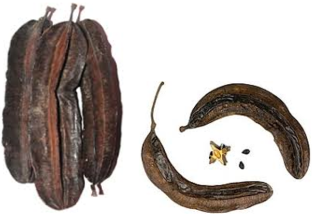
Figure 1. Tetrapleura tetraptera fruits collected from Nunya Isuikwuato, Nigeria (2024).
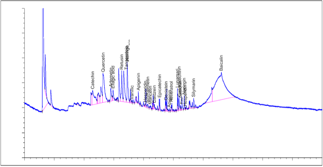
Figure 2. GC-FID chromatogram of methanol extract of Tetrapleura tetraptera.
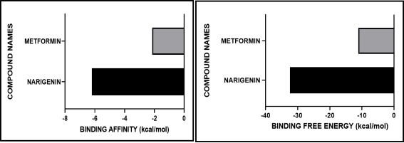
Figure 3. Comparative Binding Affinity and Binding Free Energy of Naringenin and Metformin with AMPK (PDB: 6BX6).
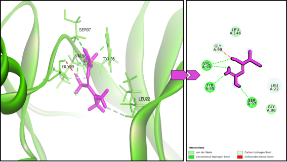
Figure 4. 3D and 2D Molecular Docking Interaction of Metformin with AMPK.
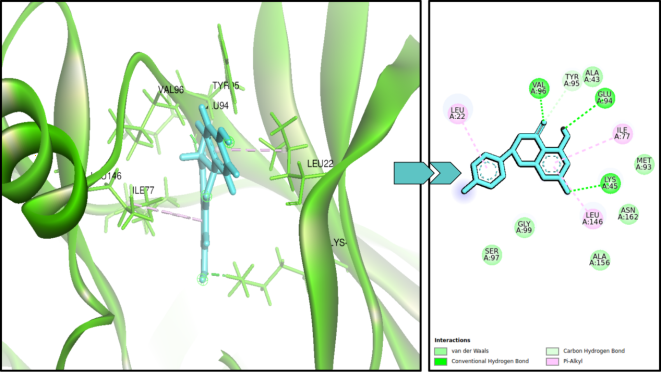
Figure 5. 3D and 2D Molecular Docking Interaction of Naringenin with AMPK.
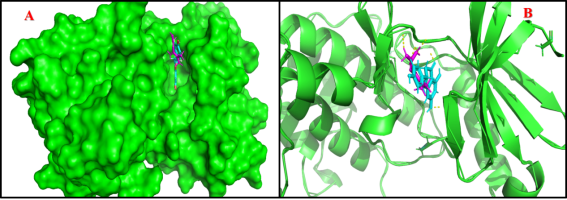
Figure 6. Surface and Ribbon Representations of AMPK Binding with Metformin and Naringenin.
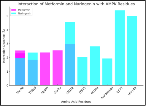
Figure 7. Interaction Distances of Metformin and Naringenin with AMPK Residues.
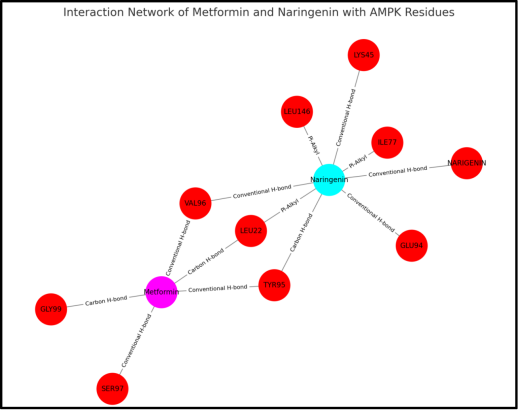
Figure 8. Interaction Network of Metformin and Naringenin with AMPK Residues.
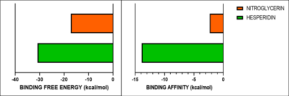
Figure 9. Comparative Binding Affinity and Binding Free Energy of Hesperidin and Nitroglycerin with Enos.
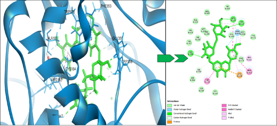
Figure 10. 3D and 2D Interaction Profile of Hesperidin with eNOS.
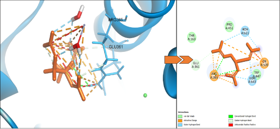
Figure 11. 3D and 2D Interaction Profile of Nitroglycerin with eNOS.
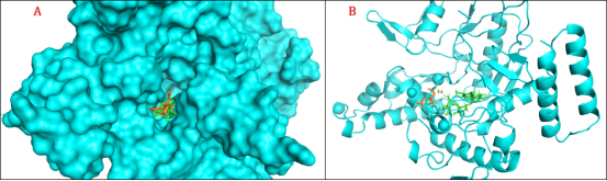
Figure 12. Binding Orientation of Hesperidin and Nitroglycerin within the eNOS Active Site.
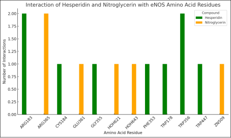
Figure 13. Interaction of Hesperidin and Nitroglycerin with eNOS Amino Acid Residues.
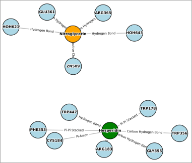
Figure 14. Interaction Network of Hesperidin and Nitroglycerin with eNOS.

Information


 Share This Article
 Twitter
 Linked In
 Facebook
 Copy
Copy Download