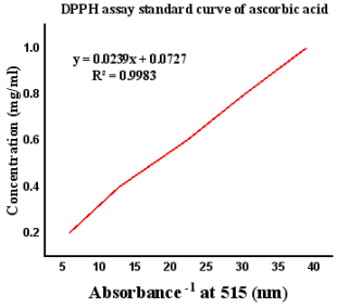
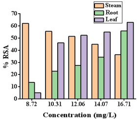
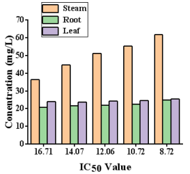
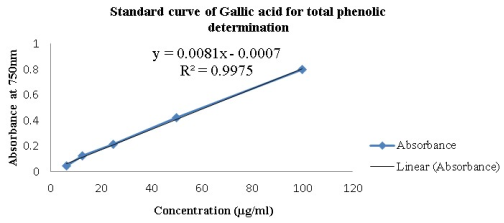
Ethiopia has a long history of using medicinal herbs for treating both human and animal illnesses. Nonetheless, not enough research has been done on the antibacterial properties and possible bioactive components of the majority of medicinal plants. Therefore, this study deals with the evaluation of phytochemical, antimicrobial, antioxidant activities, phenol content and XRF analysis of Commelina Diffusa Burm.F. plant extracts. Mean values of the antimicrobial activity, MIC, antioxidant activities, phenol content and XRF analysis were reported as mean ± standard deviation. The chloroform leaf extracts of the plant gave the highest yield 23.4% followed by methanol 22.27%. The presence of several metabolite components, including alkaloids, diterpenes, flavonoids, glycosides, phenol, protein, saponin, steroids, tannins, terpenoids, tri-terpenoids and amino acids, has been shown by qualitative phytochemical analysis of plant parts. Significant antibacterial activity against the test bacterial strains was demonstrated by steam extracts of Commelina Diffusa Burm.F. Moreover, the methanolic extract of the plant demonstrated notable antioxidant activity. The highest value of phenolic content was obtained in Commelina Diffusa Burm.F. steam methanol extract followed by leaf extract while Commelina Diffusa Burm.F. root extract shows lower phenolic content. In this study, threaten elements were determined in the Commelina Diffusa Burm.F. plant part by using XRF spectroscopy. Overall, this research contributes to the understanding of pharmacological potential of Commelina Diffusa Burm.F. and highlights the importance of further exploring its medicinal properties. The findings provide valuable insights into utilizing medicinal plants for disease treatment and support the development of natural therapeutic agents.
| Published in | Journal of Diseases and Medicinal Plants (Volume 10, Issue 3) |
| DOI | 10.11648/j.jdmp.20241003.11 |
| Page(s) | 40-51 |
| Creative Commons |
This is an Open Access article, distributed under the terms of the Creative Commons Attribution 4.0 International License (http://creativecommons.org/licenses/by/4.0/), which permits unrestricted use, distribution and reproduction in any medium or format, provided the original work is properly cited. |
| Copyright |
Copyright © The Author(s), 2024. Published by Science Publishing Group |
Antibacterial Activity, Antioxidant Activity, XRF
Plant name | Parts used | Solvent | Weight of extract obtained (g) | Yield percentage % |
|---|---|---|---|---|
Commelina Diffusa Burm.F. | Root | Methanol | 0.56 | 2.81 |
Chloroform | 0.49 | 2.46 | ||
n-hexane | 0.59 | 2.96 | ||
Distill Water | 0.22 | 1.10 | ||
Commelina Diffusa Burm.F. | Stem | Methanol | 2.71 | 13.57 |
Chloroform | 3.67 | 18.35 | ||
n-hexane | 0.82 | 4.11 | ||
Distill Water | 0.54 | 2.7 | ||
Commelina Diffusa Burm.F. | Leaf | Methanol | 4.45 | 22.27 |
Chloroform | 4.68 | 23.4 | ||
n-hexane | 3.85 | 19.26 | ||
Distill Water | 0.42 | 2.1 |
Phytochemical Analysis | Test | Methanol extract | n-hexane extract | Ethyl acetate extract | ||||||
|---|---|---|---|---|---|---|---|---|---|---|
Root | Stem | Leaf | Root | Stem | Leaf | Root | Stem | Leaf | ||
Alkaloids | Mayer’s | + | + | _ | _ | _ | _ | + | + | + |
Dragondroff’s | + | + | _ | _ | _ | _ | + | + | + | |
Wagner’s | + | + | _ | _ | _ | _ | + | + | + | |
Anthraqunies | Free and combined anthraquinones | _ | _ | _ | _ | _ | _ | _ | _ | _ |
Diterpenes | General test | _ | + | _ _ | _ | + | + | + | + | + |
Flavonoids | Lead acetate | + | + | _ | _ | _ | _ | _ | _ | _ |
Sodium hydroxide | + | + | _ | _ | _ | _ | _ | _ | _ | |
Glycosides | Keller-Killani | _ | _ | + | _ | _ | + | _ | + | _ |
Phenol | General Test | _ | _ | _ | _ | + | + | _ | + | _ |
Protein | Ninhydrin | _ | + | + | _ | _ | _ | _ | _ | |
Saponin | Frothing | _ | _ | _ | _ | _ | _ | _ | _ | _ |
Steroids | Salkowski’s | _ | + | + | _ | _ | _ | _ | _ | _ |
Tannins | Ferric chloride | _ | _ | + | _ | _ | _ | _ | _ | + |
Terpenoids | Salkowski | _ | _ | _ | _ | _ | _ | _ | _ | _ |
Tri- Terpenoids | General Test | _ | _ | + | + | + | + | _ | _ | _ |
amino acid | General Test | _ | + | + | _ | _ | _ | _ | _ | _ |
Reducing sugar | General Test | _ | _ | + | _ | _ | _ | _ | _ | _ |
Mean valuesa of inhibition zone (mm) | |||
|---|---|---|---|
Plant part | Solvents | Inhibition zone of S. auras | Inhibition zone of E. coli |
Root | Methanol | NDc | -b |
Chloroform | ND | - | |
n-hexane | - | ND | |
Distill Water | - | - | |
Steam | Methanol | 25 | 22 |
Chloroform | 23 | 19.5 | |
n-hexane | 18 | 19 | |
Distill Water | - | - | |
Leaf | Methanol | 16 | 15.5 |
Chloroform | 15.2 | 15.9 | |
n-hexane | 17 | 16.5 | |
Distill Water | - | - | |
Plant parts | Solvents | MIC of crude extracts (mg/ml) a | |
|---|---|---|---|
S. auras | E. coli | ||
Root | Methanol | NDc | -b |
Chloroform | ND | - | |
n-hexane | - | ND | |
Distill Water | - | - | |
Steam | Methanol | 2.109 | 2.234 |
Chloroform | 1.101 | 1.401 | |
n-hexane | 1.098 | 1.539 | |
Distill Water | - | - | |
Leaf | Methanol | 4.562 | 6.251 |
Chloroform | 2.914 | 3.125 | |
n-hexane | 1.243 | 1.625 | |
Distill Water | - | - | |
Extracts | Parts used | Total phenolic content (mg GvE/g) |
|---|---|---|
Methanol | Root | 53.6 |
Methanol | Stem | 61.9 |
Methanol | Leaf | 53.6 |
S. No | Root | Steam | Leaf | |||
|---|---|---|---|---|---|---|
Element | Con (ppm) | Element | Con (ppm) | Element | Con (ppm) | |
1 | Fe | 544.59 | Fe | 4767.55 | Ca | 419.13 |
2 | Ca | 218.35 | Ca | 383.87 | Fe | 273.41 |
3 | Cr | 147.63 | K | 216.95 | K | 168.58 |
4 | Zn | 84.75 | Cr | 177.54 | Cr | 155.53 |
5 | Cu | 42.52 | Zn | 121.32 | Zn | 82.09 |
6 | Sr | 31.68 | Zr | 52.47 | Sr | 38.59 |
7 | V | 29.83 | Cu | 49.46 | Cu | 37.64 |
8 | W | 28.39 | W | 44.39 | V | 32.38 |
9 | Zr | 15.23 | Sr | 39.07 | W | 27.81 |
10 | Mo | 7.69 | V | 31.26 | Zr | 14.3 |
11 | Rb | 7.13 | Rb | 9.99 | Mo | 9.08 |
12 | U | 4.74 | Mo | 8.23 | Rb | 6.96 |
13 | K | <LOD | U | 7.84 | U | 4.57 |
DMSO | Dimethyl Sulfoxide |
DPPH | 2,2-Diphenyl-1-picrylhydrazyl |
MIC | Minimum Inhibitory Concentration |
XRF | X-ray Fluorescence Spectroscopy |
| [1] | Akerele, O. Nature’s medicine bounty: don’t throw it away. World Health Forum. 14(4) (1993) 390-5. |
| [2] | Nirosha, N. & Mangalanayi, R. Antibacterial activity of leaves and stem extract of Caricapapaya L. International Journal of Advances in Pharmacy Biology and Chemistry, 2(3) (2013) 473-476. |
| [3] | Doughari, J. Studies on the antibacterial activity of root extracts of Carica papaya Linn. Africa Journal of Microbial Research, 14(2007) 037-041. |
| [4] | Pretorius, C. & Watt, E. “Purification and identification of active components of Carpobrotusedulis L. Journal of Ethropharm, 76 2001) 87-91. |
| [5] | Odoo, A. Phytochemical characterization and comparative Efficacies of crude extracts of Carica papaya. International Journal of Drug Research Technology, 2(5) (2012) 399-406. |
| [6] | Karthikeyan, A, Sudan, I. & Satheeshkumar, R. In vitro regeneration of Commelina diffusa Burm. F. Using nodal explants. World Journal of Pharmaceutical Research, 7(3) (2018) 978-986. |
| [7] | Kumari, P., Kumari, C. & Singh, P. Phytochemical Screening of Selected Medicinal Plants for Secondary Metabolites. International Journal of Life Sciences Scientific Research, 3(4) (2017) 1151-1157. |
| [8] | Eloff, J. A Sensitive and Quick Microplate Method to Determine the Minimal Inhibitory Concentration of Plant Extracts for Bacteria. PlantaMedica, 64(1998) 711-713. |
| [9] | Adetuyi, A. & Popoola, A. Journal of Science, Engineering and Technology, 8(2) (2001) 3291-3299. |
| [10] | Trease, G. & Evans, W. Pharmacognosy 11thEdn. Brailliar Tiridacanb Macmillian Publishers. (1989). |
| [11] | Sofowora, A. Medicinal Plants and Traditional Medicine in West Arica, John Wily and Sons. New York. (1982) 256. |
| [12] | Salehi-Surmaghi, M., Aynehchi, Y., Amin, GH. & Mahhmoodi, Z. DARU, 2(1992) 1-11. |
| [13] | Siddiqui, A. & Ali, M. Practical pharmaceutical chemistry.1stedition. C B S Publishers and Distributors, New Delhi. (1997) 126-131. |
| [14] | Sathish, M., Selvakumar, S., Rao, M. & Anbuselv, S. Preliminary phytochemical analysis of Dodonaeaviscosaleaves. Asian Journal of Plant Science and Research, 3(1) (2013) 43-46. |
| [15] | Segelman, AB, Fransworth, NR & Quimbi, MD. Lloydia, 32 (1969) 52-58. |
| [16] | Eloff, J. Which extractant should be used for the screening and isolation of antimicrobial components from plants? Ethanopharmacol, 60 (1998) 1-8. |
| [17] | Perez, C., Pauli, M. & Bazerque, P. An antibiotic assay by agar well-diffusion method. Acta Biologia et Medicine Experimentaalis, 15(1990) 113-115. |
| [18] | Wayne, P. Performance standards for antimicrobial susceptibility testing. (2001). Clinical Microbiology Newsletter, 23(6), 49. |
| [19] | Irshad, M., Zafaryab, M., Singh, M., Moshahid, M. & Rizvi, A. Comparative analysis of the antioxidant activity of Cassia stula extracts. International Journal of Medicinal Chemistry, (2012) 1-6. |
| [20] | Kaur, C. & Kapoor H. Anti-oxidant activity and total phenolic content of some Asian vegetables. International Journal of Food Science & Technology, 37(2002), 153-161. |
| [21] | Annalakshmi, R., Uma, R., Subash, G., Savariraj, C. & Charles, A. Evaluation of elemental content of leaves of Madhucalongifolia by X-ray fluorescence spectroscopy (XRF). Journal of Natural Product and Plant Resources, 2(4) (2012) 490-493. |
| [22] | Quang-Vinh, N. and Jong-Bang, E. Antioxidant activity of solvent extracts from Vietnamese medicinal plants. Journal of Medicinal Plants Research, 5(13) (2011) 2798. |
| [23] | Augusto, L., Josean, F., Marcelo, S., Margareth, F., Petronio, F. & Jose, M. Anti-inflammatory activity of alkaloids. Molecules, 16(2011) 8515-8534. |
| [24] | Dua, V. K., Verma, G., Singh, B., Rajan, A., Bagai, U., Agarwal, D., … Rastogi, A. (2013). Anti-malarial property of steroidal alkaloid conessine isolated from the bark of Holarrhena antidysenterica. Malaria journal, 12(2013) 1-6. |
| [25] | Benbott, A., Yahyia, A. & Belaıdi, A. Assessment of the antibacterial activity of crude alkaloids extracted from seeds and roots of the plant PeganumharmalaL. Journal of Medicinal Plants Research, 2(2012) 568-573. |
| [26] | Ameyaw, Y. & Duker-Eshun, G. The alkaloid contents of theethno-plant organs of three antimalarial medicinal plant species inthe eastern region of Ghana. International journal of chemical science, 7(2009) 48-58. |
| [27] | Akiyama, H. (2001). Antibacterial action of several tannins against Staphylococcusaureus. Journal of Antimicrobia Chemotherapy, 48(4), 487–491. |
| [28] | Kren, V., & Martinkova, L. (2001). Glycosides in Medicine: “The Role of Glycosidic Residue in Biological Activity” Curent Medicinal Chemistry, 8(11) (2001), 1303–1328. |
| [29] | Parekh, J. & Chanda, S. In-vitro Antimicrobial Activities of Extracts of Launaea procumbensRoxb. (Labiateae), VitisviniferaL. (Vitaceae) and CyperusrotundusL.(Cyperaceae). African Journal of Biomedical Research, 9 (2006) 89-93. |
| [30] | Seid, M. & Ayisha, A. Extraction and phytochemical determination of some selected traditional medicinal plants for antimicrobial susceptibility test, in Adama, Ethiopia. International Journal of Engineering, Science and Technology, 3(5) (2015) 1290-1297. |
| [31] | Bakari, S., Daoud, A., Felhi, S., Smaoui, S., Gharsallah, N. & Kadri A. Proximate analysis, mineral composition, phytochemical contents, antioxidant and antimicrobial activities and GC-MS investigation of various solvent extracts of cactus cladode. Journal of Food Science and Technology, 37(2) (2017) 286–293. |
| [32] | Felhi, S., Daoud, A., Hajlaoui, H., Mnafgui, K., Gharsallah, N. & Kadri, A. Solvent extraction effects on phytochemical constituents profiles, antioxidant and antimicrobial activities and functional group analysiSs of Ecballium elaterium seeds and peels fruits. Journal of Food Science and Technology, 37(3) (2017) 483-492. |
| [33] | Aghraz, A., Gonçalves, S., Rodríguez-Solana, R., Dra, L. A., Di Stefano, V., Dugo, G., Cicero, N., Larhsini, M., Markouk, M., & Romano, A. Antioxidant activity and enzymes inhibitory properties of several extracts from two Moroccan Asteraceae species. South African Journal of Botany, 118(2018) 58-64. |
| [34] | Alam, M. A., Syazwanie, N. F., Mahmod, N. H., Badaluddin, N. A., Mustafa, K. A., Alias, N., Aslani, F., & Prodhan, M. A. Evaluation of antioxidant compounds, antioxidant activities and capsaicinoid compounds of Chili (Capsicum sp.) germplasms available in Malaysia. Journal of Applied Research on Medicinal and Aromatic Plants, 9(2018) 46-54. |
| [35] | Soobrattee, M., Bahorun, T., Neergheen, V., Googoolye, K., & Aruoma, O. Assessment of the content of phenolics and antioxidant actions of the Rubiaceae, Ebenaceae, Celastraceae, Erythroxylaceae and Sterculaceae families of Mauritian endemic plants. Toxicology in Vitro, 22(1) (2008) 45-56. |
APA Style
Yahiya, Y. S., Temeche, A. M., Delisho, F. D., Abrar, K. A. (2024). Invitro Antibacterial, Antioxidant and XRF Analysis of Commelina Diffusa Burm.F. Plant Extracts. Journal of Diseases and Medicinal Plants, 10(3), 40-51. https://doi.org/10.11648/j.jdmp.20241003.11
ACS Style
Yahiya, Y. S.; Temeche, A. M.; Delisho, F. D.; Abrar, K. A. Invitro Antibacterial, Antioxidant and XRF Analysis of Commelina Diffusa Burm.F. Plant Extracts. J. Dis. Med. Plants 2024, 10(3), 40-51. doi: 10.11648/j.jdmp.20241003.11
AMA Style
Yahiya YS, Temeche AM, Delisho FD, Abrar KA. Invitro Antibacterial, Antioxidant and XRF Analysis of Commelina Diffusa Burm.F. Plant Extracts. J Dis Med Plants. 2024;10(3):40-51. doi: 10.11648/j.jdmp.20241003.11
@article{10.11648/j.jdmp.20241003.11,
  author = {Yonas Syraji Yahiya and Aweke Mamo Temeche and Fitusm Dejene Delisho and Kidist Ali Abrar},
  title = {Invitro Antibacterial, Antioxidant and XRF Analysis of Commelina Diffusa Burm.F. Plant Extracts
},
  journal = {Journal of Diseases and Medicinal Plants},
  volume = {10},
  number = {3},
  pages = {40-51},
  doi = {10.11648/j.jdmp.20241003.11},
  url = {https://doi.org/10.11648/j.jdmp.20241003.11},
  eprint = {https://article.sciencepublishinggroup.com/pdf/10.11648.j.jdmp.20241003.11},
  abstract = {Ethiopia has a long history of using medicinal herbs for treating both human and animal illnesses. Nonetheless, not enough research has been done on the antibacterial properties and possible bioactive components of the majority of medicinal plants. Therefore, this study deals with the evaluation of phytochemical, antimicrobial, antioxidant activities, phenol content and XRF analysis of Commelina Diffusa Burm.F. plant extracts. Mean values of the antimicrobial activity, MIC, antioxidant activities, phenol content and XRF analysis were reported as mean ± standard deviation. The chloroform leaf extracts of the plant gave the highest yield 23.4% followed by methanol 22.27%. The presence of several metabolite components, including alkaloids, diterpenes, flavonoids, glycosides, phenol, protein, saponin, steroids, tannins, terpenoids, tri-terpenoids and amino acids, has been shown by qualitative phytochemical analysis of plant parts. Significant antibacterial activity against the test bacterial strains was demonstrated by steam extracts of Commelina Diffusa Burm.F. Moreover, the methanolic extract of the plant demonstrated notable antioxidant activity. The highest value of phenolic content was obtained in Commelina Diffusa Burm.F. steam methanol extract followed by leaf extract while Commelina Diffusa Burm.F. root extract shows lower phenolic content. In this study, threaten elements were determined in the Commelina Diffusa Burm.F. plant part by using XRF spectroscopy. Overall, this research contributes to the understanding of pharmacological potential of Commelina Diffusa Burm.F. and highlights the importance of further exploring its medicinal properties. The findings provide valuable insights into utilizing medicinal plants for disease treatment and support the development of natural therapeutic agents.
},
 year = {2024}
}
TY  - JOUR
T1  - Invitro Antibacterial, Antioxidant and XRF Analysis of Commelina Diffusa Burm.F. Plant Extracts

AU  - Yonas Syraji Yahiya
AU  - Aweke Mamo Temeche
AU  - Fitusm Dejene Delisho
AU  - Kidist Ali Abrar
Y1  - 2024/07/31
PY  - 2024
N1  - https://doi.org/10.11648/j.jdmp.20241003.11
DO  - 10.11648/j.jdmp.20241003.11
T2  - Journal of Diseases and Medicinal Plants
JF  - Journal of Diseases and Medicinal Plants
JO  - Journal of Diseases and Medicinal Plants
SP  - 40
EP  - 51
PB  - Science Publishing Group
SN  - 2469-8210
UR  - https://doi.org/10.11648/j.jdmp.20241003.11
AB  - Ethiopia has a long history of using medicinal herbs for treating both human and animal illnesses. Nonetheless, not enough research has been done on the antibacterial properties and possible bioactive components of the majority of medicinal plants. Therefore, this study deals with the evaluation of phytochemical, antimicrobial, antioxidant activities, phenol content and XRF analysis of Commelina Diffusa Burm.F. plant extracts. Mean values of the antimicrobial activity, MIC, antioxidant activities, phenol content and XRF analysis were reported as mean ± standard deviation. The chloroform leaf extracts of the plant gave the highest yield 23.4% followed by methanol 22.27%. The presence of several metabolite components, including alkaloids, diterpenes, flavonoids, glycosides, phenol, protein, saponin, steroids, tannins, terpenoids, tri-terpenoids and amino acids, has been shown by qualitative phytochemical analysis of plant parts. Significant antibacterial activity against the test bacterial strains was demonstrated by steam extracts of Commelina Diffusa Burm.F. Moreover, the methanolic extract of the plant demonstrated notable antioxidant activity. The highest value of phenolic content was obtained in Commelina Diffusa Burm.F. steam methanol extract followed by leaf extract while Commelina Diffusa Burm.F. root extract shows lower phenolic content. In this study, threaten elements were determined in the Commelina Diffusa Burm.F. plant part by using XRF spectroscopy. Overall, this research contributes to the understanding of pharmacological potential of Commelina Diffusa Burm.F. and highlights the importance of further exploring its medicinal properties. The findings provide valuable insights into utilizing medicinal plants for disease treatment and support the development of natural therapeutic agents.

VL  - 10
IS  - 3
ER  -
Department of Biology, College of Natural Sciences, Arba Minch University, Arba Minch, Ethiopia
Department of Biology, College of Natural Sciences, Arba Minch University, Arba Minch, Ethiopia
Department of Biology, College of Natural Sciences, Arba Minch University, Arba Minch, Ethiopia
Department of Biology, College of Natural Sciences, Arba Minch University, Arba Minch, Ethiopia
Information