Listeria monocytogenes (L. monocytogenes) is a pathogen that frequently contaminates foods, and is the cause of listeriosis worldwide. Although L. monocytogenes is the primary species associated with human listeriosis, Listeria ivanovii (L. ivanovii) traditionally considered pathogenic mainly to ruminants has also been implicated in rare but severe infections in immunocompromised humans. The objectives of this study were to examine the ability of L. monocytogenes and L. ivanovii strains isolated from fresh vegetables and market garden produce in Abidjan, Côte d'Ivoire, to enter and multiply in JEG3 cells, to disseminate and to form LisCVs persistence vacuoles. After identification by 16sDNAr gene sequencing and serogrouping by PCR of Listeria strains isolated from fresh vegetables and market garden produce, three strains were identified as belonging to the species L. monocytogenes (2 strains, L208 and L238) and L. ivanovii (1 strain, L135). In vitro infection was carried out using JEG3 trophoblastic cells, with three reference strains (L. monocytogenes EGDe serotype 1/2a, Listeria monocytogenes CLIP80459 serotype 4b and L. ivanovii ATCC19119) serving as controls. The results showed that all the Listeria strains tested had similar characteristics in terms of their ability to penetrate, multiply and form LisCV, and higher cytotoxicity in L. monocytogenes species than in L. ivanovii in general. L. monocytogenes strains L208 and L238 showed similar invasion rates to L. monocytogenes CLIP80459.
| Published in | International Journal of Food Science and Biotechnology (Volume 10, Issue 3) |
| DOI | 10.11648/j.ijfsb.20251003.11 |
| Page(s) | 57-65 |
| Creative Commons |
This is an Open Access article, distributed under the terms of the Creative Commons Attribution 4.0 International License (http://creativecommons.org/licenses/by/4.0/), which permits unrestricted use, distribution and reproduction in any medium or format, provided the original work is properly cited. |
| Copyright |
Copyright © The Author(s), 2025. Published by Science Publishing Group |
L. monocytogenes, L. ivanovii, Infection, JEG3 Cells, Côte d’Ivoire
Cell lineage | Specifications | Origin |
|---|---|---|
JEG3 | Human epithelial cells derived from placental carcinoma | ATCC HBT-36 |
Bacterial strains | Specifications | Genetic lineage and CC |
L. monocytogenes EGDe | Reference strain (serotype 1/2a) | Lineage II, CC9 [21] |
L. monocytogenes CLIP80459 | Reference strain (serotype 4b) | Lineage I, CC4 [22] |
L. ivanovii ATCC19119 | Reference strain (serotype 5) |
Strains | Species | Genetic lineage | Serogroups | Origin | Strain accession number |
|---|---|---|---|---|---|
L135 | L. ivanovii | nd | nd | lettuce | PQ473711 |
L208 | L. monocytogenes | I | (1/2b, 3b, 4b, 4d, 4e et 7) | lettuce | PQ473712 |
L238 | L. monocytogenes | I | (1/2b, 3b, 4b, 4d, 4e et 7) | lettuce | PQ473714 |
ATCC | American Type Culture Collection |
ISO | International Organization for Standardization |
CFU | Colony-Forming Units |
CLIP | Pasteur Institute Line Collection |
| [1] | Orsi, R. H., and Wiedmann, M. (2016). Characteristics and distribution of Listeria spp., including Listeria species newly described since 2009. Applied Microbiology and Biotechnology, 100(12), 5273-5287. |
| [2] | Ajayeoba, T. A., Atanda, O. O., Obadina, A. O., Bankole, M. O., and Adelowo, O. O. (2016). The incidence and distribution of Listeria monocytogenes in ready-to-eat vegetables in South-Western Nigeria. Food Science & Nutrition, 4(1), 59-66. |
| [3] | Amusan, E. E., and Sanni, A. I. (2018). Isolation and Identification of Listeria monocytogenes in Fresh Croaker (Pseudotolithus senegalensis). IOP Conference Series: Earth and Environmental Science, 210, Article 012004. |
| [4] | Haase, J. K., Didelot, X., Lecuit, M., Korkeala, H., L. monocytogenes MLST Study Group, and Achtman, M. (2014). The ubiquitous nature of Listeria monocytogenes clones: A large-scale multilocus sequence typing study. Environmental Microbiology, 16(2), 405-416. |
| [5] | Bécavin, C., Bouchier, C., Lechat, P., Archambaud, C., Creno, S., Gouin, E., Wu, Z., Kühbacher, A., Brisse, S. and Pucciarelli, M. G. (2014). Comparison of Widely Used Listeria monocytogenes Strains EGD, 10403S, and EGD-e Highlights Genomic Differences Underlying Variations in Pathogenicity. mBio 5. |
| [6] | Jonquières, R., Bierne, H., Mengaud, J., and Cossart, P. (1998). The inlA gene of Listeria monocytogenes LO28 harbors a nonsense mutation resulting in release of internalin. Infection and Immunity, 66(7), 3420-3422. |
| [7] | Lam, G. Y., Cemma, M., Muise, A. M., Higgins, D. E., and Brumell, J. H. (2013). Host and bacterial factors that regulate LC3 recruitment to Listeria monocytogenes during the early stages of macrophage infection. Autophagy, 9(7), 985-995. |
| [8] | Portnoy, D. A. (2007). A 20‑year perspective on Listeria monocytogenes pathogenesis. In H. Goldfine & H. Shen (Eds.), Listeria monocytogenes: Pathogenesis and Host Response (chap. 1, pp. 1-12). Springer. |
| [9] | Jacquet, C., Doumith, M., Gordon, J. I., Martin, P. M. V., Cossart, P., and Lecuit, M. (2004). A molecular marker for evaluating the pathogenic potential of foodborne Listeria monocytogenes. The Journal of Infectious Diseases, 189(11), 2094-2100. |
| [10] | Nightingale, K. K., Fortes, E. D., Ho, A. J., Schukken, Y. H., Grohn, Y. T., and Wiedmann, M. (2005). Evaluation of farm management practices as risk factors for clinical listeriosis and fecal shedding of Listeria monocytogenes in ruminants. J. Am. Vet. Med. Assoc. 227, 1808-1814. |
| [11] | Van, S. A., Simpson, J. M., Ward, T. J. and Nightingale, K. K. (2010). Revelation by Single-Nucleotide Polymorphism Genotyping That Mutations Leading to a Premature Stop Codon in inlA Are Common among Listeria monocytogenes Isolates from Ready-To-Eat Foods but Not Human Listeriosis Cases. Appl. Environ. Microbiol. 76, 2783-2790. |
| [12] | Kanki, M., Naruse, H., Taguchi, M., and Kumeda, Y. (2015). Characterization of specific alleles in InlA and PrfA of Listeria monocytogenes isolated from foods in Osaka, Japan and their ability to invade Caco-2 cells. Int. J. Food Microbiol. 211, 18-22. |
| [13] | Burall, L. S., Grim, C. J., and Datta, A. R. (2017). A clade of Listeria monocytogenes serotype 4b variant strains linked to recent listeriosis outbreaks associated with produce from a defined geographic region in the US. PloS one, 12(5), e0176912. |
| [14] | Doumith, M., Jacquet, C., Gerner-Smidt, P., Graves, L. M., Loncarevic, S., Mathisen, T., Morvan, A., Salcedo, C., Torpdahl, M., Vazquez, J. A., and Martin, P. (2005). Multicenter validation of a multiplex PCR assay for differentiating the major Listeria monocytogenes serovars 1/2a, 1/2b, 1/2c, and 4b: toward an international standard. Journal of food protection, 68(12), 2648-2650. |
| [15] | Kortebi, M., Milohanic, E., Mitchell, G., Péchoux, C., Prevost, M.-C., Cossart, P. and Bierne, H. (2017). Listeria monocytogenes switches from dissemination to persistence by adopting a vacuolar lifestyle in epithelial cells. PLOS Pathog. 13, e1006734. |
| [16] | Kohler, L. J., and Roy, C. R. (2015). Biogenesis of the lysosome-derived vacuole containing Coxiella burnetii. Microbes Infect. 17, 766-771. |
| [17] | Kortebi M., (2018). Characterization of an intracellular persistence phase of the pathogen Listeria monocytogenes. Doctoral thesis from the University of Paris-Saclay. Doctoral School No. 577: Structure and Dynamics of Living Systems (SDSV). p191. |
| [18] | Portnoy, D. A. (2012). Yogi Berra, Forrest Gump, and the discovery of Listeria actin comet tails. Mol. Biol. Cell 23, 1141-1145. |
| [19] | Bierne, H., Milohanic, E., and Kortebi, M. (2018). To Be Cytosolic or Vacuolar: The Double Life of Listeria monocytogenes. Front. Cell. Infect. Microbiol. 8. |
| [20] | Radoshevich, L., and Cossart, P. (2018). To be cytosolic or vacuolar: The double life of Listeria monocytogenes. mBio, 9(1), e02174-17. |
| [21] | Orsi, R. H., Den Bakker, H. C., and Wiedmann, M. (2011). L. monocytogenes evolutionary lineages I-IV and clonal complex classification, including the placement of EGDe within CC9. PLoS Pathogens 7(5): e1000146. |
| [22] | Hain, T., Ghai, R., Billion, A., Kuenne, C. T., Steinweg, C., Izar, B., Mohamed, W., Abu Mraheil, M., Domann, E., Schaffrath, S., Kärst, U., Goesmann, A., Oehm, S., Pühler, A., Merkl, R., Vorwerk, S., Glaser, P., Garrido, P., Rusniok, C., Buchrieser, C., Goebel, W., and Chakraborty, T. (2012). Comparative genomics and transcriptomics of lineages I, II, and III strains of Listeria monocytogenes. BMC Genomics, 13, 144. |
APA Style
Francine, A. M. D., Fernand, P. S., Tiemélé, A. S., Josef, D., Boraud, A. M., et al. (2025). Intracellular Adaptation of Listeria monocytogenes and Listeria ivanovii Isolated from Fresh Produce in Côte d’Ivoire: An in Vitro Infection Study Using JEG3 Human Cells. International Journal of Food Science and Biotechnology, 10(3), 57-65. https://doi.org/10.11648/j.ijfsb.20251003.11
ACS Style
Francine, A. M. D.; Fernand, P. S.; Tiemélé, A. S.; Josef, D.; Boraud, A. M., et al. Intracellular Adaptation of Listeria monocytogenes and Listeria ivanovii Isolated from Fresh Produce in Côte d’Ivoire: An in Vitro Infection Study Using JEG3 Human Cells. Int. J. Food Sci. Biotechnol. 2025, 10(3), 57-65. doi: 10.11648/j.ijfsb.20251003.11
AMA Style
Francine AMD, Fernand PS, Tiemélé AS, Josef D, Boraud AM, et al. Intracellular Adaptation of Listeria monocytogenes and Listeria ivanovii Isolated from Fresh Produce in Côte d’Ivoire: An in Vitro Infection Study Using JEG3 Human Cells. Int J Food Sci Biotechnol. 2025;10(3):57-65. doi: 10.11648/j.ijfsb.20251003.11
@article{10.11648/j.ijfsb.20251003.11,
  author = {Aké Moussan Désirée Francine and Pekoula Senaho Fernand and Amoikon Simon Tiemélé and Deutscher Josef and Alloue Mireille Boraud and Pagliuso Alessandro and Milohanic Eliane},
  title = {Intracellular Adaptation of Listeria monocytogenes and Listeria ivanovii Isolated from Fresh Produce in Côte d’Ivoire: An in Vitro Infection Study Using JEG3 Human Cells
},
  journal = {International Journal of Food Science and Biotechnology},
  volume = {10},
  number = {3},
  pages = {57-65},
  doi = {10.11648/j.ijfsb.20251003.11},
  url = {https://doi.org/10.11648/j.ijfsb.20251003.11},
  eprint = {https://article.sciencepublishinggroup.com/pdf/10.11648.j.ijfsb.20251003.11},
  abstract = {Listeria monocytogenes (L. monocytogenes) is a pathogen that frequently contaminates foods, and is the cause of listeriosis worldwide. Although L. monocytogenes is the primary species associated with human listeriosis, Listeria ivanovii (L. ivanovii) traditionally considered pathogenic mainly to ruminants has also been implicated in rare but severe infections in immunocompromised humans. The objectives of this study were to examine the ability of L. monocytogenes and L. ivanovii strains isolated from fresh vegetables and market garden produce in Abidjan, Côte d'Ivoire, to enter and multiply in JEG3 cells, to disseminate and to form LisCVs persistence vacuoles. After identification by 16sDNAr gene sequencing and serogrouping by PCR of Listeria strains isolated from fresh vegetables and market garden produce, three strains were identified as belonging to the species L. monocytogenes (2 strains, L208 and L238) and L. ivanovii (1 strain, L135). In vitro infection was carried out using JEG3 trophoblastic cells, with three reference strains (L. monocytogenes EGDe serotype 1/2a, Listeria monocytogenes CLIP80459 serotype 4b and L. ivanovii ATCC19119) serving as controls. The results showed that all the Listeria strains tested had similar characteristics in terms of their ability to penetrate, multiply and form LisCV, and higher cytotoxicity in L. monocytogenes species than in L. ivanovii in general. L. monocytogenes strains L208 and L238 showed similar invasion rates to L. monocytogenes CLIP80459.},
 year = {2025}
}
TY  - JOUR
T1  - Intracellular Adaptation of Listeria monocytogenes and Listeria ivanovii Isolated from Fresh Produce in Côte d’Ivoire: An in Vitro Infection Study Using JEG3 Human Cells

AU  - Aké Moussan Désirée Francine
AU  - Pekoula Senaho Fernand
AU  - Amoikon Simon Tiemélé
AU  - Deutscher Josef
AU  - Alloue Mireille Boraud
AU  - Pagliuso Alessandro
AU  - Milohanic Eliane
Y1  - 2025/07/30
PY  - 2025
N1  - https://doi.org/10.11648/j.ijfsb.20251003.11
DO  - 10.11648/j.ijfsb.20251003.11
T2  - International Journal of Food Science and Biotechnology
JF  - International Journal of Food Science and Biotechnology
JO  - International Journal of Food Science and Biotechnology
SP  - 57
EP  - 65
PB  - Science Publishing Group
SN  - 2578-9643
UR  - https://doi.org/10.11648/j.ijfsb.20251003.11
AB  - Listeria monocytogenes (L. monocytogenes) is a pathogen that frequently contaminates foods, and is the cause of listeriosis worldwide. Although L. monocytogenes is the primary species associated with human listeriosis, Listeria ivanovii (L. ivanovii) traditionally considered pathogenic mainly to ruminants has also been implicated in rare but severe infections in immunocompromised humans. The objectives of this study were to examine the ability of L. monocytogenes and L. ivanovii strains isolated from fresh vegetables and market garden produce in Abidjan, Côte d'Ivoire, to enter and multiply in JEG3 cells, to disseminate and to form LisCVs persistence vacuoles. After identification by 16sDNAr gene sequencing and serogrouping by PCR of Listeria strains isolated from fresh vegetables and market garden produce, three strains were identified as belonging to the species L. monocytogenes (2 strains, L208 and L238) and L. ivanovii (1 strain, L135). In vitro infection was carried out using JEG3 trophoblastic cells, with three reference strains (L. monocytogenes EGDe serotype 1/2a, Listeria monocytogenes CLIP80459 serotype 4b and L. ivanovii ATCC19119) serving as controls. The results showed that all the Listeria strains tested had similar characteristics in terms of their ability to penetrate, multiply and form LisCV, and higher cytotoxicity in L. monocytogenes species than in L. ivanovii in general. L. monocytogenes strains L208 and L238 showed similar invasion rates to L. monocytogenes CLIP80459.
VL  - 10
IS  - 3
ER  -
Department of Food Science and Technology, Laboratory of Microbiology and Biotechnology, Nangui Abrogoua University, Abidjan, Côte d’Ivoire
Department of Food Science and Technology, Laboratory of Microbiology and Biotechnology, Nangui Abrogoua University, Abidjan, Côte d’Ivoire
Pierre Richet Institute of Bouaké / National Institute of Public Health, Côte d’Ivoire
Epigenetics and Cellular Microbiology laboratory, Micalis Institute, INRAE, AgroParisTech, Paris-Saclay University, Jouy-en-Josas, France
Department of Food Science and Technology, Laboratory of Microbiology and Biotechnology, Nangui Abrogoua University, Abidjan, Côte d’Ivoire
Epigenetics and Cellular Microbiology laboratory, Micalis Institute, INRAE, AgroParisTech, Paris-Saclay University, Jouy-en-Josas, France
Epigenetics and Cellular Microbiology laboratory, Micalis Institute, INRAE, AgroParisTech, Paris-Saclay University, Jouy-en-Josas, France
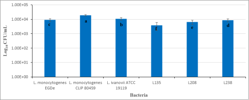
Figure 1. Inoculum (in Log10CFU/mL) of the different bacterial strains tested.
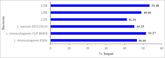
Figure 2. Invasiveness of bacterial strains.
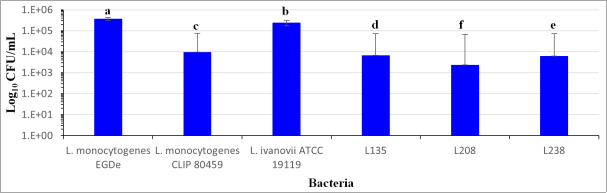
Figure 3. Long-term infection test (3 days). Log10CFU/mL at 72h p.i.
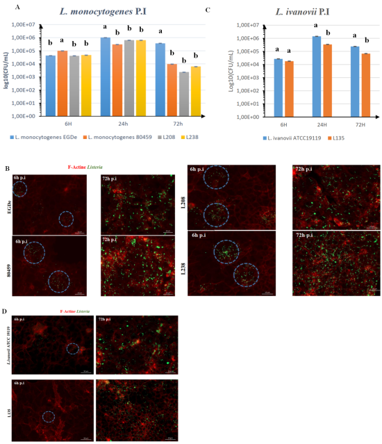
Figure 4. Bacterial dissemination. A-D. JEG3 cells were infected with L. monocytogenes (EGDe, CLIP80459, L208 and L238) and L. ivanovii (ATCC 19119 and L135) (MOI 0, 1; without 30 min exposure to gentamicin 25 µg/mL). A: Number of bacteria (L. monocytogenes) in the extracellular medium (by CFU conts). B: Micrographs of cells infected with L. monocytogenes for 6 or 72 h and visualized with the objective 20X. Images are overlays of Listeria (green) and F-actin (red) signals. Circles highlight an infection focus at 6 h p.i. Bar: 50 µm. C: Number of bacteria (L. ivanovii) in the extracellular medium (by CFU conts). D: Micrographs of cells infected with L. ivanovii for 6 or 72 h and visualized with the objective 20X. Images are overlays of Listeria (green) and F-actin (red) signals. Circles highlight an infection focus at 6 h p.i. Bar: 50 µm.
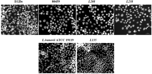
Figure 5. JEG-3 cell nuclei in white, 72 h post-infection. DAPI staining of Listeria monocytogenes (EGDe, CLIP80459, L203 and L238) infected and L. ivanovii (ATCC19119 and L135) infected JEG3 cells at 72 h p.i. Bar: 50 µm.
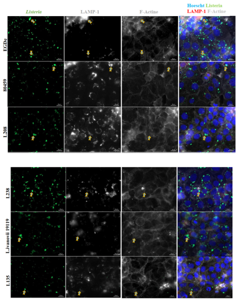
Figure 6. Formation of LisCVs at 72 h p.i. JEG3 cells were infected for 72 h and labelled by immunofluorescence. JEG3 nuclei are labelled in blue, bacteria in green, LAMP1 in red and filamentous actin in white, The arrows indicate the LisCVs. Scale bar: 20 µm.
Information

