1. Introduction
Penile gangrene is a troublesome disease with high morbidity and mortality. It is rare and attributed to the rich blood flow and collateral supply to the lower abdomen and perineum but can pose difficulties in management.
[1] | Ntia IO, Mungadi IA. The Pattern of Penile Gangrene in Sokoto, Nigeria. Afr J Urol. 2007; 13(4): 255-261. |
[2] | Ashutosh T, Neerja P, Majhail S. Fournier’s Gangrene of the Penis: A Rare Entity. J Cutan Aesthet Surg. 2010; 3(1): 41–44. https://doi.org/10.4103/0974-2077.63394 |
[16] | I-Ni Chiang, Shang-Jen Chang, Yuh-Chen Kuo, Shih-Ping Liu, Hong-Jeng Yu, Ju-Ton Hsieh, Management of Ischemic Penile Gangrene: Prompt Partial Penectomy and Other Treatment Options, The Journal of Sexual Medicine, Volume 5, Issue 11, November 2008, Pages 2725–2733. https://doi.org/10.1111/j.1743-6109.2008.00894.x |
[1, 2, 16]
Dry gangrene is usually the end result of vascular compromise. It is known that the penis have a rich arterial supply, however arterial occlusion can cause distal necrosis similar to ischemic gangrene often noted in the digits of extremities.
[3] | Khanh NP, Mitchell HS, charles AS. Severe Gangrene at the Glans Penis Requiring Penectomy as the First Major Complication of Buerger’s Disease. Am J Clin Exp Urol. 2016; 4(1): 9–11. |
[4] | Vijayan P. Gangrene of the Penis in a Diabetic Male with Multiple Amputations and Follow up. Indian J Urol. 2009 Jan-Mar; 25(1): 123–125. https://doi.org/10.4103/0970-1591.45550 |
[5] | Vikas KP, Ravimohan SM, Sudheer KD, Kim V, Girdhar SB. Priapism with Penile Gangrene: An Unusual Presentation of Multiple Myeloma. Indian J Urol. 2017 Jul-Sep; 33(3): 251–252. https://doi.org/10.4103/iju.IJU_41_17 |
[3-5]
It is estimated that around 8–18% of cancer patients have diabetes.
Etiologically, buildup of calcium deposits due to chronic renal failure, diabetes mellitus (DM), infection, thrombotic episodes and trauma have been identified. Diabetes mellitus and multiple myeloma are two overwhelming conditions for both patients and clinicians. The treatment of diabetes in the presence of cancer could be a daunting task for physicians.
Due to the increasing incidence of type 2 diabetes worldwide, a rise in the diagnosis of multiple myeloma coexisting with DM is expected.
[8] | Urvi AS, Erin M, Andriy D, Yuanhui H, Sham M, Carlyn R et al. Prevalence and Impact of Diabetes on Survival of Patients with Multiple Myeloma in Different Racial Groups. Blood Adv. 2024; 8(1): 236–247. https://doi.org/10.1182/bloodadvances.2023010815 |
[8]
A study reviewing the pattern and prevalence of penile gangrene in Sokoto, northwest Nigeria between January 1994 and December 2003 noted that patients who had penile gangrene were between the ages of 6 to 62 years with 44% presenting with more severe forms. Etiological factors identified were urethral stricture in 50%, diabetes mellitus in 13%, impacted urethral calculus in 19%, circumcision in 13%, priapism in sickle cell disease in 13% and trauma in 6%.
[1] | Ntia IO, Mungadi IA. The Pattern of Penile Gangrene in Sokoto, Nigeria. Afr J Urol. 2007; 13(4): 255-261. |
[1]
Clinicians managing such patients should be well aware of the potential impact of multiple myeloma treatment with chemotherapy non glucose metabolism in this population.
[8] | Urvi AS, Erin M, Andriy D, Yuanhui H, Sham M, Carlyn R et al. Prevalence and Impact of Diabetes on Survival of Patients with Multiple Myeloma in Different Racial Groups. Blood Adv. 2024; 8(1): 236–247. https://doi.org/10.1182/bloodadvances.2023010815 |
[9] | Mohamed AA, Yasar AA, Abubaker I. Clinical Challenges: Myeloma and Concomitant Type 2 Diabetes. South Asian J Cancer. 2013 Oct-Dec; 2(4): 290–295. https://doi.org/10.4103/2278-330X.119916 |
[8, 9]
Multiple myeloma is a malignant plasma cell disorder that accounts for approximately 10% of all hematologic cancers.
It is characterized by accumulation of clonal plasma cells, predominantly in the bone marrow.
Peripheral neuropathy is a common problem in patients with multiple myeloma and is also a common complication of type 2 diabetes.
[4] | Vijayan P. Gangrene of the Penis in a Diabetic Male with Multiple Amputations and Follow up. Indian J Urol. 2009 Jan-Mar; 25(1): 123–125. https://doi.org/10.4103/0970-1591.45550 |
[11] | Abdulla MC, Nair LK, Narayan R, Das A. Multiple Myeloma Presenting as Extensive Limb Gangrene. J. Appl. Hematol. 2014 Jul-Sep; 5(3): 107-110. |
[4, 11]
There have been in the urological literature sporadic reports of solitary cases of penile gangrene and a retrospective study of some cases associated with diabetes mellitus.
The causes of penile gangrene are diabetes mellitus, chronic renal failure, penile prosthesis, tourniquet effect created by condom appliances, thromboembolic phenomena, and hyper-coagulopathy secondary to neoplastic diseases.
[1] | Ntia IO, Mungadi IA. The Pattern of Penile Gangrene in Sokoto, Nigeria. Afr J Urol. 2007; 13(4): 255-261. |
[5] | Vikas KP, Ravimohan SM, Sudheer KD, Kim V, Girdhar SB. Priapism with Penile Gangrene: An Unusual Presentation of Multiple Myeloma. Indian J Urol. 2017 Jul-Sep; 33(3): 251–252. https://doi.org/10.4103/iju.IJU_41_17 |
[10] | Odunukwe NN, Madu JA, Nnodu OE, Okocha OE, Akingbola TS, Asuquo IM et al. Multiple Myeloma in Nigeria: A Multi-centre Epidemiological and Biomedical Study. PAM J. 2015; 22: 292. https://doi.org/10.11604/pamj.2015.22.292.7774 |
[12] | Nwabuko CC, Igbigbi EE, Chukwuonye II, Nnoli MA. Multiple Myeloma in Niger Delta, Nigeria: Complications and Outcome of Palliative Interventions. CMAR. 2017; 9: 189-196. https://doi.org/10.2147/CMAR.S126136 |
[15] | Olaomi OO. Penile Gangrene Following Cavernoglandular Shunt for Priapism: Case Report. Nig. J. of Surgical Research. 2002; 4(3-4): 112-114. https://doi.org/10.4314/njsr.v4i3.12160 |
[1, 5, 10, 12, 15]
Penile gangrene is a unique complication of severe systemic vascular disease.
[4] | Vijayan P. Gangrene of the Penis in a Diabetic Male with Multiple Amputations and Follow up. Indian J Urol. 2009 Jan-Mar; 25(1): 123–125. https://doi.org/10.4103/0970-1591.45550 |
[11] | Abdulla MC, Nair LK, Narayan R, Das A. Multiple Myeloma Presenting as Extensive Limb Gangrene. J. Appl. Hematol. 2014 Jul-Sep; 5(3): 107-110. |
[15] | Olaomi OO. Penile Gangrene Following Cavernoglandular Shunt for Priapism: Case Report. Nig. J. of Surgical Research. 2002; 4(3-4): 112-114. https://doi.org/10.4314/njsr.v4i3.12160 |
[4, 11, 15]
Weiner et al reported 57% mortality within 6 months of presentation.
The management of penile gangrene is guided by the etiology and usually conservative however surgery may be needed where conservative measures fail or in those with additional complications like infection and progression of gangrene. Surgical treatment ranges from partial penectomy with refashioning of the penile stump to total penectomy, phalloplasty, neomeatoplasty, urethroplasty. Some patients will have a fully functional penis, functional penile stump and others no penis. The risk of penile loss is high. Early presentation is advocated supported by aggressive antibiotics and urinary diversion in other to minimize loss of penis.
[1] | Ntia IO, Mungadi IA. The Pattern of Penile Gangrene in Sokoto, Nigeria. Afr J Urol. 2007; 13(4): 255-261. |
[1]
Penile gangrene is a rare condition and patients have to deal with anxiety and consequences of treatment on their self-esteem, body image, and sexual relationships. Patient may present late because they feel embarrassed and often times keep to themselves and only discuss with family and friends when they can no longer cope with the progressive deterioration. Many may withdraw from social activities due to depression, urinary symptoms or exuding foul smell from progressive gangrene. Patients require psychosocial support and partners need to show enormous understanding especially considering that their quality of life (QoL) and sexual performance will be in jeopardy. Holistic and multidisciplinary approaches in clinic with access to counseling may help patients adjust to their new situation.
2. Case Report
He is OND, a 54 year old male patient who presented to our facility with difficulty in passing urine and progressive discoloration of glans penis for a week duration. Both symptoms were insidious in onset and progressed to involve the penis up to the mid penile shaft. He also has storage and voiding lower urinary tract symptoms (LUTS), feeling of incomplete voiding and dysuria. The penile discoloration progressively worsened to involve the mid shaft of the penis with significant pain and a pain scale of 5/10. There is also associated penile discharge which was foul smelling with consequent social concerns and depression.
He is a known type 2 diabetic patient with poor control and was recently managed for diabetic ketoacidosis (DKA) by the endocrinologist. He was also diagnosed of multiple myeloma via bone marrow biopsy 6 weeks prior to presentation and had commenced his chemotherapy protocol with cyclophosphamide, subcutaneous bortezomib and other pre and post regimen including acyclovir, omeprazole, dexamethasone and fluconazole. Section of bone marrow tissue shows infiltration by malignant epithelia tumor disposed in sheets. The tumor cells are epitheloid in morphology having moderate cytoplasms with large ovoid to spindled nuclei and inconspicuous nucleoli. Immunohistochemistry shows the lesional cells to be diffusely positive for CD38 but negative for CD45, PSA, Pancytokeratin AE1/3 and CDx2. Prior to biopsy, free light chain protein electrophoresis was high suggestive and X-ray of tibia and fibula showed lytic lesions.
On examination we found a middle aged man who was on wheel chair and has lost significant weight evidenced by loss fitting clothing. He was pale but not febrile with good oxygen saturation of 100% in room air. Pulse rate was 86bpm, blood pressure was 125/70mmHg. There was also obvious foul smell when he was wheeled into the consulting room.
External genitalia revealed penile swelling involving the entire penile shaft but more from the mid shaft down to the glans penis. There was obvious macroscopic dead areas on the distal penile shaft extending to the glans penis. A circumferential ulcer was present at distal shaft of the penis extending to involve the glans. The ulcer is irregular shaped measuring about 6cm in its widest diameter. The floor is contains multiple island of necrotic tissue interspersed with slough and discharging foul smelling pus. The base is firm and the edges undermined. The surrounding skin was edematous and hyper-pigmented. The rest of the penile shaft seems engorged and firm. The external urethral meatus was ill defined and stenosed with urethrocutaneous fistula at the ventral corona as shown in
Figures 1 and 2.
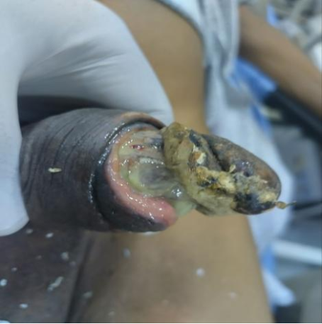
Figure 1. Showing obvious macroscopic death of distal penis and glans.
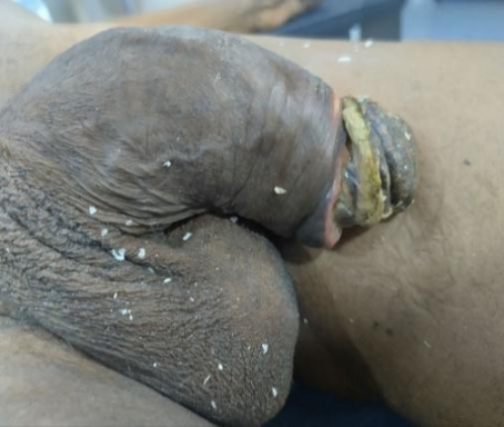
Figure 2. Firm proximal penile shaft.
We made a diagnosis penile gangrene in a known diabetic with multiple myeloma on chemotherapy to rule out carcinoma of the penis. We commenced intravenous antibiotics (rocephine, metronidazole) based on the sensitive on microscopy and culture, analgesics, sitz bath to enable proper delineation of dead and dying tissue and also to buy more time to work the patient up for surgery while limiting sepsis especially with background diabetes mellitus. He was counseled on penile amputation with refashioning of neo-urethral meatus. It was difficult and tough decision for the patient to give his consent for surgical amputation but he eventually gave his approval and surgical work up was commenced.
As part of preparation for surgery, he was counseled to keep twice daily blood sugar records while on sitz bathe and antibiotics for 2 weeks. The chart showed good blood sugar control. The hemoglobin was 7.7g/dl, WBC 11.8×10
6, other components of CBC and EUCr were within normal. ECG and ECHO were favorable and he was eventually booked for partial penile amputation with refashioning of the penis and neo meatus under regional anesthesia. Intra-operative pictures of the surgical procedures are as shown in
Figures 3 to 14. We also placed a diversion suprapubic cystostomy before we commenced partial penectomy.
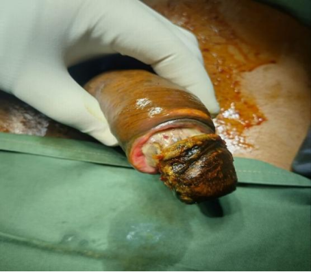
Figure 3. Patient draped to expose the surgical field.
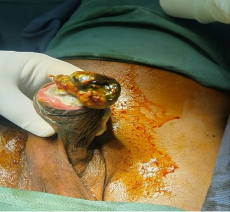
Figure 4. Showing the ventral part of the penis with obvious urethral meatal stenosis.
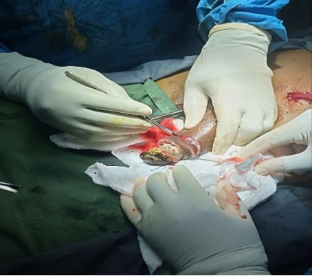
Figure 5. Incision placed 1cm proximal to the gangrenous part and extended in an oblique manner toward ventral penis.
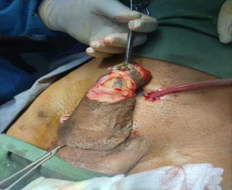
Figure 6. Prior to placement of incision we applied a tourniquet to reduce bleeding. This diagram also shows dead distal urethral covered with slough.
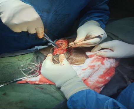
Figure 7. Incision extended obliquely and proximally to the viable part of the mid urethra.
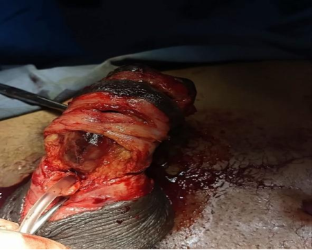
Figure 8. Penile skin degloved, viable urethra stump dissected and a feeding tube advanced to ascertain patency.
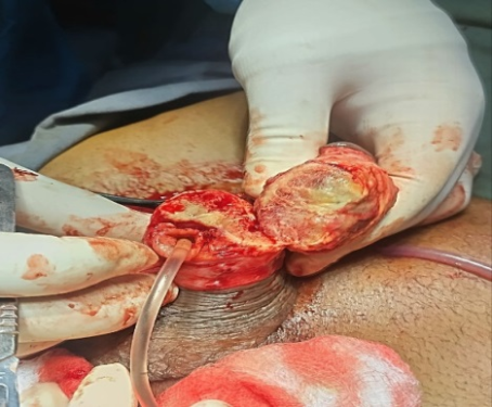
Figure 9. Partial penectomy done with the viable corpora bodies and urethral exposed.
Figure 10. Gangrenous distal penis that was sent for histological analysis.
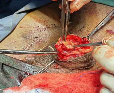
Figure 11. Amputated stump with bleeding edges and properly dissected urethra which was subsequently everted to prevent stenosis.
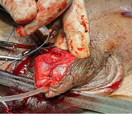
Figure 12. The tunica albuginea was repaired over the copora bodies in a water tight fashion longitudinally after the neo-urethra stump was refashioned and everted.
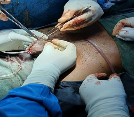
Figure 13. Closure of the penile skin. The tunica albuginea was first closed longitudinally in a water tight fashion over the copora bodies after the edges ot the urethra stump was everted to prevent stenosis.
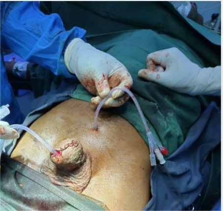
Figure 14. Size 16 French urethral catheter was inserted to keep the neo-meatus patent during healing as well as for urinary drainage and monitoring. The SPC was removed after 72hours and urethral catheter removed after 4 weeks.
The immediate postoperative condition was satisfactory. He was commenced on intravenous normal saline, meropenem, metronidazole, analgesics, vitamin C and endocrinologist review with blood sugar monitoring and oral hypoglycemic medication. Post-surgery recovery was uneventful and satisfactory and he was discharged after 10days to continue follow up as outpatient. The suprapubic catheter (SPC) was removed after 72hrs and urethral catheter removed after 4 weeks.
Histology report showed a fibrocalcific proliferation comprising of multiple foci of variably sized calcifications contained in cystic spaces with a background of fibroblastic proliferation in fascicles, whorls and collagenous matrix. There are few inflammatory cells and epitheloid/gaint cell granulomatous reactions around the areas of calcification. There is no atypia or malignancy. The histologically diagnosis was that of benign calcinosis to rule out metastatic calcifications. He is also being followed up by the hematologist, endocrinologist, and cardiologist. He remained stable with no further deterioration.
We are mindful of background type 2 diabetes mellitus and multiple myeloma which may lead to progressive gangrene of the remnant penile stump and toxic untoward effect of chemotherapy with consequent increased morbidity. More importantly is the psychosocial issues especially depression, withdrawal from social activities and poor quality of life (QoL) and sexual performance. We recommended a multidisciplinary approach involving the mental health physicians, hematologist, endocrinologist and cardiologist. Regular follow up of this index patient is inevitable and can never be overemphasized.
3. Discussion
Penile gangrene is one of the conditions encountered by urologist in clinical practice. The risk of penile loss is high especially in tropical Africa due to late presentation.
[1] | Ntia IO, Mungadi IA. The Pattern of Penile Gangrene in Sokoto, Nigeria. Afr J Urol. 2007; 13(4): 255-261. |
[2] | Ashutosh T, Neerja P, Majhail S. Fournier’s Gangrene of the Penis: A Rare Entity. J Cutan Aesthet Surg. 2010; 3(1): 41–44. https://doi.org/10.4103/0974-2077.63394 |
[13] | Weiner DM, Lowe FC. Surgical Management of Ischemic Penile Gangrene in Diabetics with End Stage Atherosclerosis. J Urol. 1996; 155: 926–929. https://doi.org/10.1016/S0022-5347(01)66348-1 |
[1, 2, 13]
Male genital gangrene commonly called Fournier’s gangrene
can be caused by urethral stricture, priapism, impacted urethral calculus, circumcision, diabetes mellitus, cancer, trauma.
[1] | Ntia IO, Mungadi IA. The Pattern of Penile Gangrene in Sokoto, Nigeria. Afr J Urol. 2007; 13(4): 255-261. |
[5] | Vikas KP, Ravimohan SM, Sudheer KD, Kim V, Girdhar SB. Priapism with Penile Gangrene: An Unusual Presentation of Multiple Myeloma. Indian J Urol. 2017 Jul-Sep; 33(3): 251–252. https://doi.org/10.4103/iju.IJU_41_17 |
[10] | Odunukwe NN, Madu JA, Nnodu OE, Okocha OE, Akingbola TS, Asuquo IM et al. Multiple Myeloma in Nigeria: A Multi-centre Epidemiological and Biomedical Study. PAM J. 2015; 22: 292. https://doi.org/10.11604/pamj.2015.22.292.7774 |
[11] | Abdulla MC, Nair LK, Narayan R, Das A. Multiple Myeloma Presenting as Extensive Limb Gangrene. J. Appl. Hematol. 2014 Jul-Sep; 5(3): 107-110. |
[12] | Nwabuko CC, Igbigbi EE, Chukwuonye II, Nnoli MA. Multiple Myeloma in Niger Delta, Nigeria: Complications and Outcome of Palliative Interventions. CMAR. 2017; 9: 189-196. https://doi.org/10.2147/CMAR.S126136 |
[1, 5, 10-12]
Surgical treatment options ranges from urethroplasty, phalloplasty, neomeatoplasty, penectomy or a combination of these options.
[1] | Ntia IO, Mungadi IA. The Pattern of Penile Gangrene in Sokoto, Nigeria. Afr J Urol. 2007; 13(4): 255-261. |
[14] | Suleiman A, Michael AN, Helina KT, Nahom GM, Yishak A, Mesfin TW. Management of Total Penile Amputation Following Circumcision in a Resource Limited Setting: A Case Report. J. Pediatr. Surg. Case Rep. 2025; 113: 102939. https://doi.org/10.1016/j.epsc.2024.102939 |
[1, 14]
Ntai et al in their review in Sokoto northwest Nigeria noted that gangrene involving the genital region was found to have a wide age range of 6 to 62 years with a mean age of 42.5 years.
[1] | Ntia IO, Mungadi IA. The Pattern of Penile Gangrene in Sokoto, Nigeria. Afr J Urol. 2007; 13(4): 255-261. |
[3] | Khanh NP, Mitchell HS, charles AS. Severe Gangrene at the Glans Penis Requiring Penectomy as the First Major Complication of Buerger’s Disease. Am J Clin Exp Urol. 2016; 4(1): 9–11. |
[1, 3]
Our index patient is 54 years, which is in keeping with the described age range in the above review. Our patient also resides in northern Nigeria which further agrees with their findings.
Our index patient had multiple etiologies such as diabetes mellitus, multiple myeloma and meatal stenosis with likelihood of a distal penile urethral stricture which apparently was not diagnosed until the penis became gangrenous. He also presented with bothersome voiding and storage lower urinary tract symptoms which may be either present prior to gangrene or as a sequel of the progressive gangrene with consequent urethral stenosis. We had dilemma determining which of the etiologies was responsible for the progressive macroscopic death. Ashutosh T et al in their case report attributed the rare condition to Fournier’s gangrene likely for a focus on the penis.
This may be true in this case because all predisposing factors in our index case could lead to a focus of infection from where progressive Fournier’s could set in and result to gangrene of the penis. The predisposing factors in our case could also lead to immunosuppression which increases the risk of infection, in this case with focus on the penis with consequent progressive gangrene.
Many studies reported that the penile gangrene may originate from a focus in the urethra like a stricture.
[1] | Ntia IO, Mungadi IA. The Pattern of Penile Gangrene in Sokoto, Nigeria. Afr J Urol. 2007; 13(4): 255-261. |
[2] | Ashutosh T, Neerja P, Majhail S. Fournier’s Gangrene of the Penis: A Rare Entity. J Cutan Aesthet Surg. 2010; 3(1): 41–44. https://doi.org/10.4103/0974-2077.63394 |
[14] | Suleiman A, Michael AN, Helina KT, Nahom GM, Yishak A, Mesfin TW. Management of Total Penile Amputation Following Circumcision in a Resource Limited Setting: A Case Report. J. Pediatr. Surg. Case Rep. 2025; 113: 102939. https://doi.org/10.1016/j.epsc.2024.102939 |
[1, 2, 14]
The gangrenous process now spreads due to underlying systemic conditions like diabetes mellitus, immunosuppression and chronic renal failure.
[4] | Vijayan P. Gangrene of the Penis in a Diabetic Male with Multiple Amputations and Follow up. Indian J Urol. 2009 Jan-Mar; 25(1): 123–125. https://doi.org/10.4103/0970-1591.45550 |
[5] | Vikas KP, Ravimohan SM, Sudheer KD, Kim V, Girdhar SB. Priapism with Penile Gangrene: An Unusual Presentation of Multiple Myeloma. Indian J Urol. 2017 Jul-Sep; 33(3): 251–252. https://doi.org/10.4103/iju.IJU_41_17 |
[10] | Odunukwe NN, Madu JA, Nnodu OE, Okocha OE, Akingbola TS, Asuquo IM et al. Multiple Myeloma in Nigeria: A Multi-centre Epidemiological and Biomedical Study. PAM J. 2015; 22: 292. https://doi.org/10.11604/pamj.2015.22.292.7774 |
[11] | Abdulla MC, Nair LK, Narayan R, Das A. Multiple Myeloma Presenting as Extensive Limb Gangrene. J. Appl. Hematol. 2014 Jul-Sep; 5(3): 107-110. |
[12] | Nwabuko CC, Igbigbi EE, Chukwuonye II, Nnoli MA. Multiple Myeloma in Niger Delta, Nigeria: Complications and Outcome of Palliative Interventions. CMAR. 2017; 9: 189-196. https://doi.org/10.2147/CMAR.S126136 |
[4, 5, 10-12]
These conditions causes artherosclerosis, peripheral angiopathy, calciphylaxis and may invariably progress to ischemia which when it occurs in the penis can lead to macroscopic death considering that the penis is has end artery,
[1] | Ntia IO, Mungadi IA. The Pattern of Penile Gangrene in Sokoto, Nigeria. Afr J Urol. 2007; 13(4): 255-261. |
[1]
The histology report confirmed benign calcinosis which is in keeping with findings by Weiner DM et al. Our index case was 54 years and immune-compromised either from multiple myeloma on chemotheraspy or diabetes mellitus which increases the likelihood of infection in the urinary tract from prostate enlargement. This must have serves as a trigger aided by the co-morbid condition including diabetes and multiple myeloma invariably leading to obvious macroscopic death on the penis.
Our index patient also had meatal stenosis with possible distal penile urethral stricture as the focus of obstruction causing bothersome storage and voiding lower urinary tract symptoms and urinary tract infection. The meatal stenosis may be a complication of progressive gangrene which could worsen the infective process triggering a vicious cycle with further deterioration. The event was rapidly progressive in our index patient as reported by Vijayan P et al
and within a week he presented with obvious gangrene and pus. We can affirm that the multiple predisposing factors acted synergistically to result in rapid progression despite initial intervention by the primary managing hematologist before referral to urologist.
Late presentation is usually characterized by significant gangrene and eventually partial or complete penile loss.
[1] | Ntia IO, Mungadi IA. The Pattern of Penile Gangrene in Sokoto, Nigeria. Afr J Urol. 2007; 13(4): 255-261. |
[2] | Ashutosh T, Neerja P, Majhail S. Fournier’s Gangrene of the Penis: A Rare Entity. J Cutan Aesthet Surg. 2010; 3(1): 41–44. https://doi.org/10.4103/0974-2077.63394 |
[1, 2]
Our index case presented late when gangrene had already set in and he eventually had a partial penectomy, refashioning of the penile stump and neo-meatus as the adopted treatment option to prevent further spread to the entire penis. As with other reported cases, he was commenced on sitz bath antibiotics, analgesics and wound care.
[1] | Ntia IO, Mungadi IA. The Pattern of Penile Gangrene in Sokoto, Nigeria. Afr J Urol. 2007; 13(4): 255-261. |
[2] | Ashutosh T, Neerja P, Majhail S. Fournier’s Gangrene of the Penis: A Rare Entity. J Cutan Aesthet Surg. 2010; 3(1): 41–44. https://doi.org/10.4103/0974-2077.63394 |
[4] | Vijayan P. Gangrene of the Penis in a Diabetic Male with Multiple Amputations and Follow up. Indian J Urol. 2009 Jan-Mar; 25(1): 123–125. https://doi.org/10.4103/0970-1591.45550 |
[13] | Weiner DM, Lowe FC. Surgical Management of Ischemic Penile Gangrene in Diabetics with End Stage Atherosclerosis. J Urol. 1996; 155: 926–929. https://doi.org/10.1016/S0022-5347(01)66348-1 |
[14] | Suleiman A, Michael AN, Helina KT, Nahom GM, Yishak A, Mesfin TW. Management of Total Penile Amputation Following Circumcision in a Resource Limited Setting: A Case Report. J. Pediatr. Surg. Case Rep. 2025; 113: 102939. https://doi.org/10.1016/j.epsc.2024.102939 |
[1, 2, 4, 13, 14]
The initial conservative approach failed which led to surgical intervention via partial penectomy. Khanh NP et al
[3] | Khanh NP, Mitchell HS, charles AS. Severe Gangrene at the Glans Penis Requiring Penectomy as the First Major Complication of Buerger’s Disease. Am J Clin Exp Urol. 2016; 4(1): 9–11. |
[3]
also reported penile amputation as a viable option for penile gangrene and the fact that our initial conservative approached failed is in keeping with their findings.
Our patient found it difficult to give his consent for surgery and had to travel to another region of the country to seek second option. This was not unusual considering the special function of the penis and religious belief for a miracle in our environment. We obliged and gave him ample time to voluntary give his consent. We involved and engaged his spouse and other family members on psychosocial support. He returned 2 weeks later and opted to allow partial penectomy. We are not unaware of the depression and poor QoL and sexual performance in this case as our patient was young and has a young spouse. Benjamin E Ayres in his report emphasized on the need for a holistic and multidisciplinary approaches in clinic with access to counseling as a means of helping patients adjust to their new situation.
We absolutely concur with this approach and further referred the index case to mental health physicians for psychological review.
Author Contributions
Obiatuegwu Kenenna: Conceptualization, Data curation, Formal Analysis, Investigation, Project administration, Supervision, Validation, Visualization, Writing – original draft, Writing – review & editing
Otabor Christopher: Funding acquisition, Validation
Magnus Felix E: Project administration, Writing – review & editing
Awuzie Chinemezu: Software, Visualization, Writing – review & editing
Okonta Emeka: Formal Analysis, Investigation, Visualization