
Venovenous and venoarterial extracorporeal membrane oxygenation (ECMO) provides life support for patients with refractory cardiopulmonary failure, but bleeding and thrombotic complications remain major causes of mortality. Conventional monitoring based on single activated clotting time (ACT) and activated partial thromboplastin time (APTT) measurements may not adequately capture intra-individual coagulation fluctuations. Previous longitudinal work linked APTT variability with bleeding and mortality, but ACT variability has rarely been evaluated concurrently. This dual-cohort retrospective study evaluated the prognostic value of ACT and APTT variability for 28-day in-hospital mortality and developed an externally validated nomogram. The derivation cohort included 300 patients from the MIMIC-IV database, and the external validation cohort included 200 patients from our ICU. Intra-individual coefficients of variation (CVs) of serial ACT and APTT measurements during ECMO support were calculated. Candidate predictors were screened using LASSO Cox regression and evaluated by Cox proportional hazards regression. Kaplan–Meier analysis compared survival across ACT-CV and APTT-CV tertiles, and model performance was assessed by discrimination, calibration, and decision curve analysis. LASSO identified age, ACT-CV, APTT-CV, lactate, and SOFA score as candidate predictors. Multivariate Cox regression showed that elevated ACT-CV (HR=2.875, 95% CI 1.692–4.891, P<0.001) and APTT-CV (HR=2.630, 95% CI 1.547–4.471, P<0.001) were independent predictors of 28-day mortality. Mortality risk increased progressively across higher ACT-CV and APTT-CV tertiles, with significantly poorer survival in patients with greater coagulation variability (all log-rank P<0.001). The five-variable nomogram achieved AUCs of 0.85 and 0.81 in the derivation and validation cohorts, respectively, outperforming SOFA and APACHE II scores, with favorable calibration and greater clinical net benefit. Intra-individual ACT and APTT variability may therefore provide accessible prognostic information for early risk stratification and individualized anticoagulation management in ECMO patients.
| Published in | Cardiology and Cardiovascular Research (Volume 10, Issue 3) |
| DOI | 10.11648/j.ccr.20261003.16 |
| Page(s) | 63-77 |
| Creative Commons |
This is an Open Access article, distributed under the terms of the Creative Commons Attribution 4.0 International License (http://creativecommons.org/licenses/by/4.0/), which permits unrestricted use, distribution and reproduction in any medium or format, provided the original work is properly cited. |
| Copyright |
Copyright © The Author(s), 2026. Published by Science Publishing Group |
Extracorporeal Membrane Oxygenation, ACT-CV, APTT-CV, Coagulation Variability, Nomogram, Prognosis
Derivation cohort(N=300) | External validation cohort(N=200) | P value | |
|---|---|---|---|
Demographic characteristics | |||
Age, median (IQR) | 59 (47, 68) | 61 (49, 70) | 0.612 |
Male, n (%) | 176 (58.7) | 113 (56.5) | 0.804 |
Ethnicity, n (%) | 0.729 | ||
White | 212 (70.7) | 145 (72.5) | |
Black | 58 (19.3) | 36 (18.0) | |
Other | 30 (10.0) | 19 (9.5) | |
Height (cm), median (IQR) | 172 (165, 178) | 170 (163, 176) | 0.587 |
Hospital LOS (days), median (IQR) | 12 (7, 21) | 11 (6, 19) | 0.426 |
Comorbidities, n (%) | |||
AIDS | 8 (2.7) | 5 (2.5) | 0.912 |
Cerebrovascular disease | 22 (7.3) | 15 (7.5) | 0.987 |
Chronic pulmonary disease | 45 (15.0) | 32 (16.0) | 0.761 |
Congestive heart failure | 38 (12.7) | 24 (12.0) | 0.823 |
Diabetes mellitus | 52 (17.3) | 34 (17.0) | 0.925 |
Mild liver disease | 19 (6.3) | 13 (6.5) | 0.941 |
Myocardial infarction | 26 (8.7) | 18 (9.0) | 0.908 |
Peripheral vascular disease | 17 (5.7) | 11 (5.5) | 0.933 |
Renal disease | 31 (10.3) | 20 (10.0) | 0.915 |
Rheumatic disease | 9 (3.0) | 6 (3.0) | 1.000 |
Dementia | 12 (4.0) | 8 (4.0) | 1.000 |
Paraplegia | 7 (2.3) | 5 (2.5) | 0.894 |
Peptic ulcer disease | 14 (4.7) | 9 (4.5) | 0.927 |
Vital signs on admission, median (IQR) | |||
Heart rate (bpm) | 102 (88, 116) | 105 (90, 118) | 0.358 |
Systolic blood pressure (mmHg) | 118 (102, 135) | 116 (100, 132) | 0.412 |
Diastolic blood pressure (mmHg) | 62 (54, 70) | 60 (52, 68) | 0.376 |
Mean blood pressure (mmHg) | 78 (68, 88) | 76 (66, 86) | 0.401 |
Respiratory rate (bpm) | 22 (18, 26) | 23 (19, 27) | 0.324 |
SpO2 (%) | 94 (90, 97) | 93 (89, 96) | 0.289 |
Laboratory parameters, median (IQR) | |||
Lactate (mmol/L) | 2.3 (1.5, 3.6) | 2.4 (1.6, 3.7) | 0.593 |
Serum albumin (g/L) | 28 (22, 34) | 27 (21, 33) | 0.625 |
Total bilirubin (μmol/L) | 22 (15, 35) | 24 (16, 38) | 0.517 |
Blood urea nitrogen (mmol/L) | 8.2 (5.1, 12.3) | 8.5 (5.3, 12.8) | 0.489 |
Serum creatinine (μmol/L) | 76 (45, 112) | 78 (47, 115) | 0.532 |
Sodium (mmol/L) | 138 (135, 142) | 139 (136, 143) | 0.316 |
Potassium (mmol/L) | 4.2 (3.8, 4.6) | 4.3 (3.9, 4.7) | 0.298 |
Chloride (mmol/L) | 102 (98, 106) | 103 (99, 107) | 0.305 |
Calcium (mmol/L) | 2.1 (1.9, 2.3) | 2.0 (1.8, 2.2) | 0.276 |
Glucose (mmol/L) | 8.9 (6.2, 12.1) | 9.1 (6.4, 12.3) | 0.453 |
Hemoglobin (g/L) | 92 (78, 105) | 90 (76, 103) | 0.387 |
Hematocrit (%) | 28 (24, 32) | 27 (23, 31) | 0.415 |
White blood cell count (×10⁹/L) | 11.2 (7.8, 15.6) | 11.5 (8.0, 15.9) | 0.362 |
Platelet count (×10⁹/L) | 186 (122, 258) | 182 (118, 252) | 0.408 |
Activated clotting time (ACT, s) | 182 (156, 214) | 185 (158, 218) | 0.327 |
Activated partial thromboplastin time (APTT, s) | 48 (36, 62) | 49 (37, 64) | 0.351 |
International normalized ratio (INR) | 1.4 (1.1, 1.7) | 1.5 (1.2, 1.8) | 0.289 |
Prothrombin time (PT, s) | 16.2 (13.5, 19.8) | 16.5 (13.8, 20.1) | 0.302 |
Blood gas analysis, median (IQR) | |||
pH | 7.32 (7.25, 7.38) | 7.31 (7.24, 7.37) | 0.418 |
PaCO2 (mmHg) | 42 (35, 50) | 43 (36, 51) | 0.374 |
PaO2/FiO2 ratio (mmHg) | 186 (124, 258) | 182 (120, 254) | 0.425 |
Bicarbonate (mmol/L) | 24 (21, 27) | 23 (20, 26) | 0.312 |
Base excess (mmol/L) | -2.1 (-5.2, 1.3) | -2.3 (-5.4, 1.1) | 0.386 |
Anion gap (mmol/L) | 12 (8, 16) | 13 (9, 17) | 0.297 |
Disease severity scores, median (IQR) | |||
SOFA score | 11 (8, 14) | 12 (8, 15) | 0.645 |
APS III score | 52 (41, 63) | 54 (43, 65) | 0.512 |
LODS score | 10 (7, 13) | 11 (8, 14) | 0.489 |
OASIS score | 42 (33, 51) | 44 (35, 53) | 0.426 |
SIRS score | 2 (1, 3) | 2 (1, 3) | 0.783 |
GCS score | 13 (10, 15) | 13 (10, 15) | 0.815 |
MELD score | 18 (12, 24) | 19 (13, 25) | 0.503 |
ECMO and treatment characteristics | |||
ECMO mode, VV/VA, n | 194 / 106 | 131 / 69 | 0.947 |
ECMO duration (h), median (IQR) | 142 (96, 215) | 138 (92, 207) | 0.735 |
Vasopressor use, n (%) | 242 (80.7) | 158 (79.0) | 0.672 |
Norepinephrine | 218 (72.7) | 142 (71.0) | 0.689 |
Epinephrine | 56 (18.7) | 38 (19.0) | 0.921 |
Dopamine | 32 (10.7) | 21 (10.5) | 0.935 |
Dobutamine | 28 (9.3) | 19 (9.5) | 0.927 |
Phenylephrine | 18 (6.0) | 12 (6.0) | 1.000 |
CRRT use, n (%) | 87 (29.0) | 56 (28.0) | 0.783 |
Mechanical ventilation, n (%) | 268 (89.3) | 178 (89.0) | 0.915 |
Core research indicators, median (IQR) | |||
ACT-CV (coefficient of variation) | 0.28 (0.21, 0.36) | 0.29 (0.22, 0.37) | 0.681 |
APTT-CV (coefficient of variation) | 0.24 (0.18, 0.33) | 0.25 (0.19, 0.34) | 0.702 |
Outcomes, n (%) | |||
Hospital mortality | 77 (25.7) | 53 (26.5) | 0.887 |
28-day mortality | 82 (27.3) | 55 (27.5) | 0.952 |
Regression coefficient | |
|---|---|
Age | 0.021049 |
ACT-CV (coefficient of variation) | 0.087521 |
APTT-CV (coefficient of variation) | 0.079164 |
Lactate (mmol/L) | 0.054002 |
SOFA score | 0.010851 |
ECMO duration | 0 |
ECMO mode (VV/VA) | 0 |
Gender | 0 |
Creatinine | 0 |
Platelet count | 0 |
pH | 0 |
Total bilirubin | 0 |
Variables | Univariate analysisHR (95% CI) | P value | Multivariate analysisHR (95% CI) | P value |
|---|---|---|---|---|
Age | 1.024 (1.003–1.046) | 0.025 | 1.021 (1.001–1.042) | 0.038 |
ACT-CV | 3.126 (1.874–5.213) | <0.001 | 2.875 (1.692–4.891) | <0.001 |
APTT-CV | 2.943 (1.761–4.920) | <0.001 | 2.630 (1.547–4.471) | <0.001 |
Lactate | 1.215 (1.084–1.362) | <0.001 | 1.182 (1.053–1.326) | 0.004 |
SOFA score | 1.107 (1.032–1.188) | 0.004 | 1.085 (1.010–1.166) | 0.025 |
Groups | HR (95% CI) | P value |
|---|---|---|
ACT-CV | ||
Low ACT-CV (<0.23) | Reference | – |
Intermediate ACT-CV (0.23–0.33) | 1.724 (1.012–2.937) | 0.045 |
High ACT-CV (>0.33) | 3.468 (2.016–5.964) | <0.001 |
APTT-CV | ||
Low APTT-CV (<0.20) | Reference | – |
Intermediate APTT-CV (0.20–0.30) | 1.681 (0.986–2.867) | 0.056 |
High APTT-CV (>0.30) | 3.105 (1.822–5.290) | <0.001 |
Subgroup stratification | HR (95% CI) of high ACT-CV | P for interaction |
|---|---|---|
Age ≤ 60 years | 2.412 (1.105–5.263) | 0.276 |
Age > 60 years | 3.084 (1.643–5.786) | 0.276 |
VV-ECMO mode | 2.761 (1.482–5.143) | 0.413 |
VA-ECMO mode | 3.015 (1.336–6.792) | 0.413 |
Lactate ≤ 2 mmol/L | 2.350 (1.044–5.293) | 0.351 |
Lactate > 2 mmol/L | 3.227 (1.751–5.944) | 0.351 |
Groups | HR (95% CI) | P value |
|---|---|---|
Low APTT-CV (<0.20) | Reference | – |
Intermediate APTT-CV (0.20–0.30) | 1.681 (0.986–2.867) | 0.056 |
High APTT-CV (>0.30) | 3.105 (1.822–5.290) | <0.001 |
Subgroup stratification | HR (95% CI) of high APTT-CV | P for interaction |
|---|---|---|
Age ≤ 60 years | 2.297 (1.062–4.966) | 0.291 |
Age > 60 years | 2.951 (1.572–5.537) | 0.291 |
VV-ECMO mode | 2.643 (1.412–4.945) | 0.447 |
VA-ECMO mode | 2.902 (1.283–6.562) | 0.447 |
Lactate ≤ 2 mmol/L | 2.284 (1.016–5.132) | 0.365 |
Lactate > 2 mmol/L | 3.096 (1.682–5.698) | 0.365 |
ECMO | Extracorporeal Membrane Oxygenation |
VV-ECMO | Venovenous Extracorporeal Membrane Oxygenation |
VA-ECMO | Venoarterial Extracorporeal Membrane Oxygenation |
ACT | Activated Clotting Time |
APTT | Activated Partial Thromboplastin Time |
CV | Coefficient of Variation |
ACT-CV | Coefficient of Variation of Activated Clotting Time |
APTT-CV | Coefficient of Variation of Activated Partial Thromboplastin Time |
BTEs | Bleeding/Thrombotic Events |
ELSO | Extracorporeal Life Support Organization |
LASSO | Least Absolute Shrinkage and Selection Operator |
SOFA | Sequential Organ Failure Assessment |
APACHE II | Acute Physiology and Chronic Health Evaluation II |
DCA | Decision Curve Analysis |
MIMIC-IV | Medical Information Mart for Intensive Care IV |
| [1] | Nunez JI, Gosling AF, O’Gara B, et al. Bleeding and thrombotic events in adults supported with venovenous extracorporeal membrane oxygenation: An ELSO registry analysis. Intensive Care Med. 2022; 48(2): 213–224. |
| [2] | Helms J, Frere C, Thiele T, et al. Anticoagulation in adult patients supported with extracorporeal membrane oxygenation: Guidance from the scientific and standardization committees on perioperative and critical care haemostasis and thrombosis of the international society on thrombosis and haemostasis. J Thromb Haemost. 2023; 21(2): 373–396. |
| [3] | Weber CF, Zacharowski K, Calatzis A, and Lindner ML. Monitoring unfractionated heparin in ECMO and cardiac surgery: The search for a valid reference standard. Front Med (Lausanne). 2026; 13: 1818646. |
| [4] | Xu Enhui, Sun Qi, Lu Juan, Xiong Ying, Wei Xinguang, and Wei Xinguang. Recent advances and future perspectives in anticoagulation management for extracorporeal membrane oxygenation. Chinese Journal of Extracorporeal Life Support. 2025; 23(2): 176–180. |
| [5] | Davidson S. Interpretation of coagulation laboratory tests for patients on ECMO. Int J Lab Hematol. 2024; 46(4): 606–612. |
| [6] | Malik MI, Aboelnazar NS, Rheault-Henry M, Vo TX, and Nagpal AD. Cumulative elevation of aPTT predicts time to major bleeding and death in ECMO: A joint longitudinal-survival model. Perfusion. 2026;: 2676591251412301. |
| [7] | Ranzani OT, Singer M, and Moreno RP. Clarifying the SOFA-2 score-reply. JAMA. 2026; 335(15): 1362–1363. |
| [8] | Marget MJ, Dunn R, and Morgan CL. Association of APACHE-II scores with 30-day mortality after tracheostomy: A retrospective study. Laryngoscope. 2023; 133(2): 273–278. |
| [9] | Lei J, Zhai J, Zhang Y, Qi J, and Sun C. Supervised machine learning models for predicting sepsis-associated liver injury in patients with sepsis: Development and validation study based on a multicenter cohort study. J Med Internet Res. 2025; 27: e66733. |
| [10] | Zou Chen. The Impact of Different Anticoagulation Timing Strategies on Prognosis and Complications in ECMO-Treated Patients. 2025. |
| [11] | Qian S, Li P, Hou X, and Dong N. Development and internal validation of the PROFIT and POSITIVE prognostic nomograms for patients undergoing VA-ECMO therapy. Sci Rep. 2025; 15(1): 20573. |
| [12] | Yang Yang, Zhu Yi, and Wu Hao. A nomogram⁃based prediction model for clinical mortality risk in VA⁃ECMO patients. Journal of Nanjing Medical University(Natural Sciences). 2026; 46(3): 425–434. |
| [13] | Xu Dacai, Ding Yao, Zhang Aijuan, et al. Best Evidence Summary for Anticoagulation Management in Adults Undergoing Extracorporeal Membrane Oxygenation. Chinese General Practice Nursing. 2025; 23(17): 3367–3372. |
| [14] | Barbaro RP, MacLaren G, Boonstra PS, et al. Extracorporeal membrane oxygenation for COVID-19: Evolving outcomes from the international extracorporeal life support organization registry. Lancet. 2021; 398(10307): 1230–1238. |
| [15] | Teng Yuan. Effects of Different Anticoagulation Monitoring Indexes on Anticoagulation and Clinical Prognosis of Adult Veno-Arterial Extracorporeal Membrane Oxygenation. 2021. |
| [16] | McMichael ABV, Ryerson LM, Ratano D, Fan E, Faraoni D, and Annich GM. 2021 ELSO adult and pediatric anticoagulation guidelines. ASAIO J. 2022; 68(3): 303–310. |
| [17] | Zeibi Shirejini S, Carberry J, McQuilten ZK, Burrell AJC, Gregory SD, and Hagemeyer CE. Current and future strategies to monitor and manage coagulation in ECMO patients. Thromb J. 2023; 21(1): 11. |
| [18] | Noitz M, Jenner D, Brooks R, et al. ECMO decannulation is associated with dynamic changes in coagulation profiles: An exploratory, nested cohort study. BMC Anesthesiol. 2026; 26(1): 137. |
| [19] | Yuan Jiankai, Duan Hongwei, and Tang Yishu. Construction of a nomogram prediction model for thrombosis risk in patients receiving ECMO supportive treatment. Journal of Modern Medicine & Health. 2026; 42(3): 597–603. |
| [20] | Zhao Qi, Chu Minjuan, and Ji Xueli. Application of a Multivariable Logistic Regression-Based Nomogram for Predicting Mortality Risk in Patients Following Extracorporeal Membrane Oxygenation. Shaanxi Medical Journal. 2026; 55(1): 68–72. |
| [21] | Guo Lanqi, Liu Songqiao, Chang Wei, Pan Chun, and Zheng Yishan. Analysis of Complications and Their Prognostic Associations in Patients Undergoing Extracorporeal Membrane Oxygenation. Modern Medical Journal. 2022; 50(10): 1227–1231. |
APA Style
Ma, S., Feng, M., Lan, J., Liu, M., Lin, Q. (2026). ACT and APTT Variability as Prognostic Markers for 28-Day In-Hospital Mortality in ECMO Patients: Development and External Validation of a Nomogram. Cardiology and Cardiovascular Research, 10(3), 63-77. https://doi.org/10.11648/j.ccr.20261003.16
ACS Style
Ma, S.; Feng, M.; Lan, J.; Liu, M.; Lin, Q. ACT and APTT Variability as Prognostic Markers for 28-Day In-Hospital Mortality in ECMO Patients: Development and External Validation of a Nomogram. Cardiol. Cardiovasc. Res. 2026, 10(3), 63-77. doi: 10.11648/j.ccr.20261003.16
AMA Style
Ma S, Feng M, Lan J, Liu M, Lin Q. ACT and APTT Variability as Prognostic Markers for 28-Day In-Hospital Mortality in ECMO Patients: Development and External Validation of a Nomogram. Cardiol Cardiovasc Res. 2026;10(3):63-77. doi: 10.11648/j.ccr.20261003.16
@article{10.11648/j.ccr.20261003.16,
  author = {Shihui Ma and Mei Feng and Jingru Lan and Manli Liu and Qingran Lin},
  title = {ACT and APTT Variability as Prognostic Markers for 28-Day In-Hospital Mortality in ECMO Patients: Development and External Validation of a Nomogram},
  journal = {Cardiology and Cardiovascular Research},
  volume = {10},
  number = {3},
  pages = {63-77},
  doi = {10.11648/j.ccr.20261003.16},
  url = {https://doi.org/10.11648/j.ccr.20261003.16},
  eprint = {https://article.sciencepublishinggroup.com/pdf/10.11648.j.ccr.20261003.16},
  abstract = {Venovenous and venoarterial extracorporeal membrane oxygenation (ECMO) provides life support for patients with refractory cardiopulmonary failure, but bleeding and thrombotic complications remain major causes of mortality. Conventional monitoring based on single activated clotting time (ACT) and activated partial thromboplastin time (APTT) measurements may not adequately capture intra-individual coagulation fluctuations. Previous longitudinal work linked APTT variability with bleeding and mortality, but ACT variability has rarely been evaluated concurrently. This dual-cohort retrospective study evaluated the prognostic value of ACT and APTT variability for 28-day in-hospital mortality and developed an externally validated nomogram. The derivation cohort included 300 patients from the MIMIC-IV database, and the external validation cohort included 200 patients from our ICU. Intra-individual coefficients of variation (CVs) of serial ACT and APTT measurements during ECMO support were calculated. Candidate predictors were screened using LASSO Cox regression and evaluated by Cox proportional hazards regression. Kaplan–Meier analysis compared survival across ACT-CV and APTT-CV tertiles, and model performance was assessed by discrimination, calibration, and decision curve analysis. LASSO identified age, ACT-CV, APTT-CV, lactate, and SOFA score as candidate predictors. Multivariate Cox regression showed that elevated ACT-CV (HR=2.875, 95% CI 1.692–4.891, P<0.001) and APTT-CV (HR=2.630, 95% CI 1.547–4.471, P<0.001) were independent predictors of 28-day mortality. Mortality risk increased progressively across higher ACT-CV and APTT-CV tertiles, with significantly poorer survival in patients with greater coagulation variability (all log-rank P<0.001). The five-variable nomogram achieved AUCs of 0.85 and 0.81 in the derivation and validation cohorts, respectively, outperforming SOFA and APACHE II scores, with favorable calibration and greater clinical net benefit. Intra-individual ACT and APTT variability may therefore provide accessible prognostic information for early risk stratification and individualized anticoagulation management in ECMO patients.},
 year = {2026}
}
TY  - JOUR
T1  - ACT and APTT Variability as Prognostic Markers for 28-Day In-Hospital Mortality in ECMO Patients: Development and External Validation of a Nomogram
AU  - Shihui Ma
AU  - Mei Feng
AU  - Jingru Lan
AU  - Manli Liu
AU  - Qingran Lin
Y1  - 2026/09/15
PY  - 2026
N1  - https://doi.org/10.11648/j.ccr.20261003.16
DO  - 10.11648/j.ccr.20261003.16
T2  - Cardiology and Cardiovascular Research
JF  - Cardiology and Cardiovascular Research
JO  - Cardiology and Cardiovascular Research
SP  - 63
EP  - 77
PB  - Science Publishing Group
SN  - 2578-8914
UR  - https://doi.org/10.11648/j.ccr.20261003.16
AB  - Venovenous and venoarterial extracorporeal membrane oxygenation (ECMO) provides life support for patients with refractory cardiopulmonary failure, but bleeding and thrombotic complications remain major causes of mortality. Conventional monitoring based on single activated clotting time (ACT) and activated partial thromboplastin time (APTT) measurements may not adequately capture intra-individual coagulation fluctuations. Previous longitudinal work linked APTT variability with bleeding and mortality, but ACT variability has rarely been evaluated concurrently. This dual-cohort retrospective study evaluated the prognostic value of ACT and APTT variability for 28-day in-hospital mortality and developed an externally validated nomogram. The derivation cohort included 300 patients from the MIMIC-IV database, and the external validation cohort included 200 patients from our ICU. Intra-individual coefficients of variation (CVs) of serial ACT and APTT measurements during ECMO support were calculated. Candidate predictors were screened using LASSO Cox regression and evaluated by Cox proportional hazards regression. Kaplan–Meier analysis compared survival across ACT-CV and APTT-CV tertiles, and model performance was assessed by discrimination, calibration, and decision curve analysis. LASSO identified age, ACT-CV, APTT-CV, lactate, and SOFA score as candidate predictors. Multivariate Cox regression showed that elevated ACT-CV (HR=2.875, 95% CI 1.692–4.891, P<0.001) and APTT-CV (HR=2.630, 95% CI 1.547–4.471, P<0.001) were independent predictors of 28-day mortality. Mortality risk increased progressively across higher ACT-CV and APTT-CV tertiles, with significantly poorer survival in patients with greater coagulation variability (all log-rank P<0.001). The five-variable nomogram achieved AUCs of 0.85 and 0.81 in the derivation and validation cohorts, respectively, outperforming SOFA and APACHE II scores, with favorable calibration and greater clinical net benefit. Intra-individual ACT and APTT variability may therefore provide accessible prognostic information for early risk stratification and individualized anticoagulation management in ECMO patients.
VL  - 10
IS  - 3
ER  -
School of Nursing, Jinan University, Guangzhou, China
Intensive Care Unit, The First Affiliated Hospital of Jinan University, Guangzhou, China
Department of Stomatology, The First Affiliated Hospital of Jinan University, Guangzhou, China
Department of Nursing, The First Affiliated Hospital of Jinan University, Guangzhou, China
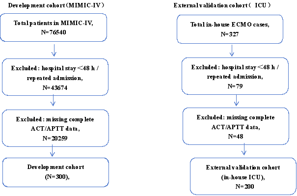
Figure 1. The flow chart of the included population.
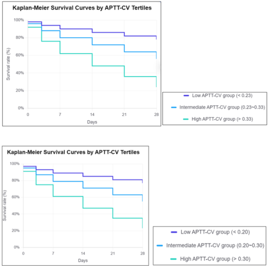
Figure 2. Kaplan Meier survival curves for 28 day mortality stratified by ACT CV tertiles. Kaplan Meier curves showing 28 day survival among ECMO patients stratified by low, intermediate and high ACT CV tertiles. Log rank test was used for group comparison. Patients in the high ACT CV group exhibited substantially poorer survival outcomes. ACT CV, coefficient of variation of activated clotting time.
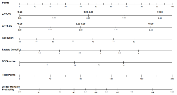
Figure 3. The prediction model was developed based on Cox proportional hazards regression and visualized as a nomogram incorporating five independent prognostic factors: age, lactate, SOFA score, ACT CV, and APTT CV for 28 day mortality among patients receiving ECMO support.
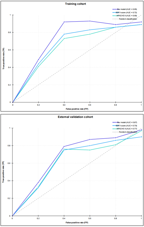
Figure 4. Receiver operating characteristic (ROC) curves of predictive models for 28 day mortality risk in patients receiving extracorporeal membrane oxygenation. (A) ROC curve of the nomogram derived from the training cohort. (B) ROC curve of the nomogram in the external validation cohort (SOFA score and APACHE Ⅱ score were applied as reference models).
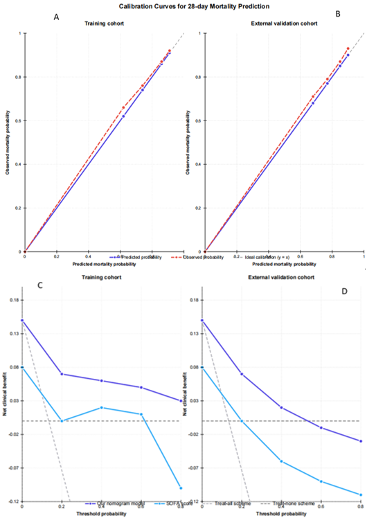
Figure 5. Calibration and decision curve analysis (DCA) curves of predictive models for 28 day mortality risk in patients receiving extracorporeal membrane oxygenation. (A) Calibration curves for predicting 28 day mortality in the training cohort. (B) External validation of calibration curves for predicting 28 day mortality in the external validation cohort. (C) Decision curve analysis (DCA) of the nomogram for predicting 28 day mortality in the training cohort. (D) DCA of the nomogram for predicting 28 day mortality in the external validation cohort (SOFA score and APACHE Ⅱ score were used as reference models).

Information



 Share This Article
 Twitter
 Linked In
 Facebook
 Copy
Copy Download