Research Article | | Peer-Reviewed

Norcantharidin Inhibits an Angiogenesis of Croquel-180 Sarcomatous Cells

Received: 11 May 2025     Accepted: 11 June 2025     Published: 10 July 2025
Views:       Downloads:
Abstract

Norcantharidin (NCTD) is a demethylated water-soluble synthetic small molecule of cantharidin (CTD), has been reported that it has anticancer activities including apoptosis and anti-angiogenesis. NCTD reduced VEGF production and the expression of integrin-β1 in tumor cells even at the low concentrations with less toxicity to the kidney or liver, and destroyed vimentin to inhibit angiogenesis in tumor cells by inducing anoikis of vascular endothelial cells. In addition, basing on recent data that the metastasis and recurrence of tumor are closely related to angiogenesis in tumor tissues, Croquel-180 sarcomatous cells, a mesenchyma-derived cells group with better angiogenesis were transplanted to Chick embryo Chorioallantoic Membrane (CAM) and the anti-angiogenic effect of NCTD was examined. When NCTD was injected at a dose of 40µg/µL and 80µg/µL, there was significant decrease in the number of microvascular branches than the control group. There was no significant difference in microvascular density (MVD) when NCTD was injected at a dose of 20µg/μL, but at a dose of 40µg/µL and 80µg/µL, MVD decreased significantly compared to the control. We reaffirmed that NCTD is a drug that can specifically inhibit angiogenesis in tumor tissues through CAM experiments, and that it is a very effective drug that can be applied to antitumor therapy targeting angiogenesis in tumor tissues. Norcantharidin is a powerful angiogenesis inhibitor capable of sufficiently inhibiting angiogenesis in tumor tissue. We confirmed that it has potential application value applicable to tumor treatment targeting endothelial cells.

Published in Biochemistry and Molecular Biology (Volume 10, Issue 3)
DOI 10.11648/j.bmb.20251003.11
Page(s) 37-44
Creative Commons

This is an Open Access article, distributed under the terms of the Creative Commons Attribution 4.0 International License (http://creativecommons.org/licenses/by/4.0/), which permits unrestricted use, distribution and reproduction in any medium or format, provided the original work is properly cited.

Copyright

Copyright © The Author(s), 2025. Published by Science Publishing Group

Keywords

Norcantharidin (NCTD), Anti-angiogenesis, Antitumor Drug, Chemotherapy

1. Introduction
With the rapid development of molecular biology, the study of cancer is intensified as it has become one of the world`s intractable diseases and it has been reported that the proliferation, invasion and metastasis of cancer cells are closely related to angiogenesis as its mechanism are revealed one by one.
Tumor cells easily enter the blood when microvascular density (MVD) in the tumor tissue is high, and the lower a degree of differentiation and the higher an ability of angiogenesis is, the better a metastasis occurs. Thus, MVD in tumor tissue was proposed as an important index for observing the angiogenesis in tumor, as it is the most necessary process in metastasis of tumor . Some researchers have found that vascular endothelial growth factor (VEGF), fibroblast growth factor (FGF), and interleukin-8(IL-8), which are angiogenesis growth factors, play an important role in angiogenesis . It has been reported that VEGFs are produced in normal cells such as smooth muscle cells and macrophages, as well as in many cancer cells such as mammary and colorectal carcinoma than normal cells .
It was reported that the balance between the anti-apoptotic protein family Bcl-2 and β1-integrins influences proapoptotic signals in cancer cells, making anoikis important in cancer and cell therapy . Vimentin, a cytoplasmic intermediate filament protein, plays an important role in the regulation of anoikis and the phenotype of epithelium-to-mesenchyma transition (EMT) . PKCγ overexpression in immortalized mammary epithelial cells results in increased levels of vimentin and β1-integrin and the cells become resistant to death by anoikis; this may generate various signaling changes associated with a preference to EMT during tumor growth . Increased Tiam1 expression leads to anoikis-resistance, increases EMT and vimentin expression as well as gives rise to more metastatic phenotype of colon tumor cells . Another family of angiogenic regulators is adhesion molecules. Blockade of various adhesion molecules, such as αvβ3 and αvβ5 integrin, induces anoikis and facilitates angiogenesis in human umbilical endothelial cells (HUVECs) . Anoikis is anchorage-related apoptosis resulting from a loss of cell–matrix interaction and was first discovered in epithelial and endothelial cells . Further-more, it also appears to act as a physiological barrier to proliferation and metastasis of tumor cells . Additionally, blocking anoikis-inducing mechanisms by an anoikis inhibitor K-ras trigger angiogenesis .
Norcantharidin (NCTD) is a demethylated water-soluble small molecule synthetic of cantharidin (CTD) as a specific inhibitor of protein phosphatase (PP1, PP2A), a signaling-related enzyme in tumor cells . Previous researchers found that NCTD, like CTD, inhibits protein phosphatase (PP 2A), which was specifically activated in malignant tumor cells, inhibits protein synthesis in tumor cells, and affects the biosynthesis of RNA and DNA to impair the division of DNA synthesis in tumor cells to inhibit the proliferation of cancer cell .
According to the literature, NCTD also inhibits the activation of Wnt target genes such as c-Jun and cyclin-D1. In human gallbladder carcinoma xenografted tumors, and NCTD-treated group decreased the expression of cyclin-D1, Bcl-2, and surviving proteins/mRNAs significantly . Similar results were noted in human gallbladder carcinoma GBC-SD cells in vitro . NCTD inhibits the growth of GBC-SD cells by increasing the rate of cell apoptosis and decreasing the expression of the proliferation related genes, such as cyclin-D1 or the apoptosis-related genes . NCTD also arrests the cell-cycle progression from G1 transition to S phase through declining cyclin D3, E, A and B transcripts and stops protein production in phytohemagglutinin (PHA-) treated peripheral blood mononuclear cells (PBMC) . The plasma VEGF levels of tumor-transplant mice, migration, and capillary-like tubes formation of HUVECs are suppressed by NCTD with potential anti-metastasis and anti-angiogenesis . One researcher confirmed that NCTD induces anoikis in colorectal cancer cell CT26 in vitro, inhibits metastasis of CT26 in vitro . In addition, Researchers found that NCTD reduced VEGF production and the expression of integrin-β1 in tumor cells even at the low concentrations with less toxicity to the kidney or liver, and destroyed vimentin to inhibit angiogenesis in tumor cells by inducing anoikis of vascular endothelial cells.
We reviewed the angiogenesis inhibitory ability of NCTD, based on the above literature, as NCTD was a potential substance capable of inhibiting the metastasis of tumor cells and had less the toxicity to human body than other anticancer drugs.
2. Materials and Methods
2.1. Materials
Research drugs: NCTD powder synthesized and purified in the Biochemistry Laboratory at the University of natural science was diluted in sterile distilled water and applied. NCTD powder has a purity of 99.2% and the analysis results are as follows. {mp: 108~110℃, m/z: 185 [M+H2O-H]-, 1H NMR (100.13 MHz, CDCl3, ppm): 3.17(2H, s, H-c), 5.03(2H, dd, J=3.0, 3.0Hz, H-b), 1.5~2.2(4H, m, 2×CH2)}.
Tumor cells; DMEM incubator containing 10% fetal bovine serum and insulin was used in the cell laboratory of Cancer Center in Academy of Medical Science, and Croquel-180 sarcomatous cells group subcultured in an incubator at 37°C, humidity and 5% CO2 was used.
Hatched chicken eggs; Each of the weight is 45-50g, the product name is “Korea-Sam Sok”, and the modification rate is over 96.2%. It was purchased from Animal Veterinary Center of Academy of Agricultural Science in Pyongyang City.
10% fetal bovine serum; It was purchased from Stem Cell Laboratory of Biotechnology Center in State Academy of Science.
DMEM culture solution; It is a product of US GIBCO company.
CO2 culture box; It is a HERA cell product.
High-speed liquid chromatography-mass spectrometer HPLC-MS (ACQUITY HPLC SQD-2)
Nuclear magnetic resonance spectrum device (BRUKER WP 100 SY)
Olympus microscope and digital camera
Airtech ultra-pure workbench (manufactured by Sojing Group Antai Company in China)
Egg test, Micropipet, Anatomy microscope
2.2. Methods
2.2.1. Cultivation and Awakening of Hatching Chicken Eggs
The surface of the hatching chicken eggs was cleaned. After immersing in a 1: 1000 washing sterilization solution for 3 minutes and drying, the long axis of the chicken eggs and the eggshell were 45° with the air chamber facing from the bottom to the right, and the set temperature was 37.8±0.5℃ and the relative humidity was 60-70%. The chicken eggs were rotated 3 times a day and light was irradiated to remove unfertilized or dying chicken eggs during the embryo development process. When the embryo was developed and fertilized on the 7th day, the eggs were removed, and the air chamber of the chicken eggs and the location of the embryo were marked under the conditions of the egg test, and the air chamber was marked as the window opening area. Immediately before operation, the air chamber must be disinfected with iodine tincture or 70% alcohol.
In the sterilized box, the following operation was performed. Make a small hole at the head of the air chamber, and then carefully peel off the egg shell with a size of 1×1cm or more at the marked place with a small needle or ophthalmic forceps. When peeling off the egg shell, the thick film in contact with the shell shouldn’t be damaged. A small gap is made in this thick membrane, and care must be taken not to damage Chick embryo Chorioallantoic Membrane (CAM) below it. In addition, through a small hole at the end of the air chamber of the rubber nipple create pressure in the air chamber of the chicken eggs so that physiological saline enters between the thick membrane and CAM . In order to form an artificial virtual air chamber (window), it is sealed with a sterilized gelatin film on the first surface. Put this in the culture cylinder and incubate for 24 hours. After incubation, the window was carefully opened, and eggs good for growth were selected and used in the experiment (Figure 1).
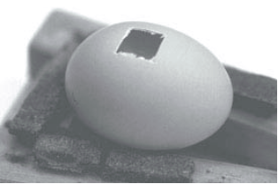
Figure 1. Windowed egg showing exposed chorioallantoic membrane (CAM).
2.2.2. Effect of NCTD on CAM Angiogenesis in Chicken Eggs
Classification of Experimental Group and Drug Injection Method.
It was divided into an experimental group in which NCTD was injected and a control group in which physiological saline was injected. According to the concentration, the experimental group was again composed of 3 groups (group 1-20μg/μL, group 2-40μg/μL, group 3-80μg/μL). Each group was given 10 egg embryos. The drug concentration of NCTD was selected in the range below 100 μg/μL based on preliminary experiments. Gelatin sponge was used as a drug carrier, and the size of the carrier was 5×5×5 mm, and the solution dropped onto the carrier was set to 20μL. The carrier is placed in the CAM with relatively few blood vessels between the two vitelline veins. The drug proceeds in micropipette, and 20 μL per chicken egg embryo in a physiological saline group, once daily, was dropped in a carrier cow for 3 days. In the experimental group, 20μL of NCTD solution diluted in concentrations of 20μg/μL, 40μg/μL, and 80μg/μL is used using a micropipette, and 20μL per each chicken egg embryo was dropped into the carrier of the chicken egg embryo of experimental group, once daily for 3 days. After injecting the drug once every day for 3 days, seal it with a gelatinous membrane and continue to wake up. The control group was injected with 0.85% physiological saline diluted at a concentration of 20μg/μL.
2.2.3. Sample Observation
On the 11th day of fertilization, the gelatinous membrane of the sealed area was peeled off, and the CAM was discharged to a size of 2×2cm around the carrier. Turn on the egg test light from the bottom of the chicken egg embryo, and determine the number of microvascular branches visible with the eye in the 5mm range around the getalatin sponge with an anatomy microscope.
How to calculate the number of angiogenesis;
Count the number of blood vessels under a microscope. First, the number of blood vessels at the edge of the carrier is divided into three types of blood vessels (small, medium, large), basing on the diameter of the blood vessel. Here, those with a diameter of 0.1 mm or less are called small blood vessels, those with a diameter of 0.1-1 mm or less are called middle blood vessels, and those with a diameter of 1 mm or more are called large blood vessels. Next, the number of angiogenesis is repeated 3 times per sample and the average number is obtained. The angiogenesis inhibition rate is calculated according to the formula below.
Angiogenesis inhibition rate (%)= (Number of Branched Microvasculatures with physiological saline group - Number of Branched Microvasculatures with drug injection group/Number of Branched Microvasculatures with physiological saline group) × 100%
2.2.4. Effect of NCTD on Angiogenesis in Tumor Tissues with Croquel-180 Sarcomatous Cells Group
Culture and inoculation of tumor cells
Using several DMEM incubators containing 10% fetal bovine serum and insulin, cultivated Croquel-180 sarcomatous cells at the same time in 37°C, humidity, and 5% CO2 incubator. Each bottle contains 90% Croquel-180 sarcoma cells. When the percentage was full up to 90%, it was digested with 0.1% pancreatic enzyme, washed twice with PBS, and then the cells were collected to make a suspension. The concentration of Croquel-180 sarcoma cells was adjusted to be 5×10⁷/ml. After selecting 7-day-old fertilized chicken embryos and opening the window in aseptic conditions according to the method described above, inoculated on CAM in the area where there were almost no blood vessels by absorbing 120 µl of Croquel-180 sarcomatous cells suspension per chicken egg embryo with micropipette. Then sealed with the sterilized gelatin colloid membrane and put it in a culture vessel and cultured for 4 days.
Laboratory Classification and Drug Injection Method
The experimental group was divided into the one in which NCTD was injected as shown in Figure 1 and a control group in which physiological saline was injected. According to the concentration, the experimental group was again composed of 3 groups (group 1-20μg/μL, group 2-40μg/μL, and group 3-80μg/μL). Each tank was made into 10 chicken egg embryos. In addition, the chicken egg embryos transplanted with Croquel- 180 sarcomatous cells group were cultured for 4 days and then a drug carrier was placed. The size of the carrier was the same as that of the above, and the carrier was placed on the CAM at the site where Croquel-180 sarcomatous cells were transplanted. The amount of solution dropped on the carrier was also set to 20μL, and a micropipette was used to drop it on the carrier. Give injection 5 times in ago every day and must be sealed with sterilized gelatin colloid membrane and continued hatching process.
Specimen fixation and observation
Specimen fixation was carried out on the 10d day after the chicken egg embryos were inoculated with Croquel-180 sarcomatous cells group. First, preliminary fixation with Formaldehyde in the air chamber was performed for 15 minutes, and the grafted tumor tissue mass was separated from the surrounding CAM. Following the pathologic tissue sample preparation process, paraffin was embedded, then consecutively bonded to a thickness of 2-4㎛, and each sample was subjected to H-E stain. For observation, three areas that was considered to have the most MVD in tumor tissues under 100-diameters was first selected, and the average value was calculated by calculating the number of microvessels in the tumor tissue under 400-diameters.
2.2.5. Statistical Method
The results are expressed as means±standard errors (SE) from at least three experiments. Statistical comparisons were based on Student's t-test or analysis of variance. Differences were considered significant at p< 0.05. All statistical analyses were done using Sigma Stat software (Jandel Scientific, San Rafael, CA).
3. Results and Discussion
The formation of capillaries around the tumor tissue and the formation of microcirculatory networks in the tumor tissues caused by tumor cells provide the material basis for tumor growth, which is called tumor angiogenesis. So, research on drugs targeting tumor blood vessels in cancer treatment is constantly being conducted, and such an anticancer treatment method is being proposed as an important anticancer treatment method in the world today as it blocks the ''life line'' on which tumor growth and metastasis depend.
Cantharidin (CTD) is a naturally occurring compound isolated from the medicinal insect blister beetle (Mylabris phalerata Pallas) . CTD is exo, exo-bicyclo [2.2.1] heptane 2, 3-dicarboxylic acid anhydride. The most important of the medicinal uses of CTD is its anticancer activities . CTD treatment could cause granulocytosis in vivo but not granulocytopenia due to most chemotherapeutics . This unique bioactivity renders CTD a promising lead compound for chemical modification to develop cancer therapeutics, but the application of CTD is limited due to its toxicity to gastrointestinal and urinary tracts . However, CTD had not been widely used in clinical treatment due to limitation of natural materials and a strong toxic effect on urinary system (LD50=1.71mg/kg), and the artificial aquaculture technology of CTD was not established. Since it was discovered in 1992 that CTD is a specific inhibitor of protein phosphatase (PP 2A), an intracellular signaling molecule, molecular studies on the anticancer activity of CTD have been actively conducted .
From this, research has been actively conducted to develop derivatives of CTD with high anti-tumor activity, low side effects, and low cost. Thus, the first synthetic of CTD developed is NCTD. NCTD is chemically known as exo-7-oxabicyclo-[2.2.1] Heptane-2, 3-dicarboxylic anhydride . NCTD is a de-methylated synthetic of CTD, which has a low manufacturing cost and anti-tumor activity similar to CTD, but exhibits minimal renal toxicity, strong tissue penetration, and relatively stable chemical substance. Unlike CTD, the synthesis of NCTD completely overcomes the limitations of the natural material source while reducing the side effects of CTD as much as possible, opening the prospect of broad application of these substances to antitumor therapy. The acute toxicity (LD50) of NCTD synthesized and purified in the Biochemistry Laboratory at the University of natural science, was 20.75mg/kg, which was a very toxic drug.
First, we conducted an experiment for angiogenesis inhibition of NCTD on CAM fertilized for 7d. NCTD was injected into a drug carrier of CAM at a dose of 20μg/μL, 40μg/μL, and 80μg/μL once a day for 3 days, and the angiogenesis inhibitory effect was observed in CAM at fertilization 11d. There was no significant difference in the number of microvascular branches that was injected at a dose of 20µg/μL than the control group, but when NCTD was injected at a dose of 40µg/µL and 80µg/µL, there was significant decrease in the number of microvascular branches than the control group (Figure 2). The above results showed that as the injection dose of NCTD increased, the number of microvascular branches in CAM was decreased, and thus the angiogenesis inhibition rate tended to increase gradually (Figure 3). In addition, it was found that the angiogenesis inhibitory effect of NCTD depends on the injection dose, but the effect was evident even at an effective concentration of 40µg/μL of NCTD.
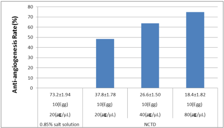
Figure 2. The angiogenesis effect of NCTD in CAM. In the figure, as the injection dose of NCTD increases, the rate of inhibition of angiogenesis in CAM is gradually increasing.
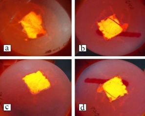
Figure 3. The angiogenesis effect of NCTD on CAM. The figure showed the microvessels formed in CAM. (A) Control group injected with physiological saline. Many microvessels were formed, and they were very remarkable. (B) Experiment group1 in which 20µg/µL of NCTD was injected. There were fewer microvessels than the control. (C) Experiment group2 in which 40μg/μL of NCTD was injected. Here, the microvessels were significantly less than the control or the case injected NCTD 20µg/μL. (D) Experiment group3 in which 80µg/µL of NCTD was injected. The microvessels were significantly less than those injected with NCTD 20µg/µL and 40µg/µL.
Since Croquel-180 sarcomatous cells are mesenchymal cells of origin, it is recognized that the angiogenesis rate is much faster than that of epithelial cells of ectodermal origin, and the number of angiogenesis is higher. Therefore, we transplanted the cells group of mesenchymal origin with very good angiogenesis and examined the angiogenesis inhibitory effect on the tissue mass.
Next, we made the cultured Croquel-180 sarcomatous cells at a concentration of 5×10⁷/ml, and then on the 7th day of fertilization inoculated at the concentration of 120µl/chicken embryo into CAM. And the survival and proliferation status of tumor cells transplanted to CAM were observed daily under an anatomy microscope. After 4 days of inoculation with Croquel-180 sarcomatous cells group, a tumor mass with a diameter of 1-2mm visible to the naked eye was formed and on the 8th day 5-7mm, on 12th day, a mass of tumor tissue of 8-9mm in diameter was formed. After 13 days of inoculation, the tumor tissue mass was not remarkably large. In CAM, Croquel-180 sarcomatous cells remarkably proliferated from day 4 to day 12 of inoculation, but gradually after day 13 (Figure 4).
Also, the blood vessels formed by Croquel-180 sarcomatous cells group, unlike the human tumor cells group, were various kinds of vessels, and the sarcomatous cells lied on the microvascular basement membrane along with the vascular endothelial cells and protruded into the vascular lumen and had an incomplete basement membrane (Figure 5).
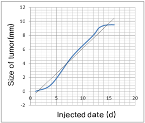
Figure 4. Growth curve of Croquel-180 sarcomatous cells group.
The Chicken egg embryos trans-planted with Croquel-180 sarcomatous cells group were cultured for 4 days, and a tumor mass (1-2mm) that could be seen with the naked eye was formed, and then a drug carrier was placed on the tumor mass, and NCTD was added at a dose of 20µg/μL, 40µg/µL and 80µg/µL, the cells were injected 5 times daily, and micro-angiogenesis in the transplanted tumor tissue was observed on the 10d day after inoculation with Croquel-180 sarcomatous cells group. There was no significant difference in microvascular density (MVD) when NCTD was injected at a dose of 20µg/μL, but at a dose of 40µg/µL and 80µg/µL, MVD decreased significantly compared to the control (Figure 6). The above results suggest that as the infusion dose of NCTD increases, the number of microvessels in tumor tissue grafted on CAM decreases, and thus the angiogenesis inhibition rate is gradually increasing (Figure 6). In addition, the angiogenesis inhibitory effect of NCTD depends on the injection dose, but the effect was evident from the effective concentration of 40µg/µL of NCTD.
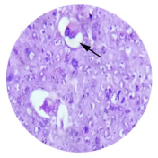
Figure 5. Microvessels in tumor tissues formed by Croquel- 180 sarcomatous cells group (H-E, 10×40).
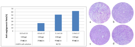
Figure 6. Effect of NCTD on angiogenesis in tumor tissue. In the left figure, as the injection dose of NCTD increases, the rate of angiogenesis inhibition in tumor tissues is gradually increasing. The right figure showed the microvessels formed in the trans- planted tumor tissue under a microscope (H-E stain, 10×10). (A) Control group infused with physiological saline. Many micro-vessels were formed. (B) Experiment group 1 in which 20µg/µL of NCTD was injected. There are fewer microvessels than the control. (C) Experiment group 2 in which 40μg/μL of NCTD was injected. There were significantly fewer microvessels than the control and experiment group1. (D) Experiment group 3 in which 80µg/µL of NCTD was injected. The number of microvessels was significantly less than the control and experiment group 2.
4. Conclusion
Norcantharidin is a powerful angiogenesis inhibitor capable of sufficiently inhibiting angiogenesis in tumor tissue. We confirmed that it has potential application value applicable to tumor treatment targeting endothelial cells.
Abbreviations

NCTD

Norcantharidin

CAM

Chorioallantoic Membrane

MVD

Microvascular Density

CTD

Cantharidin

VEGF

Vascular Endothelial Growth Factor

FGF

Fibroblast Growth Factor

EMT

Mesenchyma Transition

PHA

Phytohemagglutinin

Acknowledgments
This work was supported by grants from Laboratory Animal Center in Pyongyang University of Medical Science.
Author Contributions
Hong Chol Ri: Project administration, Revision manuscript
Dong Min Han: Experimental design, Data interpretation
Kwang Il To: Experimental design, Animal experiment
Yong Il Yun, Song-Nam Go: Software, Analyzed data, Validation
Chang-Guk Kim, Su-Chol Rim: Drafting manuscript, editing
Hong-Chol Ri, Dong-Min Han, Kwang-Il To: These Authors Contributed Equally.
Conflicts of Interest
The authors declare no conflicts of interests.
References
[1] Tonini T, Rossi F, Claudio PP. “Molecular basis of angiogenesis and cancer,” Oncogene 22 (42), 6549–6556, 2003.
[2] Ikumi Kuriwaki, Minoru Kameda, Hiroyuki Hisamichi et al., “Structure-based drug design of 1, 5-triazine and pyrimidine derivatives as novel FGFR3 inhibitors with high selectivity over VEGFR2”, Bioorganic Medicinal Chemistry 10(28), 115453-66, 2020.
[3] Li C, Shintani S, Terakado N, Klosek SK, Ishikawa T, Nakashiro K, Hamakawa H. “Microvessel density and expression of vascular endothelial growth factor, basic fibroblast growth factor, and platelet-derived endothelial growth factor in oral squamous cell carcinomas,” International Journal of Oral and Maxillofacial Surgery 34 (5), 559–565, 2005.
[4] Liu, Yu, Yang, Guoyou, Zhang, Junfeng, et al., “Anti-TNF-α monoclonal antibody reverses psoriasis through dual inhibition of inflammation and angiogenesis”, International Immunopharmacology, 1(28), 731-743, 2015.
[5] Owen JL, Iragavarapu-Charyulu V, Gunja-Smith Z, Herbert LM, Grosso JF, Lopez DM. “Upregulation of matrix metalloproteinase-9 in T lymphocytes of mammary tumor bearers: role of vascular endothelial growth factor,” Journal of Immunology 171 (8), 4340–4351, 2003.
[6] Boedefeld II WM, Bland KI, Heslin MJ. “Recent insights into angiogenesis, apoptosis, invasion, and metastasis in colorectal carcinoma” Annals of Surgical Oncology 10 (8), 839–851, 2003.
[7] C Borner& D W Andrews, “The apoptotic pore on mitochondria: are we breaking through or still stuck”, Cell Death & Differentiation, 5, 2014.
[8] Mazzoni E, AdamA, Bal de Kier Joffe E, Aguirre-Ghiso JA. “Immortalized mammary epithelial cells overexpressing protein kinase C gamma acquire a malignant phenotype and become tumorigenic in vivo,” Molecular Cancer Research 1 (10), 776–787, 2003.
[9] Meghan E Minard, Matthew H Herynk, John G Collard& Gary E Gallick, “The guanine nucleotide exchange factor Tiam1 increases colon carcinoma growth at metastatic sites in an orthotopic nude mouse model”, Oncogene, 6, 2005.
[10] Campbell NE, Kellenberger L, Greenaway J, et al., “Extracellular Matrix Proteins and Tumor Angiogenesis”, Journal of Oncology, 14(2010), 2010.
[11] Hideyoshi KAWASAKI, Tetsuya YOSHIDA, Kazuhide HORIGUCHI et al., “Characterization of Anoikis-Resistant Cells in Mouse Colonic Epithelium,” J. Vet. Med. Sci. 75(9): 1173–1180, 2013.
[12] Douma S, Van Laar T, Zevenhoven J, Meuwissen R, Van Garderen E, Peeper DS. “Suppression of anoikis and induction of metastasis by the neurotrophic receptor TrkB,” Nature 430 (7003), 1034–1039, 2004.
[13] Derouet M, Wu X, May L, Yoo BH, Sasazuki T, Shirasawa S, Rak J, Rosen KV. “Acquisition of anoikis resistance promotes the emergence of oncogenic K-ras mutations in colorectal cancer cells and stimulates their tumorigenicity in vivo,” Neoplasia 9 (7), 536–545, 2007.
[14] Timothy A. Hill, Scott G. Stewart Benjamin et al., “Heterocyclic substituted cantharidin and norcantharidin analogues—synthesis, protein phosphatase (1 and 2A) inhibition, and anti-cancer activity”, Bioorganic & Medicinal Chemistry Letters 17, 3392–3397, 2007.
[15] Jie Zhao, Xiao-Wen Guan, Shi-Wu Chen, Ling Hui, “Synthesis and biological evaluation of norcantharidin derivatives as protein phosphatase-1 inhibitors”, Bioorganic and Medicinal Chemistry Letters, vol. 20, no. 25, pp. 363–366, 2015.
[16] Xiaohua Liu, Wan Sia Heng, Paul, Qi Li, Lai Wah Chan, Novel polymeric microspheres containing norcantharidin for chemoembolization, Journal of Controlled Release, 1(116), 35-41, 2006.
[17] Y.-Z. Fan, Z.-M. Zhao, J.-Y. Fu, C.-Q. Chen, and W. Sun, “Norcantharidin inhibits growth of human gallbladder carcinoma xenografted tumors in nude mice by inducing apoptosis and blocking the cell cycle in vivo”, Hepatobiliary and Pancreatic Diseases International, vol. 9, no. 4, pp. 414–422, 2010.
[18] Y.-C. Chen, S.-C. Chang, M.-H. Wu et al., “Norcantharidin reduced cyclins and cytokines production in human peripheral blood mononuclear cells,” Life Sciences, vol. 84, no. 7-8, pp. 218–226, 2009.
[19] Y.-J. Chen, Y.-M. Tsai, C.-D. Kuo, K.-L. Ku, H.-S. Shie, and H.-F. Liao, “Norcantharidin is a small-molecule synthetic compound with anti-angiogenesis effect,” Life Sciences, vol. 85, no. 17-18, pp. 642–651, 2009.
[20] Chen YJ, Kuo CD, Tsai YM, Yu CC, Wang GS, Liao HF. “Norcantharidin induces anoikis through Jun-N-terminal kinase activation in CT26 colorectal cancer cells,” Anti-Cancer Drugs 19 (1), 55–64, 2008.
[21] Jia Hu, Er-Yong Zhang, Jiang W et. al., “Pressure shift mediated anoikis of endothelial cells in the flow field in vitro”, Journal of Biomedical Science and Engineering, 2(3), 206-212, 2010.
[22] Y.-J. Chen, C.-D. Kuo, Y.-M. Tsai, C.-C. Yu, G.-S. Wang, and H.-F. Liao, “Norcantharidin induces anoikis through Jun-Nterminal kinase activation in CT26 colorectal cancer cells,” Anti-Cancer Drugs, vol. 19, no. 1, pp. 55–64, 2008.
[23] Ribatti, D., Vacca, A., Roncali, L., and Dammacco, F, “The chick embryo chorioallantoic membrane as a model for in vivo research on angiogenesis”, Int. J. Dev. Biol. 40, 1189–1197, 1996.
[24] Chen-Hsi Hsieh, K. S. Clifford Chao, Hui-Fen Liao, Yu-Jen Chen, Norcantharidin, “Derivative of Cantharidin, for Cancer Stem Cells, Evidence-Based Complementary and Alternative Medicine”, 2013(11), 2013.
[25] Zeping Han, Baoxia Li, Juanjuan Wang, Xiangqiang Zhang, “Norcantharidin Inhibits SK-N-SH Neuroblastoma Cell Growth by Induction of Autophagy and Apoptosis”, Technology in Cancer Research & Treatment, 1(16), 2016.
[26] Patrícia Cotovio, Cristina Silva, Maria Guedes Marques et al., “Acute kidney injury by cantharidin poisoning following a silly bet on an ugly beetle”, Clin Kidney J 6, 201–203, 2013.
[27] Cohen-PT. Philp-A. Vazquez-Martin-C, “Protein phosphatase 4--from obscurity to vital functions”, FEBS Letters, 15(579), 3278-3286, 2005.
[28] Wu, Z.-W.; Yang, X.-Q.; Zhang, Y.-L, The Toxicology and Biochemical Characterization of Cantharidin on Cydia pomonella, 1(108), 2015.
Cite This Article
  • APA Style

    Ri, H., Han, D., To, K., Yun, Y. I., Go, S., et al. (2025). Norcantharidin Inhibits an Angiogenesis of Croquel-180 Sarcomatous Cells. Biochemistry and Molecular Biology, 10(3), 37-44. https://doi.org/10.11648/j.bmb.20251003.11

    Copy | Download

    ACS Style

    Ri, H.; Han, D.; To, K.; Yun, Y. I.; Go, S., et al. Norcantharidin Inhibits an Angiogenesis of Croquel-180 Sarcomatous Cells. Biochem. Mol. Biol. 2025, 10(3), 37-44. doi: 10.11648/j.bmb.20251003.11

    Copy | Download

    AMA Style

    Ri H, Han D, To K, Yun YI, Go S, et al. Norcantharidin Inhibits an Angiogenesis of Croquel-180 Sarcomatous Cells. Biochem Mol Biol. 2025;10(3):37-44. doi: 10.11648/j.bmb.20251003.11

    Copy | Download

  • @article{10.11648/j.bmb.20251003.11,
      author = {Hong-Chol Ri and Dong-Min Han and Kwang-Il To and Yong Il Yun and Song-Nam Go and Chang-Guk Kim and Su-Chol Rim},
      title = {Norcantharidin Inhibits an Angiogenesis of Croquel-180 Sarcomatous Cells
    },
      journal = {Biochemistry and Molecular Biology},
      volume = {10},
      number = {3},
      pages = {37-44},
      doi = {10.11648/j.bmb.20251003.11},
      url = {https://doi.org/10.11648/j.bmb.20251003.11},
      eprint = {https://article.sciencepublishinggroup.com/pdf/10.11648.j.bmb.20251003.11},
      abstract = {Norcantharidin (NCTD) is a demethylated water-soluble synthetic small molecule of cantharidin (CTD), has been reported that it has anticancer activities including apoptosis and anti-angiogenesis. NCTD reduced VEGF production and the expression of integrin-β1 in tumor cells even at the low concentrations with less toxicity to the kidney or liver, and destroyed vimentin to inhibit angiogenesis in tumor cells by inducing anoikis of vascular endothelial cells. In addition, basing on recent data that the metastasis and recurrence of tumor are closely related to angiogenesis in tumor tissues, Croquel-180 sarcomatous cells, a mesenchyma-derived cells group with better angiogenesis were transplanted to Chick embryo Chorioallantoic Membrane (CAM) and the anti-angiogenic effect of NCTD was examined. When NCTD was injected at a dose of 40µg/µL and 80µg/µL, there was significant decrease in the number of microvascular branches than the control group. There was no significant difference in microvascular density (MVD) when NCTD was injected at a dose of 20µg/μL, but at a dose of 40µg/µL and 80µg/µL, MVD decreased significantly compared to the control. We reaffirmed that NCTD is a drug that can specifically inhibit angiogenesis in tumor tissues through CAM experiments, and that it is a very effective drug that can be applied to antitumor therapy targeting angiogenesis in tumor tissues. Norcantharidin is a powerful angiogenesis inhibitor capable of sufficiently inhibiting angiogenesis in tumor tissue. We confirmed that it has potential application value applicable to tumor treatment targeting endothelial cells.},
     year = {2025}
    }
    

    Copy | Download

  • TY  - JOUR
    T1  - Norcantharidin Inhibits an Angiogenesis of Croquel-180 Sarcomatous Cells
    
    AU  - Hong-Chol Ri
    AU  - Dong-Min Han
    AU  - Kwang-Il To
    AU  - Yong Il Yun
    AU  - Song-Nam Go
    AU  - Chang-Guk Kim
    AU  - Su-Chol Rim
    Y1  - 2025/07/10
    PY  - 2025
    N1  - https://doi.org/10.11648/j.bmb.20251003.11
    DO  - 10.11648/j.bmb.20251003.11
    T2  - Biochemistry and Molecular Biology
    JF  - Biochemistry and Molecular Biology
    JO  - Biochemistry and Molecular Biology
    SP  - 37
    EP  - 44
    PB  - Science Publishing Group
    SN  - 2575-5048
    UR  - https://doi.org/10.11648/j.bmb.20251003.11
    AB  - Norcantharidin (NCTD) is a demethylated water-soluble synthetic small molecule of cantharidin (CTD), has been reported that it has anticancer activities including apoptosis and anti-angiogenesis. NCTD reduced VEGF production and the expression of integrin-β1 in tumor cells even at the low concentrations with less toxicity to the kidney or liver, and destroyed vimentin to inhibit angiogenesis in tumor cells by inducing anoikis of vascular endothelial cells. In addition, basing on recent data that the metastasis and recurrence of tumor are closely related to angiogenesis in tumor tissues, Croquel-180 sarcomatous cells, a mesenchyma-derived cells group with better angiogenesis were transplanted to Chick embryo Chorioallantoic Membrane (CAM) and the anti-angiogenic effect of NCTD was examined. When NCTD was injected at a dose of 40µg/µL and 80µg/µL, there was significant decrease in the number of microvascular branches than the control group. There was no significant difference in microvascular density (MVD) when NCTD was injected at a dose of 20µg/μL, but at a dose of 40µg/µL and 80µg/µL, MVD decreased significantly compared to the control. We reaffirmed that NCTD is a drug that can specifically inhibit angiogenesis in tumor tissues through CAM experiments, and that it is a very effective drug that can be applied to antitumor therapy targeting angiogenesis in tumor tissues. Norcantharidin is a powerful angiogenesis inhibitor capable of sufficiently inhibiting angiogenesis in tumor tissue. We confirmed that it has potential application value applicable to tumor treatment targeting endothelial cells.
    VL  - 10
    IS  - 3
    ER  - 

    Copy | Download

Author Information
  • Department of Pathology, Pyongyang University of Medical Sciences, Pyongyang, DPR Korea

  • Institute of Chemistry and Biology, University of Sciences, Pyongyang, DPR Korea

  • Institute of Chemistry and Biology, University of Sciences, Pyongyang, DPR Korea

  • Institute of Chemistry and Biology, University of Sciences, Pyongyang, DPR Korea

  • Institute of Chemistry and Biology, University of Sciences, Pyongyang, DPR Korea

  • Institute of Chemistry and Biology, University of Sciences, Pyongyang, DPR Korea

  • Institute of Chemistry and Biology, University of Sciences, Pyongyang, DPR Korea