Acute necrotizing encephalopathy is a rapidly progressive disease of the central nervous system that is generally described in texts as a pathology of pediatric patients, but even in people of this age range, it is rare to find a case of this disease, which presents with a fulminant tissue necrosis and can be secondary to infections, mainly of viral origin. Due to presenting very non-specific clinical manifestations, this diagnosis often goes unnoticed or is made late. ANE survivors go through three phases during the clinical course that include the prodromal stage, the period of acute encephalopathy, and the recovery stage. It is through specific radiological findings observed on computed tomography affecting the bilateral thalamus and possibly the cerebral white matter, brainstem, or cerebellum that the diagnosis can be confirmed. This report discusses the case of an 8-year-old schoolboy who begins with acute gastroenteritis, which progresses with seizures and neurological deterioration. A skull CT scan is performed with imaging findings suggestive of acute necrotizing encephalopathy. Management is established with adequate clinical response and favorable recovery of neurological symptoms. The clinical characteristics and radiological findings are the key to facilitating an early diagnosis, reducing sequelae and improving the prognosis, which provides a chance of life to people who suffer from this condition.
| Published in | American Journal of Pediatrics (Volume 10, Issue 3) |
| DOI | 10.11648/j.ajp.20241003.12 |
| Page(s) | 117-122 |
| Creative Commons |
This is an Open Access article, distributed under the terms of the Creative Commons Attribution 4.0 International License (http://creativecommons.org/licenses/by/4.0/), which permits unrestricted use, distribution and reproduction in any medium or format, provided the original work is properly cited. |
| Copyright |
Copyright © The Author(s), 2024. Published by Science Publishing Group |
Encephalopathy, Virus, Seizure, Neuroimaging
ANE | Acute Necrotizing Encephalopathy |
PICU | Pediatric Intensive Care Unit |
CT | Computed Tomography |
BEATC | Brainstem Auditory Evoked Potentials and Visual |
VEP | Evoked Potentials |
STUDIES | INCOME 05/13/24 | 05/18/24 | 05/23/24 |
|---|---|---|---|
Hemogram | Leukocytes: 8,730 Hemoglobin: 14.2 Gr/dL Hematocrit: 42.61% | Leukocytes: 9,200 Hemoglobin: 13.9 Gr/dL Hematocrit: 41.72% | Leukocytes: 8,100 Hemoglobin: 13.8 Gr/dL Hematocrit: 40.32% |
Sodium (mmol/L) | 133.2 | 135.6 | 137 |
Potassium (mmol/L) | 4.59 | 3.9 | 4.12 |
Chlorine (mmol/L) | 9.5 | 10.1 | 9.6 |
Calcium (mmol/L) | 1.77 | 1.56 | 1.87 |
Creatinine (mg/dL) | 0.67 | 0.65 | 0.54 |
BUN (mg/dL) | 9.81 | 6.81 | 10.4 |
Urea (mg/dL) | twenty-one | 22 | 23 |
IgM | 100 | ||
IgG | 889 | ||
IgA | 125 | ||
ANA | Negative | ||
Varicella Zoster Antibody IgG-IgM | Negative | ||
GPT (UI/L) | 10 | 13 | 12 |
GOT (UI/L) | twenty-one | 28 | 25 |
C3 | 83 | ||
C4 | 27.8 | ||
Hepatitis B – Ag Surface | Negative | ||
Urinalysis + Gram | Not pathological | ||
Dengue IgM | Negative | ||
CSF cytochemistry | Color before centrifuging: (Colorless) Appearance before centrifuging: (Transparent) Color after centrifuging: (Colorless) Appearance after centrifuging: (Transparent) pH: (8.0) Density: (1.010) Glucose: (48) LDH: (81) Total Proteins: (99.1) Bacteria: (Not observed) Leukocytes: (Not observed) Bacteria: (Not observed) Yeasts: (Not observed) POLYMORPHONUCLEAR: (Absent) | ||
CSF culture | Negative after 48 hours | ||
|---|---|---|---|
PH | 7,313 | 7.38 | 7,425 |
PCO2 (mmHg) | 26.4 | 30.1 | 32.5 |
HCO3 (mmol/L) | 16.9 | 19.3 | 22.1 |
B.E. | -6.9 | -4.1 | -23 |
PO2 (mmHg) | 162 | 103 | 106 |
LDH (mg/dL) | 476 | 352 | 248 |
CRP (mg/dL) | Minor 6 | Minor 6 | Minor 5 |
AC ANCAS | Negative | ||
AC PANCAS | Negative | ||
AC CANCA | Negative | ||
ANTI SSB | Negative | ||
ANTISM | Negative | ||
ANTI RNP | Negative | ||
ANTINUCLEAR ANTIBODIES | Negative | ||
TOTAL EXTRACTABLE NUCLEAR ANTIBODIES | Negative | ||
ENAS | Negative | ||
ANTI RO | Negative | ||
Electroencephalogram | Abnormal due to the inadequate organization of the background rhythms expected for age. | ||
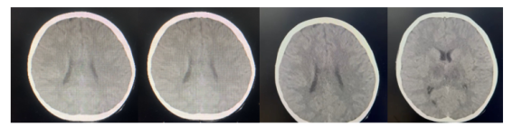
Brain Magnetic Resonance | Highly suggestive of hypoxic-ischemic lesions bilaterally and symmetrically involving the thalamus, inferior colliculi and vermis in the midline. Subtle changes of diffuse cortical cerebellar atrophy for the patient's age. | ||
| [1] | Alper G. Acute necrotizing encephalopathy mimicking ADEM. In Waubant E, Lotze T, eds. Pediatric demyelinating diseases of the central nervous system and their mimics. Cham, Switzerland; 2017; p. 11-5. |
| [2] |
Liu HH, Chan OW, Lin JJ, WanPingWu P, Chang CC. COVID-19-associated acute necrotizing encephalopathy in a 3-year-old child. Pediatr Neonatol. 2023 Nov; 64(6): 688-689.
https://doi.org/10.1016/j.pedneo.2023.04.007 Epub 2023 Jun 16. PMID: 37380549; PMCID: PMC10271917. |
| [3] |
Holla VV, Gohel AB, Kartik N, Netravathi M. Acute Necrotizing Encephalopathy as a Complication of Chikungunya Infection. Neurol India. 2021 Mar-Apr; 69(2): 490-492.
https://doi.org/10.4103/0028-3886.314525 PMID: 33904484. |
| [4] |
Mizuguchi M, Ichiyama T, Imataka G, Okumura A, Goto T, Sakuma H, Takanashi JI, Murayama K, Yamagata T, Yamanouchi H, Fukuda T, Maegaki Y. Guidelines for the diagnosis and treatment of acute encephalopathy in childhood. Brain Dev. 2021 Jan; 43(1): 2-31.
https://doi.org/10.1016/j.braindev.2020.08.001 Epub 2020 Aug 20. PMID: 32829972. |
| [5] |
Acute necrotizing encephalopathy of childhood. Diagnostic imaging: Case presentation. Saab S., Bautista J., Serrano S., Rodríguez N Rev. Colomb. Radiol. 2015; 26(2): 4223-7.
https://contenido.acronline.org/Publicaciones/RCR/RCR26-2/10_Encefalopatia.pdf |
| [6] | Lee YJ, Hwang SK, Kwon S. Acute necrotizing encephalopathy in children: A long way to go. J Korean Med Sci. 2019; 34(19): e143, |
| [7] | Sakrani N, Almazrouei S, Mohan S, Ramsi, M. Adenovirus as a rare cause of acute necrotising encephalitis. BMJ Case Reports. 2019; 12. [posted 2019 Dec; cited 2021 Sep]: [3p.]. Available from: |
| [8] | EL Jackson, W. Eilbert, and AM Hathcock, Acute necrotizing encephalopathy: a case report, American Journal of Emergency Medicine, |
| [9] |
Neilson DE, Adams MD, Orr CM, Schelling DK, Eiben RM, Kerr DS, Anderson J, Bassuk AG, Bye AM, Childs AM, Clarke A, Crow YJ, Di Rocco M, Dohna-Schwake C, Dueckers G, Fasano AE, Gika AD, Gionnis D, Gorman MP, Grattan-Smith PJ, Hackenberg A, Kuster A, Lentschig MG, Lopez-Laso E, Marco EJ, Mastroyianni S, Perrier J, Schmitt-Mechelke T, Servidei S, Skardoutsou A, Uldall P, van der Knaap MS, Goglin KC, Tefft DL, Aubin C, de Jager P, Hafler D, Warman ML. Infection-triggered familial or recurrent cases of acute necrotizing encephalopathy caused by mutations in a component of the nuclear pore, RANBP2. Am J Hum Genet. 2009 Jan; 84(1): 44-51.
https://doi.org/10.1016/j.ajhg.2008.12.009 PMID: 19118815; PMCID: PMC2668029. |
| [10] |
Sarigecili E, Ucar HK, Havali C, Cansu A, Aydin K. Acute necrotizing encephalopathy associated with RANBP2 mutation: value of MRI findings for diagnosis and intervention. Acta Neurol Belg. 2023 Apr; 123(2): 571-582.
https://doi.org/10.1007/s13760-022-02166-x Epub 2022 Dec 26. PMID: 36572756; PMCID: PMC9792159. |
| [11] | Kansagra SM, Gallentine WB Cytokine storm of acute necrotizing encephalopathy. Pediatric Neurology. 2011; 45 (6): 400–402. |
| [12] |
Manara R, Franzoi M, Cogo P, Battistella PA. Acute necrotizing encephalopathy: combined therapy and favorable outcome in a new case. Childs Nerv Syst. 2006 Oct; 22(10): 1231-6.
https://doi.org/10.1007/s00381-006-0076-9 Epub 2006 Mar 14. PMID: 16816978. |
| [13] |
Cole J, Evans E, Mwangi M, Mar S. Acute Disseminated Encephalomyelitis in Children: An Updated Review Based on Current Diagnostic Criteria. Pediatr Neurol. 2019 Nov; 100: 26-34.
https://doi.org/10.1016/j.pediatrneurol.2019.06.017 Epub 2019 Jul 3. PMID: 31371120. |
| [14] |
Wu X, Wu W, Pan W, Wu L, Liu K, Zhang HL. Acute necrotizing encephalopathy: an underrecognized clinicoradiologic disorder. Mediators Inflamm. 2015; 2015: 792578.
https://doi.org/10.1155/2015/792578 Epub 2015 Mar 22. PMID: 25873770; PMCID: PMC4385702. |
| [15] | Lee EP, Lin JJ, Chang HP, Yen CW, Hsieh MS, Chan OW, et al. Ferritin as an effective predictor of neurological outcomes in children with acute necrotizing encephalopathy. Pediatr Neurol [Internet]. 2024; 152: 162–8. Available at: |
| [16] | Tautiva-Rojas, María, & Bogantes-Ledezma, Sixto. (2021). Encefalitis aguda necrotizante, diagnóstico retador en una niña con deterioro neurológico súbito. Acta Médica Costarricense, 63(4), 223-228. |
APA Style
Algarín, R. A. S., Caballero, A. M. M., Villadiego, E. A. T., Ruiz, R. R., Montañez, A. Á. (2024). Acute Necrotizing Encephalopathy in Children: A Case Report in Barranquilla-Colombia. American Journal of Pediatrics, 10(3), 117-122. https://doi.org/10.11648/j.ajp.20241003.12
ACS Style
Algarín, R. A. S.; Caballero, A. M. M.; Villadiego, E. A. T.; Ruiz, R. R.; Montañez, A. Á. Acute Necrotizing Encephalopathy in Children: A Case Report in Barranquilla-Colombia. Am. J. Pediatr. 2024, 10(3), 117-122. doi: 10.11648/j.ajp.20241003.12
AMA Style
Algarín RAS, Caballero AMM, Villadiego EAT, Ruiz RR, Montañez AÁ. Acute Necrotizing Encephalopathy in Children: A Case Report in Barranquilla-Colombia. Am J Pediatr. 2024;10(3):117-122. doi: 10.11648/j.ajp.20241003.12
@article{10.11648/j.ajp.20241003.12,
  author = {Ricardo Andrés Sánchez Algarín and Angelica Maria Mendoza Caballero and Emanuel Alexis Troncoso Villadiego and Richard Romero Ruiz and Adolfo Álvarez Montañez},
  title = {Acute Necrotizing Encephalopathy in Children: A Case Report in Barranquilla-Colombia
},
  journal = {American Journal of Pediatrics},
  volume = {10},
  number = {3},
  pages = {117-122},
  doi = {10.11648/j.ajp.20241003.12},
  url = {https://doi.org/10.11648/j.ajp.20241003.12},
  eprint = {https://article.sciencepublishinggroup.com/pdf/10.11648.j.ajp.20241003.12},
  abstract = {Acute necrotizing encephalopathy is a rapidly progressive disease of the central nervous system that is generally described in texts as a pathology of pediatric patients, but even in people of this age range, it is rare to find a case of this disease, which presents with a fulminant tissue necrosis and can be secondary to infections, mainly of viral origin. Due to presenting very non-specific clinical manifestations, this diagnosis often goes unnoticed or is made late. ANE survivors go through three phases during the clinical course that include the prodromal stage, the period of acute encephalopathy, and the recovery stage. It is through specific radiological findings observed on computed tomography affecting the bilateral thalamus and possibly the cerebral white matter, brainstem, or cerebellum that the diagnosis can be confirmed. This report discusses the case of an 8-year-old schoolboy who begins with acute gastroenteritis, which progresses with seizures and neurological deterioration. A skull CT scan is performed with imaging findings suggestive of acute necrotizing encephalopathy. Management is established with adequate clinical response and favorable recovery of neurological symptoms. The clinical characteristics and radiological findings are the key to facilitating an early diagnosis, reducing sequelae and improving the prognosis, which provides a chance of life to people who suffer from this condition.
},
 year = {2024}
}
TY  - JOUR
T1  - Acute Necrotizing Encephalopathy in Children: A Case Report in Barranquilla-Colombia

AU  - Ricardo Andrés Sánchez Algarín
AU  - Angelica Maria Mendoza Caballero
AU  - Emanuel Alexis Troncoso Villadiego
AU  - Richard Romero Ruiz
AU  - Adolfo Álvarez Montañez
Y1  - 2024/07/23
PY  - 2024
N1  - https://doi.org/10.11648/j.ajp.20241003.12
DO  - 10.11648/j.ajp.20241003.12
T2  - American Journal of Pediatrics
JF  - American Journal of Pediatrics
JO  - American Journal of Pediatrics
SP  - 117
EP  - 122
PB  - Science Publishing Group
SN  - 2472-0909
UR  - https://doi.org/10.11648/j.ajp.20241003.12
AB  - Acute necrotizing encephalopathy is a rapidly progressive disease of the central nervous system that is generally described in texts as a pathology of pediatric patients, but even in people of this age range, it is rare to find a case of this disease, which presents with a fulminant tissue necrosis and can be secondary to infections, mainly of viral origin. Due to presenting very non-specific clinical manifestations, this diagnosis often goes unnoticed or is made late. ANE survivors go through three phases during the clinical course that include the prodromal stage, the period of acute encephalopathy, and the recovery stage. It is through specific radiological findings observed on computed tomography affecting the bilateral thalamus and possibly the cerebral white matter, brainstem, or cerebellum that the diagnosis can be confirmed. This report discusses the case of an 8-year-old schoolboy who begins with acute gastroenteritis, which progresses with seizures and neurological deterioration. A skull CT scan is performed with imaging findings suggestive of acute necrotizing encephalopathy. Management is established with adequate clinical response and favorable recovery of neurological symptoms. The clinical characteristics and radiological findings are the key to facilitating an early diagnosis, reducing sequelae and improving the prognosis, which provides a chance of life to people who suffer from this condition.

VL  - 10
IS  - 3
ER  -
Pediatrics Department, Pediatric Specialized Care Center, Barranquilla, Colombia
Biography: Ricardo Andrés Sanchez Algarín, general practitioner from Universidad Libre of Barranquilla, specialist in Epidemiology from Universidad Autonoma of Bucaramanga, currently a 2nd year resident of the Pediatrics Postgraduate Program at Universidad Simon Bolivar, and pursuing a diploma in pediatric and neonatal intensive care.

Pediatrics Department, Pediatric Specialized Care Center, Barranquilla, Colombia
Biography: Angelica Maria Mendoza Caballero, doctor graduated from the Simón Bolivar University of Colombia, specialist in Pediatrics from the same institution and Master in clinical medicine from the University of Alcalá in Spain, currently taking the 2024 Boston Children's Global Pediatric Resident Digital Program and reviewer of American Journal of Pediatrics (AJP).

Pediatrics Department, Pediatric Specialized Care Center, Barranquilla, Colombia
Pediatric Intensive Care Units, Specialized Care Center, Barranquilla, Colombia
Pediatric Neurology Department, Pediatric Specialized Care Center, Barranquilla, Colombia
Information