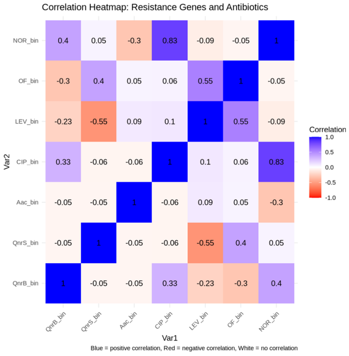
Introduction: Antibiotic resistance, particularly to quinolones, represents a major public health concern in Burkina Faso. In recent decades, plasmid-mediated quinolone resistance (PMQR) mechanisms have emerged, especially among Gram-negative bacteria. These mechanisms include, among others, qnr genes (qnrA, qnrB, qnrS). This study aimed to investigate the presence of quinolone resistance determinants in Gram-negative bacilli isolated from pus and vaginal samples at Saint Camille Hospital of Ouagadougou (HOSCO). Methodology: A total of 19 strains of Escherichia coli isolated from pus and vaginal swabs were collected for bacteriological and molecular analysis. Four antibiotics, namely ciprofloxacin (CIP), norfloxacin (NOR), ofloxacin (OF) and levofloxacin (LEV) were used for sensitivity testing and molecular analysis focused on the detection of qnrA, qnrB, and qnrS type genes. Results: Resistance rates to CIP, NOR, OF, and LEV were 57.89%, 52.63%, 52.63%, and 31.37%, respectively. Molecular analysis revealed the presence of qnrB, and qnrS genes in 28.47% of the isolates, for each gene. The qnrA gene was not detected in any isolate. The analysis of the genetic support of resistance genes revealed that 50% of the qnrS genes were plasmid-borne, while only 25% of the qnrB genes were associated with plasmids. La Correlation analysis between resistance genes and antibiotics showed a moderate positive correlation between qnrB and NOR/LEV, thereby suggesting the involvement of qnrB in resistance to these antibiotics. Conclusion: These findings highlight the need for continuous surveillance of antibiotic resistance in clinical isolates.
| Published in | American Journal of Biomedical and Life Sciences (Volume 13, Issue 5) |
| DOI | 10.11648/j.ajbls.20251305.11 |
| Page(s) | 90-97 |
| Creative Commons |
This is an Open Access article, distributed under the terms of the Creative Commons Attribution 4.0 International License (http://creativecommons.org/licenses/by/4.0/), which permits unrestricted use, distribution and reproduction in any medium or format, provided the original work is properly cited. |
| Copyright |
Copyright © The Author(s), 2025. Published by Science Publishing Group |
Quinolones, Qnr Genes, Burkina Faso
Genes | Primer Sequences (5' → 3') | Amplicon Size (bp) | References |
|---|---|---|---|
qnrA | For: ATTTCTCACGCCAGGATTTG | 516 bp | [21] |
Rev: GATCGGCAAAGGTTAGGTCA | |||
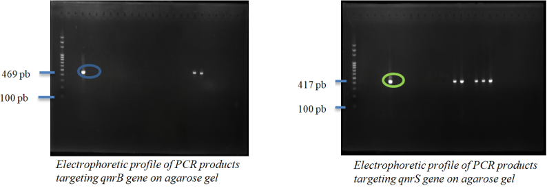
qnrB | For: GATCGTGAAAGCCAGAAAGG | 469 bp | [21] |
Rev.: ACGATGCCTGGTAGTTGTCC | |||
qnrS | For: ACGACATTCGTCAACTGCAA | 417 bp | [21] |
Rev: TAAATTGGCACCCTGTAGGC |
Species | Pus samples | Vaginal swabs |
|---|---|---|
E. coli | 7 | 3 |
K. pneumoniae | 3 | 2 |
P. aeruginosa | 3 | 0 |
P. mirabilis | 1 | 0 |
Identification Number | Sex | Bacterial Species | CIP | LEV | OF | NOR |
|---|---|---|---|---|---|---|
Pus 2 | M | K. pneumoniae | S | S | R | S |
Pus 11 | F | P. aeruginosa | R | S | S | R |
Pus 10 | M | E. coli | R | R | R | R |
Pus 1 | M | E. coli | R | R | R | S |
PV32 | F | K. pneumoniae | R | S | R | R |
PUS 5931 | F | E. coli | R | R | R | R |
PUS42 | F | E. coli | R | R | R | R |
PV2 | M | E. coli | R | S | S | R |
Pus 9 | M | P. mirabilis | S | R | R | S |
Pus 8 | F | K. pneumoniae | R | R | R | R |
Pus 5 | F | E. coli | S | S | S | S |
Pus 4 | M | P. aeruginosa | R | S | S | R |
Pus 3 | M | E. coli | R | S | R | R |
Pus 12 | F | E. coli | R | S | R | R |
PV32 | F | E. coli | S | S | S | S |
PV12 | F | K. pneumoniae | S | S | S | S |
PV 1 | M | E. coli | S | S | S | S |
Pus 7 | M | K. pneumoniae | S | S | S | S |
Pus 6 | F | P. aeruginosa | S | S | S | S |
Genes | Presence/Absence | Frequency | Total | Percentage |
|---|---|---|---|---|
qnrA | Négative | 14 | 14 | 100 |
qnrB | Négative | 10 | 14 | 71.4 |
qnrB | Positive | 4 | 14 | 28.6 |
qnrS | Négative | 10 | 14 | 71,4 |
qnrS | Négative | 4 | 14 | 28,6 |
Genes | Positive | Negative |
|---|---|---|
P-qnrS | 2 (50%) | 2 (50%) |
p-qnrB | 1 (25%) | 3 (75%) |
HOSCO | Saint Camille Hospital of Ouagadougou |
PMQR | Plasmid-Mediated Quinolone Resistance |
CIP | Ciprofloxacin |
NOR | Norfloxacin |
LEV | Levofloxacin |
OF | Ofloxacin |
DNA | Deoxyribonucleic Acid |
EMB | Eosin Methylene Blue |
CASFM | French Society for Microbiology's Antibiogram Committee |
| [1] | S. Swedan, E. A. Alabdallah, et Q. Ababneh, « Resistance to aminoglycoside and quinolone drugs among Klebsiella pneumoniae clinical isolates from northern Jordan », Heliyon, vol. 10, no 1, p. e23368, déc. 2023, |
| [2] | G. A. Jacoby, « Mechanisms of Resistance to Quinolones », Clinical Infectious Diseases, vol. 41, no Supplement_2, p. S120‑S126, juill. 2005, |
| [3] | Y. Jiang et al., « Plasmid-mediated quinolone resistance determinants qnr and aac(6’)-Ib-cr in extended-spectrum -lactamase-producing Escherichia coli and Klebsiella pneumoniae in China », Journal of Antimicrobial Chemotherapy, vol. 61, no 5, p. 1003‑1006, mars 2008, |
| [4] | L. Meradi, A. Djahoudi, A. Abdi, M. Bouchakour, J.-D. Perrier Gros Claude, et M. Timinouni, « Résistance aux quinolones de types qnr, aac (6′)-Ib-cr chez les entérobactéries isolées à Annaba en Algérie », Pathologie Biologie, vol. 59, no 4, p. e73‑e78, août 2011, |
| [5] | F. D. Salah et al., « Distribution of quinolone resistance gene (qnr) in ESBL-producing Escherichia coli and Klebsiella spp. in Lomé, Togo », Antimicrob Resist Infect Control, vol. 8, p. 104, juin 2019, |
| [6] | P. Nordmann et L. Poirel, « Emergence of plasmid-mediated resistance to quinolones in Enterobacteriaceae », Journal of Antimicrobial Chemotherapy, vol. 56, no 3, p. 463‑469, sept. 2005, |
| [7] | J. A. Collins et N. Osheroff, « Gyrase and Topoisomerase IV: Recycling Old Targets for New Antibacterials to Combat Fluoroquinolone Resistance », ACS Infect Dis, vol. 10, no 4, p. 1097‑1115, avr. 2024, |
| [8] | K. J. Aldred, R. J. Kerns, et N. Osheroff, « Mechanism of Quinolone Action and Resistance », Biochemistry, vol. 53, no 10, p. 1565‑1574, mars 2014, |
| [9] | S.-G. Kim, B.-E. Kim, J. H. Lee, et D.-W. Kim, « Novel Qnr Families as Conserved and Intrinsic Quinolone Resistance Determinants in Aeromonas spp. », J Microbiol Biotechnol, vol. 34, no 6, p. 1276‑1286, juin 2024, |
| [10] | M. Goudarzi, M. Azad, et S. S. Seyedjavadi, « Prevalence of Plasmid-Mediated Quinolone Resistance Determinants and OqxAB Efflux Pumps among Extended-Spectrum β -Lactamase Producing Klebsiella pneumoniae Isolated from Patients with Nosocomial Urinary Tract Infection in Tehran, Iran », Scientifica, vol. 2015, p. 1‑7, 2015, |
| [11] | M. Goudarzi et M. Fazeli, « Quinolone Resistance Determinants qnr, qep, and aac(6’)-Ib-cr in Extended-Spectrum Β-Lactamase Producing Escherichia coli Isolated From Urinary Tract Infections in Tehran, Iran », Shiraz E-Med J, vol. 18, no 5, Art. no 5, 2017, |
| [12] | L. Jamali et al., « Prévalence des gènes de résistance plasmidique aux quinolones chez des entérobactéries communautaires isolées au Maroc », ISSN 2351-8014 Vol. 11 No. 2 Nov. 2014, pp. 387-399. |
| [13] | N. Guessennd et al., « Résistance aux quinolones de type qnr chez les entérobactéries productrices de bêta-lactamases à spectre élargi à Abidjan en Côte d’Ivoire », Pathologie Biologie, vol. 56, no 7, p. 439‑446, nov. 2008, |
| [14] | P. Nordmann et H. Mammeri, « Résistance plasmidique aux quinolones », Antibiotiques, vol. 9, no 4, p. 246‑253, déc. 2007, |
| [15] | M. Gharbi, M. A. S. Abbas, S. Hamrouni, et A. Maaroufi, « First Report of aac(6′)-Ib and aac(6′)-Ib-cr Variant Genes Associated with Mutations in gyrA Encoded Fluoroquinolone Resistance in Avian Campylobacter coli Strains Collected in Tunisia », Int J Mol Sci, vol. 24, no 22, p. 16116, nov. 2023, |
| [16] | M. J. Pons, C. Gomes, et J. Ruiz, « QnrVC, a new transferable Qnr-like family », Enferm Infecc Microbiol Clin, vol. 31, no 3, p. 191‑192, mars 2013, |
| [17] | B. Javadi, K. Kafshdouzan, S. H. Emadi Chashmi, et O. Pazhand, « Plasmid-mediated quinolone resistance in Escherichia coli isolates from commercial broiler chickens in Semnan, Iran », Iran J Microbiol, vol. 16, no 2, p. 193‑200, avr. 2024, |
| [18] | U. O. Kibwana et al., « Fluoroquinolone resistance among fecal extended spectrum βeta lactamases positive Enterobacterales isolates from children in Dar es Salaam, Tanzania », BMC Infect Dis, vol. 23, no 1, p. 135, mars 2023, |
| [19] | S. Ferjani, M. Saidani, F. S. Amine, et I. Boutiba-Ben Boubaker, « Prevalence and Characterization of Plasmid-Mediated Quinolone Resistance Genes in Extended-Spectrum β-Lactamase-Producing Enterobacteriaceae in a Tunisian Hospital », Microbial Drug Resistance, vol. 21, no 2, p. 158‑166, avr. 2015, |
| [20] | J. Strahilevitz, G. A. Jacoby, D. C. Hooper, et A. Robicsek, « Plasmid-Mediated Quinolone Resistance: a Multifaceted Threat », Clinical Microbiology Reviews, vol. 22, no 4, p. 664‑689, oct. 2009, |
| [21] | A. Robicsek et al., « Fluoroquinolone-modifying enzyme: a new adaptation of a common aminoglycoside acetyltransferase », Nat Med, vol. 12, no 1, p. 83‑88, janv. 2006, |
| [22] | « Quinolone resistance (qnrA) gene in isolates of Escherichia coli collected from the Al-Hillah River in Babylon Province, Iraq », Pharmacia, vol. 68, no 1, p. 1‑7, janv. 2021, |
| [23] | C. L. Ventola, « The Antibiotic Resistance Crisis », P T, vol. 40, no 4, p. 277‑283, avr. 2015. |
| [24] | A. S. Ouedraogo, H. Jean Pierre, A. L. Bañuls, R. Ouédraogo, et S. Godreuil, « Emergence and spread of antibiotic resistance in West Africa: contributing factors and threat assessment », Médecine et Santé Tropicales, vol. 27, no 2, p. 147‑154, mai 2017, |
| [25] | H. S. Sader, M. Castanheira, et R. K. Flamm, « Antimicrobial Activity of Ceftazidime-Avibactam against Gram-Negative Bacteria Isolated from Patients Hospitalized with Pneumonia in U.S. Medical Centers, 2011 to 2015 », Antimicrob Agents Chemother, vol. 61, no 4, p. e02083-16, mars 2017, |
| [26] | Salmanov AG, Vitiuk AD, Hrynchuk SY, Bober AS, Hrynchuk OB, Berestooy OA, Chernega TV, Rud vo. vaginal cuff infection after hysterectomy in ukraine. Wiad Lek. 2021; 74(2): 196-201. PMID: 33813471. |
| [27] | I. A. Elegbe, A. A. Adefioye, et I. Elegbe, « Aerobic urethral flora of women with infertility and gynecologic problems », Int J Gynaecol Obstet, vol. 21, no 3, p. 241‑245, juin 1983, |
| [28] | C. A. Devi, A. Ranjani, D. Dhanasekaran, N. Thajuddin, et T. Ramanidevi, « Surveillance of multidrug resistant bacteria pathogens from female infertility cases », African Journal of Biotechnology, vol. 12, no 26, Art. no 26, 2013, |
| [29] | Ghiasi, H. Fazaeli, N. Kalhor, M. Sheykh-Hasan, et R. Tabatabaei-Qomi, « Assessing the prevalence of bacterial vaginosis among infertile women of Qom city », Iran J Microbiol, vol. 6, no 6, p. 404‑408, déc. 2014. |
| [30] | S. Abrar, A. Vajeeha, N. Ul-Ain, et S. Riaz, « Distribution of CTX-M group I and group III β-lactamases produced by Escherichia coli and klebsiella pneumoniae in Lahore, Pakistan », Microbial Pathogenesis, vol. 103, p. 8‑12, févr. 2017, |
| [31] | M. Bouchakour et al., « Plasmid-mediated quinolone resistance in expanded spectrum beta lactamase producing enterobacteriaceae in Morocco », The Journal of Infection in Developing Countries, vol. 4, no 12, Art. no 12, août 2010, |
| [32] | H. Ejaz et al., « The Rising Tide of Antibiotic Resistance: A Study on Extended-Spectrum Beta-Lactamase and Carbapenem-Resistant Escherichia coli and Klebsiella pneumoniae », Journal of Clinical Laboratory Analysis, vol. 38, no 10, p. e25081, 2024, |
| [33] | Hrabák J, Študentová V, Jakubů V, Adámková V, Dvořáková L, Balejova M, Bergerová T, Chmelařová E, Ježek P, Kabelíková P, Kolář M, Paterová P, Tejkalová R, Papagiannitsis C, Žemličková H. Prevalence study on carbapenemase-producing Escherichia coli and Klebsiella pneumoniae isolates in Czech hospitals--results from Czech Part of European Survey on Carbapenemase--Producing Enterobacteriaceae (EuSCAPE). Epidemiol Mikrobiol Imunol. 2015 Jun; 64(2): 87-91. PMID: 260996. |
| [34] | J. Silva-Sanchez et al., « Prevalence and characterization of plasmid-mediated quinolone resistance genes in extended-spectrum β-lactamase-producing Enterobacteriaceae isolates in Mexico », Microb Drug Resist, vol. 17, no 4, p. 497‑505, déc. 2011, |
| [35] | H. Y. Kang, M. D. Tamang, S. Y. Seol, et J. Kim, « Dissemination of Plasmid-mediated qnr, aac(6’)-Ib-cr, and qepA Genes Among 16S rRNA Methylase Producing Enterobacteriaceae in Korea », J Bacteriol Virol, vol. 39, no 3, p. 173, 2009, |
| [36] | C. M. Nsofor, M. Y. Tattfeng, et C. A. Nsofor, « High prevalence of qnrA and qnrB genes among fluoroquinolone-resistant Escherichia coli isolates from a tertiary hospital in Southern Nigeria », Bulletin of the National Research Centre, vol. 45, no 1, p. 26, janv. 2021, |
APA Style
Nikiema, R., Dabire, A. M., Kouta, O. F., Bambara, E. B. L., Ouedraodo, N., et al. (2025). Characterization of Quinolone Resistance Genes in Gram-Negative Bacilli Isolated from Pus and Vaginal Swabs at Saint Camille Hospital, Ouagadougou (HOSCO), Burkina Faso. American Journal of Biomedical and Life Sciences, 13(5), 90-97. https://doi.org/10.11648/j.ajbls.20251305.11
ACS Style
Nikiema, R.; Dabire, A. M.; Kouta, O. F.; Bambara, E. B. L.; Ouedraodo, N., et al. Characterization of Quinolone Resistance Genes in Gram-Negative Bacilli Isolated from Pus and Vaginal Swabs at Saint Camille Hospital, Ouagadougou (HOSCO), Burkina Faso. Am. J. Biomed. Life Sci. 2025, 13(5), 90-97. doi: 10.11648/j.ajbls.20251305.11
AMA Style
Nikiema R, Dabire AM, Kouta OF, Bambara EBL, Ouedraodo N, et al. Characterization of Quinolone Resistance Genes in Gram-Negative Bacilli Isolated from Pus and Vaginal Swabs at Saint Camille Hospital, Ouagadougou (HOSCO), Burkina Faso. Am J Biomed Life Sci. 2025;13(5):90-97. doi: 10.11648/j.ajbls.20251305.11
@article{10.11648/j.ajbls.20251305.11,
  author = {Rabietou Nikiema and Amana Metuor Dabire and Olawoumi Fabrice Kouta and Eliada Benoit Lionel Bambara and Nicolas Ouedraodo and Rhaina Olivia Badini and Pegd-Wende Rose Bonkoungou},
  title = {Characterization of Quinolone Resistance Genes in Gram-Negative Bacilli Isolated from Pus and Vaginal Swabs at Saint Camille Hospital, Ouagadougou (HOSCO), Burkina Faso
},
  journal = {American Journal of Biomedical and Life Sciences},
  volume = {13},
  number = {5},
  pages = {90-97},
  doi = {10.11648/j.ajbls.20251305.11},
  url = {https://doi.org/10.11648/j.ajbls.20251305.11},
  eprint = {https://article.sciencepublishinggroup.com/pdf/10.11648.j.ajbls.20251305.11},
  abstract = {Introduction: Antibiotic resistance, particularly to quinolones, represents a major public health concern in Burkina Faso. In recent decades, plasmid-mediated quinolone resistance (PMQR) mechanisms have emerged, especially among Gram-negative bacteria. These mechanisms include, among others, qnr genes (qnrA, qnrB, qnrS). This study aimed to investigate the presence of quinolone resistance determinants in Gram-negative bacilli isolated from pus and vaginal samples at Saint Camille Hospital of Ouagadougou (HOSCO). Methodology: A total of 19 strains of Escherichia coli isolated from pus and vaginal swabs were collected for bacteriological and molecular analysis. Four antibiotics, namely ciprofloxacin (CIP), norfloxacin (NOR), ofloxacin (OF) and levofloxacin (LEV) were used for sensitivity testing and molecular analysis focused on the detection of qnrA, qnrB, and qnrS type genes. Results: Resistance rates to CIP, NOR, OF, and LEV were 57.89%, 52.63%, 52.63%, and 31.37%, respectively. Molecular analysis revealed the presence of qnrB, and qnrS genes in 28.47% of the isolates, for each gene. The qnrA gene was not detected in any isolate. The analysis of the genetic support of resistance genes revealed that 50% of the qnrS genes were plasmid-borne, while only 25% of the qnrB genes were associated with plasmids. La Correlation analysis between resistance genes and antibiotics showed a moderate positive correlation between qnrB and NOR/LEV, thereby suggesting the involvement of qnrB in resistance to these antibiotics. Conclusion: These findings highlight the need for continuous surveillance of antibiotic resistance in clinical isolates.
},
 year = {2025}
}
TY  - JOUR
T1  - Characterization of Quinolone Resistance Genes in Gram-Negative Bacilli Isolated from Pus and Vaginal Swabs at Saint Camille Hospital, Ouagadougou (HOSCO), Burkina Faso

AU  - Rabietou Nikiema
AU  - Amana Metuor Dabire
AU  - Olawoumi Fabrice Kouta
AU  - Eliada Benoit Lionel Bambara
AU  - Nicolas Ouedraodo
AU  - Rhaina Olivia Badini
AU  - Pegd-Wende Rose Bonkoungou
Y1  - 2025/10/27
PY  - 2025
N1  - https://doi.org/10.11648/j.ajbls.20251305.11
DO  - 10.11648/j.ajbls.20251305.11
T2  - American Journal of Biomedical and Life Sciences
JF  - American Journal of Biomedical and Life Sciences
JO  - American Journal of Biomedical and Life Sciences
SP  - 90
EP  - 97
PB  - Science Publishing Group
SN  - 2330-880X
UR  - https://doi.org/10.11648/j.ajbls.20251305.11
AB  - Introduction: Antibiotic resistance, particularly to quinolones, represents a major public health concern in Burkina Faso. In recent decades, plasmid-mediated quinolone resistance (PMQR) mechanisms have emerged, especially among Gram-negative bacteria. These mechanisms include, among others, qnr genes (qnrA, qnrB, qnrS). This study aimed to investigate the presence of quinolone resistance determinants in Gram-negative bacilli isolated from pus and vaginal samples at Saint Camille Hospital of Ouagadougou (HOSCO). Methodology: A total of 19 strains of Escherichia coli isolated from pus and vaginal swabs were collected for bacteriological and molecular analysis. Four antibiotics, namely ciprofloxacin (CIP), norfloxacin (NOR), ofloxacin (OF) and levofloxacin (LEV) were used for sensitivity testing and molecular analysis focused on the detection of qnrA, qnrB, and qnrS type genes. Results: Resistance rates to CIP, NOR, OF, and LEV were 57.89%, 52.63%, 52.63%, and 31.37%, respectively. Molecular analysis revealed the presence of qnrB, and qnrS genes in 28.47% of the isolates, for each gene. The qnrA gene was not detected in any isolate. The analysis of the genetic support of resistance genes revealed that 50% of the qnrS genes were plasmid-borne, while only 25% of the qnrB genes were associated with plasmids. La Correlation analysis between resistance genes and antibiotics showed a moderate positive correlation between qnrB and NOR/LEV, thereby suggesting the involvement of qnrB in resistance to these antibiotics. Conclusion: These findings highlight the need for continuous surveillance of antibiotic resistance in clinical isolates.

VL  - 13
IS  - 5
ER  -
Biochemistry-Microbiology-Molecular Biology and Genetics, Joseph Ki-Zerbo University, Ouagadougou, Burkina Faso
Department of Biochemistry-Microbiology, University Joseph Ki-Zerbo, Ouagadougou, Burkina Faso
Biochemistry-Microbiology-Molecular Biology and Genetics, Joseph Ki-Zerbo University, Ouagadougou, Burkina Faso
Biochemistry-Microbiology-Molecular Biology and Genetics, Joseph Ki-Zerbo University, Ouagadougou, Burkina Faso
National Malaria Research and Training Centre (CNRFP), Ouagadougou, Burkina Faso
Biochemistry-Microbiology-Molecular Biology and Genetics, Joseph Ki-Zerbo University, Ouagadougou, Burkina Faso
Biochemistry-Microbiology-Molecular Biology and Genetics, Joseph Ki-Zerbo University, Ouagadougou, Burkina Faso
Information