Research Article | | Peer-Reviewed

Gastroprotective and Antigastric Ulcer Effects of the Methanol Fraction of Cleistopholis Patens Leaves on Two Ulcer Models in Rats

Received: 14 March 2025     Accepted: 26 March 2025     Published: 4 July 2025
Views:       Downloads:
Abstract

Background: Cleistopholis patens leaves are used to treat stomach disorders in Nigeria. This research investigated the gastroprotective and antigastric ulcer effects of the methanol fraction of Cleistopholis patens leaves (MFCPL) in rats. Methods: In vitro experiments involved DPPH-radical scavenging and H+/K+-ATPase inhibitory activities. Two ulcer models, diclofenac sodium (DS) (using 50 mg/kg b.w. DS orally) and acidified ethanol (AE) (using 0.3 M HCl + 60% ethanol orally), were used. The experimental rats (n=24) were grouped into 6 of 4 groups for each of the DS and AE ulcer models. In the DS model, group 1 served as the normal group, whereas group 2 was ulcer-induced without treatment. Group 3 received 20 mg/kg b.w. omeprazole, whereas groups 4, 5 and 6 received 50, 100 and 200 mg/kg b.w. MFCPL, respectively, for 14 days. For the AE ulcer model, 7 days after pretreatment with MFCPL, all groups except group 1 received acidified ethanol and were sacrificed after 1 hr. The ulcer indices, gastric mucosa morphology and serum antioxidant status of the rats were evaluated. Additionally, GC‒MS analyses of MFCPL were carried out. Results: The MFCPL had H+, K+-ATPase-inhibiting and DPPH-radical-scavenging activities, with half-maximal inhibitory concentrations (IC50) of 50.10 ± 10.52 μg/ml and 16.72 ± 2.47 μg/ml, respectively. Compared with the ulcer control, the administration of MFCPL led to a significant (p < 0.05) reduction in total ulcer counts and gastric volumes with high pH. Compared with those in the control group, the number of gastric lesions in the treated group was markedly lower than that in the control group. The antioxidant status of the treated rats improved. GC‒MS analyses of the MFCPL revealed 20 bioactive compounds. Conclusion: MCPL has an antigastric ulcer effect attributable to its H+/K+-ATPase inhibition and antioxidant activities due to its rich bioactive components and hence can be used to manage ulcers and their associated biochemical aberrations.

Published in Advances in Biochemistry (Volume 13, Issue 3)
DOI 10.11648/j.ab.20251303.11
Page(s) 61-78
Creative Commons

This is an Open Access article, distributed under the terms of the Creative Commons Attribution 4.0 International License (http://creativecommons.org/licenses/by/4.0/), which permits unrestricted use, distribution and reproduction in any medium or format, provided the original work is properly cited.

Copyright

Copyright © The Author(s), 2025. Published by Science Publishing Group

Keywords

Cleistopholis Patens, Antigastric Ulcer, H+/K+-ATPase, Antioxidants, GC‒MS

1. Introduction
Approximately half of the world's population is affected by Helicobacter pylori (H. pylori) colonization, which is still one of the most common causes of peptic ulcer illness . H. pylori is more prevalent in underdeveloped countries, particularly those in Africa, Central America, Central Asia, and Eastern Europe . Increased generation of reactive oxygen species (ROS), such as superoxide, hydroxyl, and other ROS, damages a variety of tissues and ultimately results in the formation of ulcers and related complications . A peptic ulcer is a lesion of the digestive tract caused by acid that is characterized by denuded mucosa that extends into the submucosa or muscularis propria . A gastric ulcer is found in the stomach, whereas a duodenal ulcer is found in the first part of the intestines. Nonsteroidal anti-inflammatory drug (NSAID) use, alcohol and cigarette use, H. pylori infection, and Zollinger–Ellison syndrome can all cause peptic ulcers .
Gastric acid, a digestive fluid produced within the stomach lining, is composed of hydrochloric acid, potassium chloride, and sodium chloride . By triggering digestive enzymes, which jointly breakdown the lengthy chains of amino acids in proteins, gastric acid plays a crucial in the digestion of proteins . The gastric hydrogen, potassium ATPase, a member of the P2-type ATPase family, is an important membrane protein responsible for the secretion of stomach acid. Since H+/K+-ATPase is the last enzyme involved in acid secretion, blocking the pump may be a more effective way to reduce acid production than using a receptor antagonist . An important pathogenic component of gastric mucosa injury is oxidative stress. Plants with antioxidant capacity function as the main bioactive mechanism in an herbal reservoir for the treatment of ulcers .
Cleistopholis patens Benth (family: Annonaceae) is a 20–30 m tall, sun-loving plant that may be found in different parts of Africa. The common name for Cleistopholis patens is "salt and oil tree"; however, in Igbo and Yoruba, the names are “Ojo” and “Apako” or “Oke”, respectively. It is an herb that is used to cure infertility, malaria, headaches, and other ailments . Traditional healers in south-eastern Nigeria have utilized Cleistopholis patens leaf extracts to treat gastrointestinal ailments and cardiovascular diseases . Hepatitis has been treated in Ghana via leaf infusions comprising lemon grass, papaya, or other plants . In Nigeria and other parts of Africa, root bark and leaves are used to treat stomach problems, typhoid fever, and urogenital infections. Owing to the low efficiency and serious side effects of currently available medications, treating peptic ulcers is still difficult.
Thus, this work was carried out for the first time to assess the antigastric ulcer potential of the methanol fraction of Cleistopholis patens leaf extract in two distinct models, namely, diclofenac sodium (NSAID) and ethanol-induced stomach damage in experimental rats. First, the antioxidant capacities of several Cleistopholis patens leaf extract fractions were evaluated in vitro via 2,2-diphenyl-1-picrylhydrazyl (DPPH) radical scavenging activity, as were their inhibitory effects on H+/K+-ATPase. The MFCPL with the highest DPPH and H+/K+-ATPase activity was then employed for additional research. Gastric juice was used for the ulcer indices, whereas serum was used for the lipid peroxidation markers and antioxidant parameters. Moreover, the gastroprotective efficacy of the MFCPL was assessed via pathological sections of stomach tissue from several groups. GC‒MS analysis was conducted on the MFCPL to identify the bioactive components responsible for their activity.
2. Materials and Methods
2.1. Plant Materials
Fresh leaves of Cleistopholis patens were obtained from Amalla Egazi in Udenu L.G.A. of Enugu State, Nigeria. A botanist from the Bioresources Diversity and Conservation Programme, Nsukka, named Alfred Ozioko, verified the collection. The herbarium has received a voucher specimen with the number Intercedd/839 (Figure 1).
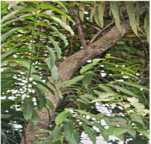
Figure 1. Cleistopholis patens leaves.
2.2. Equipment
The equipment used in this study was as follows: Centrifuge, micropipette, water bath, and thermometer (New–Life Laboratory, England). A stop watch (Paris), Refrigerator (Restpoint Company Ltd., England), spectrophotometer (Merck, Germany), ordinary centrifuge (Model 800D China), digital weighing balance (PEC Medicals USA), and pH meter (United Kingdom) were used.
2.3. Experimental Animals
Fresh sheep stomach mucosal scrapings were acquired at the Obollo-Afor market slaughterhouse in the Udenu local government area of Enugu State, Nigeria. Swiss albino male rats (48), weighing between 150 and 300 g each, were acquired from the University of Nigeria, Nsukka's Faculty of Veterinary Medicine's animal house. Before the experiment started, the rats were maintained on a standard diet (commercial grower's mash) and water for one week to help them acclimate to the new conditions. This study was reviewed and approved by the Committee on Ethics and Biosafety, Faculty of Biological Sciences, University of Nigeria, Nsukka, and was given the ethical clearance number UNN/FBS/EC/1084.
2.4. Preparation of the Methanol Fraction of Cleistopholis Patens Leaves (MFCPL)
After two weeks of shade drying, fresh Cleistopholis patens leaves were ground into powder via a machine grinder. Five hundred grams of finely ground Cleistopholis patens leaves were macerated for 72 hours and stirred periodically in 5 liters of 100% methanol. To effectively remove fine residues, the mixture was first filtered with muslin cloth and then again with Whatman No. 1 filter paper. After being dried using a rotary evaporator at 45°C, the methanol extract was refrigerated at 40°C. A variety of solvents, including n-hexane, chloroform, ethyl acetate, and methanol, were used to fractionate the crude methanol extract of Cleistopholis patens leaves according to their polarity . The different fractions were used to determine the most potent fraction on the basis of their DPPH-radical scavenging activity and inhibitory effect on H+/K+-ATPase activity. Compared with the other fractions, the methanol fraction of Cleistopholis patens leaves (MFCPL) was found to have the highest DPPH-radical scavenging activity and inhibitory effect on H+/K+-ATPase activity; hence, it was selected for further studies.
2.5. Phytochemical Screening of the Crude Methanol Extract and Fractions of Cleistopholis Patens Leaves (CMEFCPL)
The crude extract and fractions of C. patens leaves (CMEFCPL) were subjected to phytochemical analysis in accordance with the protocol of Harborne .
2.6. Determination of the DPPH Radical Scavenging Activity of the CMEFCPL
Braca et al. was used to evaluate the DPPH radical scavenging activity of the methanol extract of Cleistopholis patens leaves and their fractions . A 1.0 ml methanol mixture containing different amounts of each sample extract (20–640 μg/ml) was mixed with 0.5 ml of 0.076 mM DPPH in methanol. After the mixture was shaken well, it was left to stand at room temperature in the dark for half an hour. Because ascorbic acid has strong reducing power and weak metal-chelating ability at various doses, it was employed as the reference compound in this triplicate experiment. After that, each sample's absorbance at 517 nm was measured, and the percentage of inhibitory activity was calculated as follows:
% inhibition activity=Absorbance of control-Absorbance of sample/standardAbsorbance of control x 100
The inhibition curves were prepared, and the 1C50 values were determined.
2.7. Determination of the Effect of the CMEFCPL on H+/K+-ATPase Activity
Sheep stomach mucosal scrapings were used to prepare proton potassium ATPase, and the enzyme activity was measured . Briefly, clean test tubes (4) were used for each sample, while an additional two test tubes were used for the sample and reagent blanks. A known volume (0.1 ml) of the enzyme extract (H+/K+-ATPase) from the mucosal homogenate of the sheep stomach was added to all test tubes except the sample blank and reagent blank. Subsequently, 0.1 ml of varying extract concentrations (50–400 mg/ml) were introduced into distinct test tubes. Similarly, 0.1 ml of various omeprazole (20–160 mg/ml) concentrations were applied to additional test tubes (standard controls). Thereafter, 0.2 ml of Tris buffer (pH 7.4) and 0.2 ml of magnesium chloride (2 mM) were added to all test tubes. Subsequently, 0.2 ml of potassium chloride was added to all test tubes. Thereafter, 0.2 ml of ATP was added to all the test tubes and allowed to incubate for 30 seconds. The reaction was stopped by adding 1.0 ml of trichloroacetic acid (TCA). This also precipitated proteins in the samples. The test tubes were centrifuged at 3000 rpm for 10 minutes. Thereafter, 1.0 ml of each supernatant was transferred to another clean test tube before the addition of 1.5 ml of distilled water. A drop of nitric acid was added to all test tubes in addition to 2.5 ml of vanadate reagent. Finally, the sample blank was mixed with 0.1 ml of enzyme extract only after the reaction had stopped. At a wavelength of 470 nm, spectrophotometric measurements were obtained. Each sample's inhibitory ATPase activity was determined via the following formula:
inhibitory ATPase activity=Control-Mean of absorbance of each sample-Sample BlankControl x 100
2.8. Determination of the Median Lethal Dose (LD50) of the Methanol Extract of Cleistopholis Patens Leaves
The median lethal dose (LD50) of the methanol extract of Cleistopholis patens leaves was determined according to Lorke .
2.9. Experimental Design
For the NSAID-induced gastric ulcer model, groups 2 to 6 were orally administered 50 mg/kg b.w diclofenac sodium to induce gastric ulcer injury before oral treatment with various dosages of MFCPL (curative design) for 7 days, whereas for the preventive design, the rats were given various dosages of MFCPL for 14 days before induction of gastric injury with 50 mg/kg b.w diclofenac sodium. For each design (curative and preventive), a total of twenty-four (24) experimental rats were grouped into six groups (6) of four rats (4): group 1 (normal control), group 2 (ulcer-induced without treatment or positive control), group 3 (received 20 mg/kg b.w omeprazole after ulcer induction), group 4 (received 50 mg/kg b.w MFCPL after ulcer induction), and group 5 (received 100 mg/kg b.w MFCPL after ulcer induction), whereas group 6 received 200 mg/kg b.w MFCPL after ulcer induction. In the acidified ethanol model, gastric injury was induced with acidified ethanol (0.3 M HCl + 60% ethanol) after pretreatment with graded dosages of MFCPL. Briefly, a total of twenty-four (24) experimental rats were grouped into six (6) of four (4) rats each as follows: group 1 (normal control), group 2 (0.3 M HCl + 60% ethanol or positive control), group 3 (received 20 mg/kg b.w omeprazole before administration with acidified ethanol), group 4 (received 50 mg/kg b.w. of MFCPL before administration with acidified ethanol), and group 5 (received 100 mg/kg b.w. of MFCPL before administration with acidified ethanol), whereas group 6 received 200 mg/kg b.w. of MFCPL before administration with acidified ethanol. The treatment lasted for 7 consecutive days before the induction of ulcers with acidified ethanol. On the 7th day, 1 hr after the last dose, all the groups except the normal control group received acidified ethanol (0.3 M HCl + 60% ethanol) orally to induce gastric mucosal injury, which lasted for 1 hr, after which the animals were sacrificed, and samples were collected for analyses.
2.10. Collection of Gastric Juice
After every rat was sacrificed, its stomach was quickly removed, and it was then split apart along the larger curvature so that the contents could be measured for pH and volume. Following gentle rinsing with phosphate-buffered saline (PBS) to eliminate any blood clots, the gastric tissue samples were analysed macroscopically to determine the gastric ulcer index .
2.10.1. Gross Gastric Lesion Evaluation
A digital camera was used to take pictures of each stomach to perform a gross examination for any indications of gastric mucosal ulcers, which manifested as lengthy, hemorrhagic lesions that ran parallel to the stomach's long axis. For each group, the total number of ulcerated areas was determined and averaged .
2.10.2. Ulcer Indices Assessment
The volume of gastric juice was measured via a calibrated syringe . The pH of the stomach juice was measured with a digital pH meter. In summary, 1.0 ml of gastric juice was mixed with 1.0 ml of distilled water to create an aliquot, and a pH meter (HI 9021) was used to determine the pH of the mixture. Using a digital pH meter (HI 9021) and 0.1 N NaOH solutions, samples of gastric contents were analysed for hydrogen ion concentration .
2.10.3. Biochemical Analyses of the Serum
The activities of superoxide dismutase (SOD), catalase (CAT), and glutathione peroxidase and the levels of the lipid peroxidation product malondialdehyde (MDA) in the serum were determined as described by Durak et al. , Aebi , Paglia and Valentine ), and Wallin et al. , respectively.
2.10.4. GC‒MS Analysis of the MFCPL
A mass spectrophotometer (Agilent 19091-433HP, USA) and a 7890A gas chromatograph system were used for the GC‒MS analysis. A 5675C Inert MSD with a Triple-Axis detector interfaced with an HP-5 MS-fused silica column (5% phenyl methyl siloxane 30.0 m × 250 μm, film thickness 0.25 μm) formed part of the setup. Helium gas was used as the carrier, and its flow rate was set at 1.0 millilitres per minute. A 1 μl injector operating in split mode with a split ratio of 1:50 and an injection temperature of 300°C are examples of additional GC‒MS conditions. The temperature of the ion source was 250°C, the contact temperature was 300°C, the pressure was 16.2 psi, and the duration was 1.8 mm.
After being held at 36°C for five minutes, the temperature of the column rose at a rate of 4°C/min to 150 °V. The temperature was increased to 250°C at a rate of 20°C per minute and held there for five minutes. The elution took 47.5 minutes in total. The relative percent amount of each component was calculated by summing the areas of each component's average peak. The supplier supplied MS Solution software, which was used to manage the system and gather data.
2.10.5. Statistical Analysis
Version 20 of the Statistical Product for Service Solution (SPSS) was used to analyse the data, and the results are reported as the mean ± standard deviation of the mean. One-way analysis of variance (ANOVA) was used to determine whether there were significant differences in the results, and p < 0.05 was considered the accepted level of significance.
3. Results
3.1. Phytochemical Composition of the CMEFCPL
The phytochemical compositions of both the crude extract and the fractions of Cleistopholis patens leaves revealed various amounts of steroids, terpenoids, glycosides, reducing sugars, alkaloids, total phenols, tannins and flavonoids, except in the chloroform fraction, where alkaloids were not detected (Table 1).
Table 1. Quantitative phytochemical composition of Cleistopholis patens leaves (mg/100 g).

Phytochemicals

Crude Extract

Chloroform Fraction

Ethylacetate Fraction

Methanol Fraction

Steroid

3.13 ± 0.20c

0.86 ± 0.25a

4.06 ± 0.07d

2.37 ± 0.18b

Terpenoids

64.03 ± 3.42b,c

28.88 ± 2.92a

68.87 ± 24.10 c

44.02 ± 6.17a,b

Glycoside

5.31 ± 0.26a,b

6.09 ± 0.03 c

4.99 ± 0.11 a

5.43 ± 0.28 b

Reducing Sugar

456.16 ± 88.38 b

296.74 ± 1.96 a

504.89 ± 29.72 b

317.03 ± 34.10 a

Alkaloids

70.97 ± 6.97 b

ND

76.81 ± 15.99 b

70.00 ± 1.44 b

Total phenols

354.57 ± 4.14 c

96.77 ± 2.42 a

374.46 ± 7.92 d

243.01 ± 11.47 b

Tannins

15.25 ± 0.55 c

6.68 ± 0.49 a

9.21 ± 0.33 b

17.06 ± 1.26 d

Flavonoids

255.10 ± 41.20 b

142.08 ± 40.63 a

321.30 ± 12.19b,c

387.50 ± 43.59 c

The findings are presented as the means ± SDs (n = 3). (ND: not detected)
At p < 0.05, mean values throughout the column with distinct letters as superscripts are deemed significant.
3.2. Acute Toxicity Study Results
The acute toxicity test of the methanol extract of Cleistopholis patens leaves revealed no mortality or adverse reactions at a dose range of 10–5000 mg/kg body weight within the test population (Table 2).
Table 2. Results of acute toxicity tests on Cleistopholis patens leaves.

Phase/Groups

Dosage of extract (mg/kg b.w)

Mortality rate (%)

Phase I

Group 1

10

0/3

Group 2

100

0/3

Group 3

1000

0/3

Phase II

Group 1

1600

0/3

Group 2

2900

0/3

Group 3

5000

0/3

n = 3
3.3. DPPH Radical Scavenging Activity of the CMEFCPL
The MFCPL had the highest DPPH radical scavenging activity (IC50 =16.72±2.4 μg/ml), followed by the ethyl acetate fraction (IC50 = 17.04±0.79 μg/ml). The chloroform fraction (IC50 = 22.99±0.87 μg/ml) and crude extract (IC50 = 30.96±5.37 μg/ml) had the lowest scavenging activities (Table 3).
Table 3. DPPH radical scavenging activities of methanol extracts and fractions of Cleistopholis patens leaves.

Concentration (μg/ml)

Crude

Chloroform

Ethylacetate

Methanol

Standard (ascorbic acid)

20

58.06 ± 6.41a

64.88 ± 1.02a

66.02 ± 2.80a

65.45 ± 2.72a

93.20±2.49bc

40

53.98 ± 11.84a

65.81 ± 2.64ab

74.48 ± 3.58b

71.90 ± 4.96a

94.15±0.61bc

80

66.38 ± 3.13ab

69.24 ± 2.28b

73.48 ± 2.80b

73.69 ± 4.41a

93.15±1.63bc

160

64.23 ± 9.79a

68.24 ± 0.66ab

73.33 ± 2.82b

75.05 ± 13.71a

90.43±.63ab

320

77.78 ± 1.08bc

74.12 ± 3.13c

82.72 ± 1.38c

91.76 ± 1.58b

90.43±1.07ab

640

84.80 ± 3.45c

77.49 ± 2.57c

94.27 ± 1.40d

91.11 ± 1.02b

89.43±2.26a

The findings are presented as the means ± SDs (n = 3).
At p < 0.05, mean values throughout the column with distinct letters as superscripts are deemed significant. The IC50 values were as follows: crude = 30.96±5.37 μg/ml, chloroform = 22.99±0.87 μg/ml, ethylacetate = 17.04±0.79 μg/ml, methanol = 16.72±2.47 μg/ml and standard drug (ascorbic acid) = 3.05±0.21 μg/ml.
3.4. Hydrogen Potassium ATPase Inhibitory Activities of the CMEFCPL
The hydrogen potassium ATPase inhibitory activities of the CMEFCPL as well as the standard drug (omeprazole) revealed that the methanol fraction had the highest H+, K+-ATPase inhibitory activity, followed by the ethyl acetate fraction, compared with the standard drug (omeprazole). The crude extract and chloroform fractions had the lowest inhibitory effects on H+, K+-ATPase (Table 4).
Table 4. Hydrogen potassium ATPase inhibitory activities of the CMEFCPL.

Concentration (μg/ml)

Std drug

Crude

Chloroform

Ethylacetate

Methanol

50

53.85 ± 2.54a

43.72 ± 7.79a

10.64 ± 0.184c

62.82 ± 6.17a

52.31 ± 7.25 a

100

60.00 ± 4.36ab

59.23 ±7.25ab

5.77 ± 0.54b

57.44 ± 13.78a

53.72 ± 7.43 a

200

65.90 ± 1.81bc

53.46 ±2.72ab

59.10 ± 0.18d

57.18 ± 7.62 a

75.13 ± 14.50 a

400

70.77 ± 4.71c

66.28 ± 3.80b

1.67 ± 0.18a

65.00 ± 1.27 a

72.18 ± 1.27 a

The findings are presented as the means ± SDs (n = 3).
At p < 0.05, mean values throughout the column with distinct letters as superscripts are deemed significant. (IC50 values of STD Drug = 14.55±2.36 μg/ml, Crude = 86.10±1.40 μg/ml, Chloroform = 5,518,734.10 ± 716810.78 μg/ml, Ethylacetate = 60.83±11.81 μg/ml and Methanol =50.10±10.52 μg/ml).
3.5. Gross Appearance of the Gastric Mucosa of Rats Pretreated with MFCPLs (Preventive Study)
The diagram in Figure 2 revealed that there was observable dose depedent reduction in gastric lesions of the pretreated rats (groups 3-6) than in those of the untreated animals (group 2) upon induction of ulcers. The arrows point at the gastric lesions.
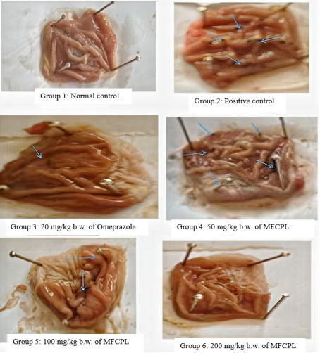
Figure 2. Gross appearance of the gastric mucosa of rats pretreated with MFCPL (preventive design).
3.6. Gastroprotective Effect of the MFCPL on Gastric Ulcer Indices (Preventive Study)
Administration of MFCPLs before the induction of ulcers (preventive study) significantly reduced the total ulcer count in the pretreated groups compared with that in the untreated animals (p < 0.05). Similarly, the gastric volume of group 2 was increased, although not significantly (p > 0.05), compared with that of the pretreated animals. The gastric pH of the untreated animals was significantly lower (p < 0.05) than that of the pretreatment animals (Table 5).
Table 5. Gastroprotective effect of the MFCPL on gastric ulcer indices (preventive study).

Groups

Total ulcer

G. Volume

G. pH

Group 1

0.00 ± 0.00a

0.23 ± 0.01 b

4.50 + 0.12 e

Group 2

43.50 ± 4.30f

0.47 ± 0.01 a

2.48 + 0.85 a

Group 3

4.50 ± 2.08b

0.25 ± 0.02 a

4.22 + 0.13d,e

Group 4

20.50 ± 1.29e

0.38 ± 0.08a,b

3.20 + 0.08 b

Group 5

14.25 ± 2.22d

0.35 ± 0.01a,b

3.50 + 0.02b,c

Group 6

8.75 ± 1.70c

0.30 ± 0.02 a

3.90 + 0.08c,d

The findings are presented as the means ± SD (n = 4).
At p < 0.05, mean values throughout the column with distinct letters as superscripts are deemed significant.
Group 1: Normal control
Group 2: Positive control (ulcer induced with 50 mg/kg b.w. diclofenac sodium without treatment)
Group 3: Standard control (pretreated with 20 mg/kg b.w. omeprazole before ulcer induction)
Group 4: Pretreatment with 50 mg/kg b.w. MFCPL before ulcer induction
Group 5: Pretreatment with 100 mg/kg b.w. MFCPL before ulcer induction
Group 6: Pretreatment with 200 mg/kg b.w. MFCPL before ulcer induction
3.7. Gross Appearance of the Gastric Mucosa of Rats Treated with the MFCPL After Ulcer Induction (Curative Study)
The diagram in Figure 3 shows that, compared with that in Group 2, there was an observable reduction in the number of gastric lesions in the treated rats (groups 3--6) after the induction of ulcers. The gastric lesions are indicated by arrows.
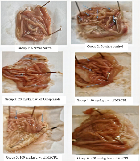
Figure 3. Gross appearance of the gastric mucosa of ulcer-induced rats treated with the MFCPL (curative design).
3.8. Effects of the MFCPL on Gastric Ulcer Indices (Curative Study)
Compared with no treatment, the administration of MFCPL following the induction of ulcers significantly decreased the total ulcer count and gastric volume of the treated animals (p<0.05). The gastric pH of the untreated group was significantly lower (p < 0.05) than that of the treated groups (Table 6).
Table 6. Effects of the MFCPL on gastric ulcer indices (curative study).

Groups

Total Ulcer

G. Volume

G. pH

Group 1

0.00 ± 0.00a

0.23 ± 0.01 a

4.55 ± 0.10 c

Group 2

36.75 ±7.27d

0.45 ± 0.06 b

2.78 ± 0.48 a

Group 3

2.50 ± 2.65a

0.30 ± 0.08 a

4.25 ± 0.48b,c

Group 4

14.00 ± 6.68c

0.35 ± 0.06 a

3.30 ± 1.34a,b

Group 5

10.25 ± 5.12b,c

0.33 ± 0.13 a

3.40 ± 0.08a,b

Group 6

6.00 ± 1.83a,b

0.30 ± 0.08 a

3.73 ± 0.05a,b

The findings are presented as the means ± SD (n = 4).
At p < 0.05, mean values throughout the column with distinct letters as superscripts are deemed significant.
Group 1: Normal control
Group 2: Positive control (ulcer induced with 50 mg/kg b.w. diclofenac sodium without treatment)
Group 3: Standard control (ulcer-induced + 20 mg/kg b.w. omeprazole)
Group 4: ulcers induced before the administration of 50 mg/kg b.w. MFCPL
Group 5: ulcers induced before the administration of 100 mg/kg b.w. MFCPL
Group 6: ulcers induced before the administration of 200 mg/kg b.w. MFCPL
3.9. Gross Appearance of the Gastric Mucosa of Rats Pretreated with MFCPL (Acidified Ethanol Model)
The diagram in Figure 4 shows that in response to the administration of MFCPL before the induction of ulcers with acidified ethanol, there was an observable reduction in the number of gastric lesions in the pretreated rats (groups 3-6) compared with those in group 2. The gastric lesions are indicated by arrows.
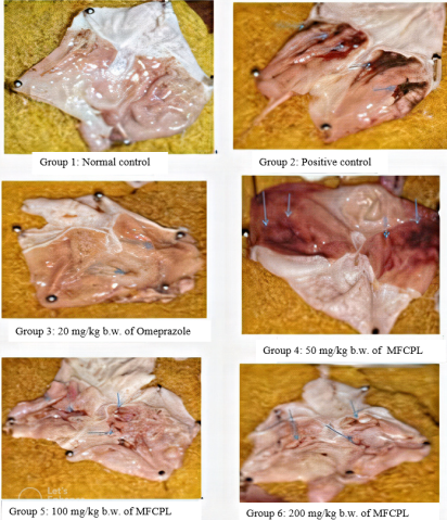
Figure 4. Gross appearance of the gastric mucosa of ulcer-induced rats pretreated with MFCPL (acidified ethanol model).
3.10. Gastroprotective Effect of the MFCPL on Gastric Ulcer Indices (Acidified Ethanol Model)
Pretreatment with the MFCPL before ulcer induction via acidified ethanol significantly reduced the total ulcer count and gastric volume (p<0.05) in the treatment groups compared with those in the untreated animals. Moreover, the gastric pH of group 2 was lower than that of the treatment groups (Table 7).
Table 7. Gastroprotective effects of the MFCPL on gastric ulcer indices (acidified ethanol model).

Groups

TOTAL ULCER

G. VOLUME

G. pH

Group 1

0.00 ± 0.00a

0.36 ± 0.01 d

4.25 ± 0.24 e

Group 2

42.25 ± 2.06e

0.47 ± 0.01 e

2.10 ± 0.82 a

Group 3

3.00 ±1.41b

0.25 ± 0.01 a

4.20 ± 0.16 e

Group 4

22.25 ± 2.22d

0.36 ± 0.04c,d

2.60 ± 0.08 b

Group 5

10.75 + 1.26c

0.33 ± 0.01 c

3.35 ± 0.25c

Group 6

5.25 ± 1.710b

0.30 ± 0.01 b

3.80 ± 0.16d

The findings are presented as the means ± SD (n = 4).
At p < 0.05, mean values throughout the column with distinct letters as superscripts are deemed significant.
Group 1: Normal control
Group 2: Positive control (ulcer induced with acidified ethanol (0.3 M HCl + 60% ethanol) without treatment)
Group 3: Standard control (ulcer-induced + 20 mg/kg b.w. omeprazole)
Group 4: 50 mg/kg b.w. of MFCPL + acidified ethanol (0.3 M HCl + 60% ethanol)
Group 5: 100 mg/kg b.w. of MFCPL + acidified ethanol (0.3 M HCl + 60% ethanol)
Group 6: 200 mg/kg b.w. of MFCPL + acidified ethanol (0.3 M HCl + 60% ethanol)
3.10.1. Effects of the MFCPL on Lipid Peroxidation Markers, Malondialdehyde (MDA) and Antioxidant Enzymes
In both the preventive and curative designs, there was a significant (p<0.05) increase in the MDA levels of the untreated animals, with corresponding decreases in the SOD, CAT and GPx activities. However, pretreatment of the test groups with MFCPL resulted in a reduction (p<0.05) in the malondialdehyde concentration, with corresponding increases in the SOD, CAT and GPx activities (Table 8).
Table 8. Effects of the MFCPL on lipid peroxidation markers, malondialdehyde (MDA) and antioxidant enzymes.

GROUPS

MDA (mg/dl)

SOD (U/mg)

CAT (U/mg)

GPx (U/mg)

Preventive

Curative

Preventive

Curative

Preventive

Curative

Preventive

Curative

Group 1

0.67 ± 0.19a

0.67 ± 0.19a

11.33 ± 0.05d

11.33 ± 0.06d

7.91 ± 1.80c

7.91 ± 1.80c

0.56 ± 0.04d

0.54 ± 0.05d

Group 2

3.27 ± 0.13d

3.79 ± 0.09e

4.56 ± 0.52a

4.55 ± 0.58a

1.93 ± 0.10a

2.02 ± 0.02a

0.13 ± 0.03a

0.24 ± 0.22a,b

Group 3

0.58 ± 0.33 a

0.91 ± 0.17 b

11.04 ± 0.35d

11.00 ± 0.47d

7.12 ± 1.28b,c

7.18 ± 1.14b

0.53 ± 0.02d

0.42 ± 0.19b,c,d

Group 4

3.04 ± 0.08d

3.35 ± 0.18d

5.66 ± 1.16b

5.35 ± 1.18a,b

2.16 ± 0.11a

2.16 ± 0.11a

0.17 ± 0.01a

0.16 ± 0.01a

Group 5

2.19 ± 0.16c

2.09 ± 0.14c

6.28 ± 0.80b

6.40 ± 0.98b

3.25 ± 0.10 a

3.24 ± 0.07 a

0.33 ± 0.02b

0.33 ± 0.04a,b,c

Group 6

1.02 ± 0.01b

1.11 ± 0.13b

8.60 ± 0.94c

8.93 ± 0.94c

5.86 ± 0.88b

6.21± 0.89b

0.44 ± 0.03c

0.46 ± 0.03c,d

The findings are presented as the means ± SD (n = 4).
At p < 0.05, mean values throughout the column with distinct letters as superscripts are deemed significant.
Preventive design
Group 1: Normal control
Group 2: Positive control (ulcer induced with 50 mg/kg b.w. diclofenac sodium without treatment)
Group 3: Standard control (pretreated with 20 mg/kg b.w. omeprazole before ulcer induction)
Group 4: Pretreatment with 50 mg/kg b.w. MFCPL before ulcer induction
Group 5: Pretreatment with 100 mg/kg b.w. MFCPL before ulcer induction
Group 6: Pretreatment with 200 mg/kg b.w. MFCPL before ulcer induction
Curative design
Group 1: Normal control
Group 2: Positive control (ulcer induced with 50 mg/kg b.w. diclofenac sodium without treatment)
Group 3: Standard control (ulcer-induced + 20 mg/kg b.w. omeprazole)
Group 4: ulcers induced before the administration of 50 mg/kg b.w. MFCPL
Group 5: ulcers induced before the administration of 100 mg/kg b.w. MFCPL
Group 6: ulcers induced before the administration of 200 mg/kg b.w. MFCPL
3.10.2. Chemical Constituents Identified in MFCPL Via GC‒MS
GC‒MS analyses of MFCPL revealed the presence of various bioactive compounds at different compositions, including pentanoic acid, alpha-D-galactopyranoside (22,47%), hexadecenoic acid (16.17%), decanoic acid, 3-methyl-tridecanoic acid, 2,4-methylene-beta-sedoheptitol (9.45%), beta-D-glucopyranoside (8.73%), pentanoic acid (6.47%), dodecanoic acid, glycine, N-methyl-N-(1-oxododecyl) (4.55%), octadecanoic acid (3.86%), glycerin (3.49%), acetic acid, butyl ester (3.21%), oleic acid, and 1-hexadecene (1.44%), among others (Table 9). The corresponding chromatogram is shown in Figure 5.
Table 9. Chemical Constituents Identified in the MFCPL via Gas Chromatography‒Mass Spectrometry.

Compounds

Peak

Retention time (min)

% Peak Area

Molecular weight (g/mol)

Molecular formular

Acetic acid, butyl ester

1

3.346

3.27

16.16

C6H12O2

Glycerin

2

5.893

2.15

92.09382

C3H8O3

Glycerin

3

5.946

3.49

92.09382

C3H8O3

Dodecane

4

7.175

1.21

170.34

C12H26

Tridecane

5

7.981

1.48

184.4

C13H28

Tetradecane

6

8.728

2.20

198.39

C14H30

Dodecanoic acid,

7

9.851

4.55

200.3178

C12H24O2

1-Hexadecene,

8

10.045

1.67

224.42

C16H32

Hexadecane

9

10.087

2.96

226.445

C₁₆H₃₄

Methyl hexanoic acid

10

10.398

22.47

130.187

C7H14O2

Pentanoic acid

11

10.457

6.47

102.13

C5H10O2

Alpha.-D-Galactopyranoside

12

10.486

8.73

194.18

C7H14O6

Decanoic acid

13

10.851

9.45

172.26

C10H20O2

Tetradecanoic acid

14

11.081

2.41

228.37

C14H28O2

1-Pentadecanethiol

15

11.781

2.87

286.56

C18H38S

Hexadecanoic acid, methyl ester

16

12.022

1.27

270.45

C17H34O2

Hexadecanoic acid

17

12.245

16.17

256.43

C₁₆H₃₂O₂

1,2 Benzenedicarboxylic acid

18

12.298

1.89

166.14

C8H6O4

Oleic Acid,

19

13.327

1.44

282.46

C18H34O2

Octadecanoic acid

20

13.475

3.86

284.48

C18H36O2

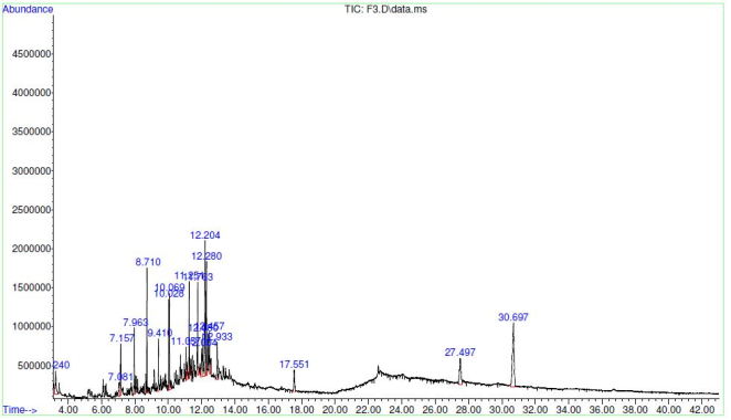
Figure 5. Gas Chromatogram of the MFCPL.
4. Discussion
Phytochemical screening of both crude extracts and fractions of Cleistopholis patens leaves revealed various amounts of steroids, terpenoids, glycosides, reducing sugars, alkaloids, total phenols, tannins and flavonoids. However, alkaloids were not detected in the chloroform fraction. The results of the phytochemical screening were consistent with those of previous studies . The antioxidant qualities of natural substances contribute to the protection of the stomach mucosa via radical scavenging processes. The results of this investigation are in line with those of Bhoumik et al., who reported that because phenolic compounds have the capacity to donate hydrogen, they are important compounds with scavenging properties . The flavonoid content was greater in the methanol fraction than in the ethyl acetate and other fractions. This result is in line with that of Liu et al., who suggested that the abundance of rich phytoconstituents such as tannins, flavonoids, and total polyphenols may be responsible for the gastroprotective effectiveness of the C. ignea aerial parts extract . Flavonoids can protect against stomach injury and are effective antioxidants in addition to maintaining capillary and mucus membrane integrity . Hence, the presence of high amounts of flavonoids in the MFCPL could contribute to its gastroprotective activities. Even at the greatest dosage, the Cleistopholis patens leaf methanol extract did not have any harmful effects. This finding is consistent with the findings of Onaolapo . Hence, the MFCPL is considered safe for human consumption at the dose used for the study.
Since reactive oxygen species play a role in the mechanism of ulcer induction, DPPH was used to test the antioxidant activities of Cleistopholis patens leaf extract and its various fractions. Additionally, their inhibitory effects on H+, K+-ATPase were measured. The DPPH scavenging activities of the crude methanol extract and different fractions of Cleistopholis patens leaves revealed that the methanol fraction had the highest DPPH scavenging activity, with an IC50 of 16.72 ± 2.47 μg/ml, followed by the ethyl acetate fraction, with an IC50 of 17.04 ± 0.79 μg/ml. However, these activities were less than the standard (ascorbic acid) IC50 of 3.09 μg/ml. The free radical scavenging activity of the MFCPL in this study was greater than that reported by Daniels and Temikotan , who reported 40% inhibition at 100 mg/ml. The DPPH scavenging activity of the MFCPL revealed that it can serve as an antioxidant and could contribute to the neutralization of free radicals, which are implicated in disease conditions, including ulcers. The ability of phenolics, such as flavonoids, polyphenols, tannins, and phenolic terpenes, to scavenge free radicals to provide antioxidant effects in plant products has been proven . Phenolic compounds have antioxidant effects because of their redox characteristics, which help dissolve peroxides and quench singlet and triplet oxygen. Viral infections, autoimmune illnesses, inflammation, and digestive system issues such as ulcers and gastrointestinal inflammation all seem to be caused by oxidative damage, which is currently thought to be the main mechanism underlying many neurological and other conditions that affect humans . The antioxidant capacity of MFCPL could contribute to increasing the excess reactive oxygen species, which have been implicated in ulcers . Hence, the high amounts of flavonoids found in the MFCPL might have played an important role in reducing the degree of ulceration in the gastric region of the treated animals in this study.
Hydrogen potassium ATPase in vitro inhibitory activities of the crude extract and fractions of Cleistopholis patens leaves revealed that the MFCPL had the highest H+, K+ - ATPase inhibitory activity, with an IC50 of 50.10 ± 10.52 μg/ml, necessitating its use for further analysis. The H+ and K+-ATPase inhibitory activities of the MFCPL were found to be comparable but greater than those reported by Onasanwo et al., who reported an IC50 of 43.8 μg/ml using Kolaviron from Garcinia kola Heckel .
The pH of gastric acid in the human stomach lumen is generally between 1.5 and 3.5, and it is controlled by the proton pump H+/K+ ATPase . On the other hand, overproduction of this enzyme may harm mucous membranes and result in stomach ulcers. The primary cause of gastric ulcers is the breakdown of the mucosal barrier, which consists of the mucosal coat and surface epithelium that borders the stomach. This damage could result from a decrease in mucus synthesis, an increase in stomach acid production, or a reduction in mucosal blood flow . The H+/K+ ATPase enzyme is known to transport H+ against a concentration gradient in stomach parietal cells. Additionally, the proton pump serves as the last and most common method for stimulating acid production . The H+/K+ ATPase inhibitory properties of naturally occurring phenolic compounds, including flavonoids and quercetin, have been established . These authors also reported that phenolic hydroxyl groups contribute to enzyme inhibition. The results of this study confirm the previous findings, which show that the MFCPL has a wide variety of potential applications in the treatment of gastrointestinal problems by decreasing the activity of H+/K+-ATPase.
The NSAID (preventive and curative) and acidified ethanol models of ulcer induction revealed that group 2 (ulcer-induced without treatment) had the highest total ulcer counts. These results confirmed that NSAIDs and alcohol disrupt the mucosal membrane of parietal cells, thereby causing gastric ulceration. These findings are in accordance with the findings of Bjarnason et al., who reported that all conventional NSAIDs have topical effects and inhibit cyclooxygenase 1 (COX1) and cyclooxygenase 2 (COX2), thereby preventing the synthesis of prostaglandin E2 (PGE2) . Similarly, Ketuly et al. confirmed that ethanol can lead to ulceration of the gastric mucosa when administered orally, which was in agreement with the findings of this study .
Pharmacological medicines that are frequently utilized include nonsteroidal anti-inflammatory medications such as diclofenac sodium. Because of the decrease in endogenous prostaglandin synthesis, which is known to be cytoprotective in the stomach mucosa, they cause gastric ulcers . Ethanol is a drug that contributes to more severe gastric ulcers because it promotes significant disturbances in the stomach mucosa . Acid "back diffusion," histamine release, sodium and potassium efflux, calcium influx, free radical production, leukotriene production, decreased gastric mucosal blood flow, decreased mucus production in the gastric lumen, decreased endogenous glutathione and prostaglandin levels, increased ischemia, and increased gastric vascular permeability are the mechanisms by which alcohol causes gastric ulcers . However, the MFCPL from this study mimics the activities of omeprazole, a proton pump inhibitor with accelerated gastric ulcer healing activity, as observed in the experimental group compared with the untreated animals.
In both models, the stomach volumes of the ulcer-induced nontreated animals were significantly greater (p < 0.05) than those of the treatment groups. The deposition of intraluminal hydrochloric acid in the stomach mucosa and increased gastric acid output could be the causes of the increase in stomach volume. According to earlier research, the primary aggressive agent causing stomach mucosal damage is gastric acid secretion . When the MFCPL was given to the treatment groups, their stomach volumes were significantly (p < 0.05) lower than those of the untreated animals. This could be because the MFCPL inhibits the activity of H+/K+ ATPase.
Compared with the treatment groups, group 2 (positive control) presented a substantial (p < 0.05) reduction in gastric pH. There is a chance that increased stomach acid output is the cause of this decrease in pH. These findings are comparable to those of previous reports . An imbalance between the defensive and aggressive components of the stomach mucosa results in gastric ulcers. The primary aggravating factor in gastric mucosal damage is gastric acid production . Compared with no MFCPL, MFCPL has the capacity to protect the gastric mucosa against ulceration due to an increase in the acidity of the gastric region since it decreased the acidity (increased pH) of the treated groups compared with the untreated animals. MFCPL decreased gastrointestinal acidity in both models, possibly because antihistamine medications, such as cimetidine, inhibit H2 receptors in the stomach and decrease the acidity of the gastric juice .
The main byproduct of lipid peroxidation that can be used to diagnose stomach damage caused by reactive oxygen species is malondialdehyde . Compared with those in the experimental groups treated with MFCPL, the concentrations of the lipid peroxidation marker, malondialdehyde (MDA) in the ulcer-induced animals (group 2) were significantly greater (p<0.05). Shetty et al. reported increased MDA concentrations upon the induction of ulcers via indomethacin, which was reduced upon the administration of Benicasa hispida extract, which was comparable to the findings of this study . Similar findings were also reported by AL-Wajeeh et al., who reported that the administration of Vitex pubescens leaf extract to ulcerated rats caused stomach mucosal injury provoked by ethanol, resulting in a decrease in MDA levels . The abundant flavonoid and phenolic components in the extract, which have the ability to absorb free radicals and cause lipid peroxidation, may explain the reduced amount of MDA in the groups treated with the MFCPL.
The stomach wall mucosa has been shown to be significantly defended by antioxidant enzymes from a variety of necrotic agents. Oxidative stress may play a significant role in the development and course of stomach ulcers . The cytotoxicity of free radicals to the gastrointestinal membrane decreases with increasing activity of antioxidant enzymes such as catalase (CAT), glutathione peroxidase (GPx), and superoxide dismutase (SOD) . In the present study, the induction of ulcers with NSAIDs (diclofenac sodium) led to a significant (p ≤ 0.05) decrease in the levels of all the antioxidant enzymes (SOD, CAT, GPx) in group 2 (ulcer-induced without treatment). Reactive oxygen species (ROS) produced during ulcer induction as a result of oxidative stress may be the cause of this decrease in antioxidant enzymes. Reactive oxygen species, such as the hydroxyl radical (·OH), hydrogen peroxide (H2O2), and superoxide anion radical (O2·), significantly affect the etiology of peptic ulcers .
The activity of antioxidant enzymes significantly (p < 0.05) increased in the experimental groups after they received the MFCPL. In line with the present study, there was an observable increase in the activities of antioxidant enzymes such as CAT and SOD in animals pretreated with V. pubescens extract compared with those in the ulcer control . The increase in the activities of glutathione peroxidase (GPx), catalase (CAT), and superoxide dismutase (SOD) may be related to the polyphenolic chemicals found in MFCPL, including flavonoids, alkaloids, and phenols. Researchers have discovered that phytochemicals, including flavonoids and phenolics, have free radical scavenging properties, which can help prevent peptic ulcer disease . The findings of this investigation are consistent with those of da Silva et al., who reported that citrus flavanone glycoside accelerates the healing of acetic acid-induced stomach ulcers in rats . Antioxidants are responsible for preventing the stomach mucosa from developing ulcers because they have the capacity to guard tissue against harm via a radical scavenging process .
This study revealed that, compared with no MFCPL, MFCPL has protective and therapeutic effects on the gastric region by increasing the level of antioxidant enzymes, with a corresponding reduction in the MDA concentration.
Twenty bioactive chemicals were identified by GC‒MS studies of MFCPLs, some of which may have contributed to the reported antiulcer efficacy of these substances. Some of the bioactive compounds identified in MFCPLs were also found in aqueous extracts of Lophira lanceolata leaves, which were proven to have antiulcer activity . Glycerine, which has also been identified in MFCPLs when combined with Alchemilla vulgaris (Lady's mantle), has been proven to have an antiulcer effect . Hence, the presence of glycerine in the MFCPLE might have played a vital role in its antiulcer activity. Esters of hexadecenoic acid have been reported to have antioxidant properties . This may have contributed to the antioxidant activities of the MFCPLs and thereby increased the number of free radicals, leading to ulcers. The antimicrobial and anticancer activities of hexadecanoic acid have been reported . Sudha et al. reported that hexadecenoic acid, a methyl ester, has hypocholesterolemic and anticoronative activities . The oleic acid identified in the present study has been reported to have anti-inflammatory, antiandrogenic, cancer preventive and dermatitigenic properties .
GC–MS analyses of various plant extracts revealed that most of the herbal extracts with medicinal properties contained some of the bioactive compounds present in the MFCPL. For example, octadecanoic acid has been reported to have antibacterial activity . Hence, MFCPLs taken orally might play a role in preventing ulcer progression in ulcer patients by inhibiting H. pylori. Traditional healers in Eastern Cape frequently utilize octadecanoic acid to cure diseases such as laryngitis, vaginal infections, TB, dysentery, and diabetes mellitus . The main antioxidant and antibacterial component extracted from Myristica fragrans (nutmeg) is tetradecanoic acid , as does the antibacterial agent from P. capillacea . Tetradecanoic acid also has hypocholesterolemic, nematocidal, and inhibitory effects on cell division. These therapeutic properties of the compounds identified in MFCPLE might have contributed immensely to their antiulcer activities as well as their ability to ameliorate biochemical aberrations associated with ulcers.
5. Conclusions
In the present study, MFCPL significantly inhibited the activity of proton pump ATPase, suggesting that it may have antigastric ulcer effects. This could be attributed to its rich bioactive components, such as polyphenolic compounds, coupled with other compounds with antioxidant capacity, as shown by the phytochemical and GC‒MS analyses. Hence, MFCPL can be used to manage ulcers and their associated biochemical aberrations.
Abbreviations

MFCPL

Methanol Fraction of Cleistopholis Patens Leaves

H. pylori

Helicobacter pylori

ROS

Reactive Oxygen Species

NSAID

Nonsteroidal Anti-inflammatory Drug

DPPH

2,2-diphenyl-1-picrylhydrazyl

CMEFCPL

Crude Methanol Extract And Fractions of Cleistopholis Patens Leaves

LD50

Median Lethal Dose

ANOVA

One-way Analysis of Variance

Acknowledgments
The authours acknowledge the efforts of Mr. Ikechukwu Anthony Agbo, Chief technologist of Faculty of Veterinary Medicine, University of Nigeria, Nsukka who gave technical support in handling the experimental animals.
Author Contributions
Nnamani Ikenna Ikenna: Conceptualization, Data curation, Formal Analysis, Investigation, Methodology, Resources, Software, Validation, Writing – original draft, Writing – review & editing
Parker Joshua: Conceptualization, Data curation, Formal Analysis, Investigation, Methodology, Project administration, Software, Supervision, Validation, Writing – original draft, Writing – review & editing
Ikechukwu Okoro: Data curation, Formal Analysis, Investigation, Methodology, Validation, Writing – original draft, Writing – review & editing
Innocent Okagu: Conceptualization, Data curation, Formal Analysis, Investigation, Methodology, Project administration, Validation, Writing – original draft, Writing – review & editing
Victor Ogugua: Conceptualization, Data curation, Formal Analysis, Funding acquisition, Investigation, Methodology, Project administration, Software, Supervision, Validation, Writing – original draft, Writing – review & editing
Funding
This work is not supported by any external funding.
Data Availability Statement
The data is available from the corresponding author upon reasonable request.
Conflicts of Interest
The authors declare no conflicts of interest.
References
[1] Siddique O, Ovalle A, Siddique AS, Moss SF. Helicobacter pylori infection: an update for the internist in the age of increasing global antibiotic resistance. Am. J. Med. 2018; 131(5): 473-479. Available at:
[2] Hooi JK, Lai WY, Ng WK, Suen MM, Underwood FE, Tanyingoh D, Malfertheiner P, Graham DY, Wong VW, Wu JC, Chan FK. Global prevalence of Helicobacter pylori infection: systematic review and meta-analysis. Gastroenterology. 2017; 153(2): 420-429. Available at:
[3] Okechukwu PN, Ekeuku SO, Sharma M, Nee CP, Chan HK, Mohamed N, Froemming GR. In vivo and in vitro antidiabetic and antioxidant activity of spirulina. Pharmacogn. Mag. 2019; 15 (Suppl 1): S17-S29. Available at:
[4] Narayanan M, Reddy KM, Marsicano E. Peptic ulcer disease and Helicobacter pylori infection. Missouri medicine. 2018; 115(3): 219. Available at:
[5] Liu B, Feng X, Zhang J, Wei Y, Zhao X. Preventive effect of Anji white tea flavonoids on alcohol-induced gastric injury through their antioxidant effects in Kunming mice. Biomolecules. 2019 4; 9(4): 137. Available at:
[6] Zhernakova A, Kurilshikov A, Bonder MJ, Tigchelaar EF, Schirmer M, Vatanen T, Mujagic Z, Vila AV, Falony G, Vieira-Silva S, Wang J. Population-based metagenomics analysis reveals markers for gut microbiome composition and diversity. Science. 2016 29; 352(6285): 565-569. Available at:
[7] Marieb E, Keller S. Essentials of human anatomy and physiology. Harlow: Pearson Education, Limited, 2021. Available at:
[8] Spechler SJ. Proton pump inhibitors: what the internist needs to know. Medical Clinics. 2019; 103(1): 1-14. Available at:
[9] Palle S, Kanakalatha A, Kavitha CN. Gastroprotective and antiulcer effects of Celastrus paniculatus seed oil against several gastric ulcer models in rats. J. Diet. Suppl. 2018; 15(4): 373-385. Available at:
[10] Onaolapo AJ. Entomotoxic activity of powder of Cleistopholis patens benth against the indian meal moth, plodia interpunctella (hübner) (lepidoptera: pyralidae). Int. J. Entomol. Res. 2017; 5(2): 33-42. Available at:
[11] Udem SC, Ezeonuegbu UC, Obidike RI. Experimental studies on the hypolipidemic and haematological properties of aqueous leaf extract of Cleistopholis patens Benth. & Diels. (Annonacae) in hypercholesterolemic rats. Ann Med Health Sci Res. 2011; 1(1): 115-122. Available at:
[12] Ouattara ZA, Yapi TA, Békro YA, Mamyrbékova-Békro AJ, Paoli M, Tomi P, Casanova J, Bighelli A, Tomi F. Composition and chemical variability of root bark oil from ivoirian Cleistopholis patens. Nat. Prod. Commun. 2018; 13(6): 6-9. Available at:
[13] Still WC, Kahn M, Mitra A. Rapid chromatographic technique for preparative separations with moderate resolution. J. Org. Chem. 1978; 43(14): 2923-2925. Available at:
[14] Harborne JB. (1973). Phytochemical methods: a guide to modern techniques of plant analysis. Dordrecht, Springer Netherlands; 1973. Available at:
[15] Braca A, De Tommasi N, Di Bari L, Pizza C, Politi M, Morelli I. Antioxidant principles from bauhinia tarapotensis. J. Nat Prod. 2001; 64(7): 892-895. Available at:
[16] Reyes-Chilpa R, Baggio CH, Alavez-Solano D, Estrada-Muñiz E, Kauffman FC, Sanchez RI, Mesia-Vela S. Inhibition of gastric H+, K+-ATPase activity by flavonoids, coumarins and xanthones isolated from Mexican medicinal plants. J. Ethnopharmacol. 2006; 105(1-2): 167-172. Available at:
[17] Lorke D. Determination of acute toxicity. Arch Toxicol. 1983; 53: 275-279.
[18] ‌Guth PH, Aures D, Paulsen G. Topical aspirin plus HCl gastric lesions in the rat: cytoprotective effect of prostaglandin, cimetidine, and probanthine. Gastroenterology. 1979; 76(1): 88-93. Available at:
[19] Mózsik G, Moron F, Jávor T. Cellular mechanisms of the development of gastric mucosal damage and of gastrocytoprotection induced by prostacyclin in rats. A pharmacological study. Prostaglandins, Leukotrienes and Medicine. 1982 1; 9(1): 71-84. Available at:
[20] Amr AR, Maysa ME. Anti-ulcer effect of cinnamon and chamomile aqueous extracts in rat models. J Am Sci. 2010; 6(12): 209-216. Available at:
[21] Ketuly KA, Abdulla MA, Hadi HA, Mariod AA, Abdel-Wahab SI. Anti-ulcer activity of the 9alpha-bromo analogue of Beclomethasone dipropionate against ethanol-induced gastric mucosal injury in rats. J Med Plants Res. 2011; 5(4): 514-20. Available at:
[22] Durak I, Canbolat O, Kavutçu M, Öztürk HS, Yurtarslani Z. Activities of total, cytoplasmic, and mitochondrial superoxide dismutase enzymes in sera and pleural fluids from patients with lung cancer. J. Clin. Lab. Anal. 1996; 10(1): 17-20. Available at:
[23] Aebi HE. Catalase. In: Bergmeyer HU, editor. Methods of enzymatic analysis. Weinhem, Verlag Chemie; 1983. p. 273–286.
[24] Paglia DE, Valentine WN. Studies on the quantitative and qualitative characterization of erythrocyte glutathione peroxidase. J Lab Clin Med. 1967; 70(1): 158-69. Available at:
[25] Wallin B, Rosengren B, Shertzer HG, Camejo G. Lipoprotein oxidation and measurement of thiobarbituric acid reacting substances formation in a single microtiter plate: its use for evaluation of antioxidants. Anal. Biochem. 1993; 208(1): 10-15. Available at:
[26] Osuntokun OT. Evaluation of Inhibitory Zone Diameter (IZD), phytochemical screening, elemental composition and proximate analysis of crude cleistopholis patens (Benth) on infectious clinical isolates. J Mol Biomark Diagn. 2018; 9(385): 2. Available at:
[27] Daniels A, Temikotan T. Fatty acid profile, antioxidant and antibacterial effect of the ethyl acatate extract of cleistopholis patens. Bull. Sci. Res. 2021; 3: 21-31. Available at:
[28] Bhoumik D, Masresha B, Mallik A. Antiulcer properties of herbal drugs: a review. Int J Biomed Res. 2017; 8(3): 116-24. Available at:
[29] Harborne, J. B., & Williams, C. A. (2000). Advances in flavonoid research since 1992. Phytochem. 55(6), 481-504. Available at: http://dx.doi.org/10.1016/S0031-9422(00)00235-1
[30] Rahman MA, Moon SS. Antioxidant polyphenol glycosides from the plant Draba nemorosa. Bull. Korean Chem. Soc. 2007; 28(5): 827-31. available at:
[31] Aruoma OI. Methodological considerations for characterizing potential antioxidant actions of bioactive components in plant foods. Mutat. Res. - Fundam. Mol. Mech. Mutagen. 2003; 523: 9-20. Available at:
[32] Abd El-Dayem AM, El-Agaimy MA. Nonwood forest products, physico-chemical characteristics and fatty acid composition of three seed oils. In WOCMAP I-Medicinal and Aromatic Plants Conference: part 1 of 4 333 1992 Jul 19 (pp. 287-294). Available at:
[33] Onasanwo SA, Singh N, Olaleye SB, Palit G. Anti-ulcerogenic and proton pump (H+, K+ ATPase) inhibitory activity of Kolaviron from Garcinia kola Heckel in rodents. Indian J. Exp. Biol. 2011; 49: 461-468. Available at:
[34] Murakami S, Arai I, Muramatsu M, Otomo S, Baba K, Kido T, Kozawa M. Inhibition of gastric H+, K+-ATPase and acid secretion by cassigarol A, a polyphenol from Cassia garrettiana Craib. Biochem. Pharmacol. 1992; 44(1): 33-37. Available at:
[35] Bjarnason I, Scarpignato C, Holmgren E, Olszewski M, Rainsford KD, Lanas A. Mechanisms of damage to the gastrointestinal tract from nonsteroidal anti-inflammatory drugs. Gastroenterology. 2018; 154(3): 500-14. Available at:
[36] Robert A, Nezamis JE, Lancaster C, Hanchar AJ. Cytoprotection by prostaglandins in rats: Prevention of gastric necrosis produced by alcohol, HCl, NaOH, hypertonic NaCl, and thermal injury. Gastroenterology. 1979; 77(3): 433-43. Available at:
[37] Hiruma-Lima CA, Batista LM, de Almeida AB, de Pietro Magri L, dos Santos LC, Vilegas W, Brito AR. Antiulcerogenic action of ethanolic extract of the resin from Virola surinamensis Warb. (Myristicaceae). J. Ethnopharmacol. 2009; 122(2): 406-9. Available at:
[38] Glavin GB, Szabo S. Experimental gastric mucosal injury: laboratory models reveal mechanisms of pathogenesis and new therapeutic strategies. The FASEB journal. 1992; 6(3): 825-831. Available at:
[39] Bento EB, de Brito Júnior FE, de Oliveira DR, et al. Antiulcerogenic activity of the hydroalcoholic extract of leaves of Annona muricata Linnaeus in mice. Saudi J. Biol. Sci. 2018; 25(4): 609-621 available at:
[40] Abebaw M, Mishra B, Gelayee DA. Evaluation of anti-ulcer activity of the leaf extract of Osyris quadripartita Decne. (Santalaceae) in rats. J. Exp. Pharmacol. 2017; 16: 1-1. Available at:
[41] Eswaran MB, Surendran S, Vijayakumar M, Ojha SK, Rawat AK, Rao CV. Gastroprotective activity of Cinnamomum tamala leaves on experimental gastric ulcers in rats. J. Ethnopharmacol.. 2010; 128(2): 537-540. Available at:
[42] Kwiecien S, Brzozowski T, Konturek SJ. Effects of reactive oxygen species action on gastric mucosa in various models of mucosal injury. J Physiol Pharmacol. 2002; 53(1): 39-50. Available at:
[43] Shetty BV, Arjuman A, Jorapur A, Samanth R, Yadav SK, Valliammai N, Tharian AD, Sudha K, Rao GM. Effect of extract of Benincasa hispida on oxidative stress in rats with indomethacin induced gastric ulcers. Indian J Physiol Pharmacol. 2008; 52(2): 178-182. Available at:
[44] Saeed AL-Wajeeh N, Halabi MF, Hajrezaie M. et al. Correction: the gastroprotective effect of vitex pubescens leaf extract against ethanol-provoked gastric mucosal damage in sprague-dawley rats. Plos one. 2017; 12(5): e0179072. Available at:
[45] Hajrezaie M, Golbabapour S, Hassandarvish P, et al. Acute toxicity and gastroprotection studies of a new schiff base derived copper (II) complex against ethanol-induced acute gastric lesions in rats. PloS one. 2012; 7(12): e51537. Available at:
[46] Tuluce Y, Ozkol H, Koyuncu I, Ine H. Gastroprotective effect of small centaury (Centaurium erythraea L) on aspirin-induced gastric damage in rats. Toxicol. Ind. Health. 2011; 27(8): 760-768. Available at:
[47] Ighodaro OM, Akinloye OA. First line defence antioxidants-superoxide dismutase (SOD), catalase (CAT) and glutathione peroxidase (GPX): Their fundamental role in the entire antioxidant defence grid. Alex. J. Med. 2018; 54(4): 287-293. Available at:
[48] Karaoğlan ES, Albayrak A, Kutlu Z, Bayır Y. Gastroprotective and antioxidant effects of Eremurus spectabilis Bieb. methanol extract and its isolated component isoorientin on indomethacin induced gastric ulcers in rats. Acta Cir. Bras. 2018; 33: 609-618. Available at:
[49] da Silva LM, Pezzini BC, Somensi LB et al. Hesperidin, a citrus flavanone glycoside, accelerates the gastric healing process of acetic acid-induced ulcer in rats. Chem Biol Interact. 2019; 308: 45-50. Available at:
[50] Tachakittirungrod S, Okonogi S, Chowwanapoonpohn S. Study on antioxidant activity of certain plants in Thailand: Mechanism of antioxidant action of guava leaf extract. Food Chem. 2007; 103(2): 381-8. Available at:
[51] Boniface MT, Ngozi OE, Eiza YP, Mathew AO, Adejoh IP. Gas Chromatography‒Mass Spectrometry (GC‒MS) Profiling and Anti-ulcer Activity of the Aqueous Extract of Lophira Lanceolata Leaves. Int. J. Adv. Res. Biol. Sci. 2020; 7(8): 48-55. Available at:
[52] Shrivastava R, John GW. Treatment of aphthous stomatitis with topical Alchemilla vulgaris in glycerine. Clin. Drug Investig. 2006; 26: 567-573. Available at:
[53] Mamani R, Alhaji NM. GC‒MS analysis of phytocomponents in methanolicextract of Coleus aromaticus. Journal of Pharmacognosy and Phytochemistry. 2019; 8(4): 106-109. Available at:
[54] Klavina L, Springe G, Nikolajeva V, Martsinkevich I, Nakurte I, Dzabijeva D, Steinberga I. Chemical composition analysis, antimicrobial activity and cytotoxicity screening of moss extracts (moss phytochemistry). Molecules. 2015; 20(9): 17221-17243. Available at:
[55] Sudha T, Chidambarampillai S, Mohan VR. GC‒MS analysis of bioactive components of aerial parts of Fluggea leucopyrus Willd. (Euphorbiaceae). J. Appl. Pharm. Sci. 2013; 3(5): 126-130. Available at:
[56] Okoro I, Joshua P, Okagu I, Amah C, Ikenna N, Chiaka-Onyemeze N, Anaduaka E, Obasi D, Okoroh P, Orieke D, Ogugua V. Proximate analyses, in vitro antioxidant activity and GC-MS analyses of fermented Pentaclethra macrophylla seeds. Life Research Journal. 2024; 7(3): 12. Available at:
[57] El-Din SM, El-Ahwany AM. Bioactivity and phytochemical constituents of marine red seaweeds (Jania rubens, Corallina mediterranea and Pterocladia capillacea). J Taibah Univ Sci. 2016; 10(4): 471-484. Available at:
[58] Omoruyi BE, Afolayan AJ, Bradley G. Chemical composition profiling and antifungal activity of the essential oil and plant extracts of Mesembryanthemum edule (L.) bolus leaves. Afr. J. Tradit. Complement. Altern. Med. 2014; 11(4): 19-30. Available at:
[59] Narasimhan B, Dhake AS. Antibacterial principles from Myristica fragrans seeds. Journal of medicinal food. 2006 Sep 1; 9(3): 395-399. Available at:
[60] Gupta AD, Bansal VK, Babu V, Maithil N. Chemistry, antioxidant and antimicrobial potential of nutmeg (Myristica fragrans Houtt). J. Genet. Eng. & Biotechnol. 2013; 11(1): 25-31. Available at:
[61] Agoramoorthy G, Chandrasekaran M, Venkatesalu V, Hsu MJ. Antibacterial and antifungal activities of fatty acid methyl esters of the blind-your-eye mangrove from India. Braz. J. Microbiol. 2007; 38: 739-742. Available at:
Cite This Article
  • APA Style

    Ikenna, N., Joshua, P., Okoro, I., Okagu, I., Ogugua, V. (2025). Gastroprotective and Antigastric Ulcer Effects of the Methanol Fraction of Cleistopholis Patens Leaves on Two Ulcer Models in Rats. Advances in Biochemistry, 13(3), 61-78. https://doi.org/10.11648/j.ab.20251303.11

    Copy | Download

    ACS Style

    Ikenna, N.; Joshua, P.; Okoro, I.; Okagu, I.; Ogugua, V. Gastroprotective and Antigastric Ulcer Effects of the Methanol Fraction of Cleistopholis Patens Leaves on Two Ulcer Models in Rats. Adv. Biochem. 2025, 13(3), 61-78. doi: 10.11648/j.ab.20251303.11

    Copy | Download

    AMA Style

    Ikenna N, Joshua P, Okoro I, Okagu I, Ogugua V. Gastroprotective and Antigastric Ulcer Effects of the Methanol Fraction of Cleistopholis Patens Leaves on Two Ulcer Models in Rats. Adv Biochem. 2025;13(3):61-78. doi: 10.11648/j.ab.20251303.11

    Copy | Download

  • @article{10.11648/j.ab.20251303.11,
      author = {Nnamani Ikenna and Parker Joshua and Ikechukwu Okoro and Innocent Okagu and Victor Ogugua},
      title = {Gastroprotective and Antigastric Ulcer Effects of the Methanol Fraction of Cleistopholis Patens Leaves on Two Ulcer Models in Rats
    },
      journal = {Advances in Biochemistry},
      volume = {13},
      number = {3},
      pages = {61-78},
      doi = {10.11648/j.ab.20251303.11},
      url = {https://doi.org/10.11648/j.ab.20251303.11},
      eprint = {https://article.sciencepublishinggroup.com/pdf/10.11648.j.ab.20251303.11},
      abstract = {Background: Cleistopholis patens leaves are used to treat stomach disorders in Nigeria. This research investigated the gastroprotective and antigastric ulcer effects of the methanol fraction of Cleistopholis patens leaves (MFCPL) in rats. Methods: In vitro experiments involved DPPH-radical scavenging and H+/K+-ATPase inhibitory activities. Two ulcer models, diclofenac sodium (DS) (using 50 mg/kg b.w. DS orally) and acidified ethanol (AE) (using 0.3 M HCl + 60% ethanol orally), were used. The experimental rats (n=24) were grouped into 6 of 4 groups for each of the DS and AE ulcer models. In the DS model, group 1 served as the normal group, whereas group 2 was ulcer-induced without treatment. Group 3 received 20 mg/kg b.w. omeprazole, whereas groups 4, 5 and 6 received 50, 100 and 200 mg/kg b.w. MFCPL, respectively, for 14 days. For the AE ulcer model, 7 days after pretreatment with MFCPL, all groups except group 1 received acidified ethanol and were sacrificed after 1 hr. The ulcer indices, gastric mucosa morphology and serum antioxidant status of the rats were evaluated. Additionally, GC‒MS analyses of MFCPL were carried out. Results: The MFCPL had H+, K+-ATPase-inhibiting and DPPH-radical-scavenging activities, with half-maximal inhibitory concentrations (IC50) of 50.10 ± 10.52 μg/ml and 16.72 ± 2.47 μg/ml, respectively. Compared with the ulcer control, the administration of MFCPL led to a significant (p Conclusion: MCPL has an antigastric ulcer effect attributable to its H+/K+-ATPase inhibition and antioxidant activities due to its rich bioactive components and hence can be used to manage ulcers and their associated biochemical aberrations.
    },
     year = {2025}
    }
    

    Copy | Download

  • TY  - JOUR
    T1  - Gastroprotective and Antigastric Ulcer Effects of the Methanol Fraction of Cleistopholis Patens Leaves on Two Ulcer Models in Rats
    
    AU  - Nnamani Ikenna
    AU  - Parker Joshua
    AU  - Ikechukwu Okoro
    AU  - Innocent Okagu
    AU  - Victor Ogugua
    Y1  - 2025/07/04
    PY  - 2025
    N1  - https://doi.org/10.11648/j.ab.20251303.11
    DO  - 10.11648/j.ab.20251303.11
    T2  - Advances in Biochemistry
    JF  - Advances in Biochemistry
    JO  - Advances in Biochemistry
    SP  - 61
    EP  - 78
    PB  - Science Publishing Group
    SN  - 2329-0862
    UR  - https://doi.org/10.11648/j.ab.20251303.11
    AB  - Background: Cleistopholis patens leaves are used to treat stomach disorders in Nigeria. This research investigated the gastroprotective and antigastric ulcer effects of the methanol fraction of Cleistopholis patens leaves (MFCPL) in rats. Methods: In vitro experiments involved DPPH-radical scavenging and H+/K+-ATPase inhibitory activities. Two ulcer models, diclofenac sodium (DS) (using 50 mg/kg b.w. DS orally) and acidified ethanol (AE) (using 0.3 M HCl + 60% ethanol orally), were used. The experimental rats (n=24) were grouped into 6 of 4 groups for each of the DS and AE ulcer models. In the DS model, group 1 served as the normal group, whereas group 2 was ulcer-induced without treatment. Group 3 received 20 mg/kg b.w. omeprazole, whereas groups 4, 5 and 6 received 50, 100 and 200 mg/kg b.w. MFCPL, respectively, for 14 days. For the AE ulcer model, 7 days after pretreatment with MFCPL, all groups except group 1 received acidified ethanol and were sacrificed after 1 hr. The ulcer indices, gastric mucosa morphology and serum antioxidant status of the rats were evaluated. Additionally, GC‒MS analyses of MFCPL were carried out. Results: The MFCPL had H+, K+-ATPase-inhibiting and DPPH-radical-scavenging activities, with half-maximal inhibitory concentrations (IC50) of 50.10 ± 10.52 μg/ml and 16.72 ± 2.47 μg/ml, respectively. Compared with the ulcer control, the administration of MFCPL led to a significant (p Conclusion: MCPL has an antigastric ulcer effect attributable to its H+/K+-ATPase inhibition and antioxidant activities due to its rich bioactive components and hence can be used to manage ulcers and their associated biochemical aberrations.
    
    VL  - 13
    IS  - 3
    ER  - 

    Copy | Download

Author Information
  • Abstract
  • Keywords
  • Document Sections

    1. 1. Introduction
    2. 2. Materials and Methods
    3. 3. Results
    4. 4. Discussion
    5. 5. Conclusions
    Show Full Outline
  • Abbreviations
  • Acknowledgments
  • Author Contributions
  • Funding
  • Data Availability Statement
  • Conflicts of Interest
  • References
  • Cite This Article
  • Author Information