Abstract
Background: Anogenital distance (AGD) is a hormone dependent anatomical landmark that serves as a measure of perineal growth. Previous studies have shown that it is shorter in children with undescended testis (UDT) when compared with those with normal external genitalia (NEG). In sub-Saharan Africa, however, there is paucity of information regarding the relationship between AGD and UDT, hence the need for this study. Objective: To determine whether AGD parameters are shorter in children with UDT when compared to those with normal external genitalia. Method: Three AGD parameters were measured using a digital caliper in 86 children (43 with UDT and 43 with normal external genitalia). These parameters include anoscrotal distance (ASD), anopenal distance 1 (APD1) and anopenal distance 2 (APD2). Data was collected over a 12-month period and analyzed using Statistical Package for Social Sciences (IBM SPSS) version 25 software. Results: Forty-three (43) boys with UDT were matched against the same number of boys with normal external genitalia in our study. There were no statistically significant differences among the two groups in the mean age, height, weight, body-mass index (BMI), gestational age and birth weight. The mean anoscrotal distance (ASD) and ASD index were 39.05±8.14 mm and 2.23±1.17mm/kg for the test group and 44.92±7.81mm and 2.79±1.07mm/kg for the control group respectively. The differences in the mean ASD and the mean ASD index between the test and the control groups were statistically significant with p < 0.05. The mean anopenile distance 1 (APD1) and anopenile distance 2 (APD2) were 77.33±13.09mm and 96.20±8.49mm for the test group and 80.63±10.15 mm and 97.48±12.86mm for the control group respectively. There were no statistically significant differences in the APD1 and APD2 between the 2 groups. Conclusion: The study showed that boys with UDT had consistently shorter AGD parameters than those with well descended testis.
Keywords
Anogenital Distance, Anogenital Distance Index, Digital Caliper, Normal External Genitalia, Undescended Testis
1. Introduction
Anogenital distance (AGD) is the distance from the centre of the anus to the junction between the smooth perineal skin and the rugated skin of the scrotum or to the anterior or posterior border of the base of penis in males. In females it is the distance from the centre of the anus to the posterior convergence of the fourchette or base of clitoris
| [1] | Thankamony A, Lek N, Carroll D, Williams M, Dunger DB, Acerini CL, Ong KK, Hughes IA. Anogenital distance and penile length in infants with hypospadias or cryptorchidism: comparison with normative data. Environ Health Perspect. 2014; 122(2): 207-210. https://doi.org/10.1289/ehp.1307178 |
| [2] | Salazar-Martinez E, Romano-Riquer P, Yanez-Marquez E, Longnecker MP, Hernandez-Avila M. Anogenital distance in human male and female newborns: a descriptive, cross-sectional study. Environ Health. 2004; 3(1): 8-13. https://doi.org/10.1186/1476-069X-3-8 |
[1, 2]
. Anogenital distance is a sexually dimorphic biomarker that is normally longer in males than in females due to the difference on exposure and effect of testosterone during embryonic development. It is a marker of perineal growth and forms from the growth and development of genital tubercle, genital swellings and cloacal membrane
| [1] | Thankamony A, Lek N, Carroll D, Williams M, Dunger DB, Acerini CL, Ong KK, Hughes IA. Anogenital distance and penile length in infants with hypospadias or cryptorchidism: comparison with normative data. Environ Health Perspect. 2014; 122(2): 207-210. https://doi.org/10.1289/ehp.1307178 |
| [2] | Salazar-Martinez E, Romano-Riquer P, Yanez-Marquez E, Longnecker MP, Hernandez-Avila M. Anogenital distance in human male and female newborns: a descriptive, cross-sectional study. Environ Health. 2004; 3(1): 8-13. https://doi.org/10.1186/1476-069X-3-8 |
[1, 2]
.
The development of the male external genitalia and AGD is an androgen dependent process, and the amount of androgen available for the growth of foetal tissues can be interrupted by some environmental chemicals acting as endocrine disrupters. This can lead to testicular dysgenesis syndrome that may be marked by the presence of undescended testis (UDT), hypospadias and reduced AGD in childhood; testicular cancer and infertility in adults
. The severity of the genital anomaly depends on the extent of the hormone disruption by these chemicals and it varies from individual to individual; and from one region to the other
| [5] | Toppari J, Virtanen HE, Main KM, Skakkebaek NE. Cryptorchidism and hypospadias as a sign of testicular dysgenesis syndrome (TDS): environmental connection. Birth Defects Res A Clin Mol Teratol. 2010; 88(10): 910-917. https://doi.org/10.1002/bdra.20707 |
| [6] | Boisen KA, Chellakooty M, Schmidt IM, Kai CM, Damgaard IN, Suomi AM et al. Hypospadias in a cohort of 1072 Danish newborn boys: prevalence and relationship to placental weight, anthropometrical measurements at birth, and reproductive hormone levels at three months of age. J Clin Endocrinol Metab. 2005; 90(7): 4041-4046. https://doi.org/10.1210/jc.2005-0302 |
[5, 6]
.
Undescended testis occurs when one or both testes fail to migrate to the floor of the testis after birth
| [7] | Madden NP. Testis hydrocoele and varicocoele. In: Thomas DFM, Duffy PG, Rickwood AMK, eds. Essentials of Paediatric Urology. 2nd ed. London: Informa Healthcare; 2008. 247-256. |
| [8] | Hutson JM. Undescended testis, torsion, and varicocele. In: Grosfeld JL, O’Neill JA, Coran AG, Fonkalsrud EW, Caldamone AA, eds. Pediatric Surgery. 6th ed. Vol 1. Philadelphia: Mosby Elsevier; 2006. 1193-1205. |
| [9] | Lee JJ, Dairiki Shortliffe LM. Undescended testes and testicular tumors. In: Holcomb GW. Murphy JP, Ostlie DJ, St. Peter SD eds. Aschcraft’s Pediatric Surgery. 6th ed. New York: Elsevier Saunders; 2014. 689-699. |
[7-9]
. Investigations have shown that infants exposed to environmental endocrine disrupting agents during intrauterine life may develop shortened AGD as well as UDT
. Many authors in Western countries have demonstrated that male caucasian patients with UDT have decreased AGD when compared to the normal population
| [1] | Thankamony A, Lek N, Carroll D, Williams M, Dunger DB, Acerini CL, Ong KK, Hughes IA. Anogenital distance and penile length in infants with hypospadias or cryptorchidism: comparison with normative data. Environ Health Perspect. 2014; 122(2): 207-210. https://doi.org/10.1289/ehp.1307178 |
| [10] | Jain VG, Singal AK. Shorter anogenital distance correlates with undescended testis: a detailed genital anthropometric analysis in human newborns. Hum Reprod. 2013; 28(9): 2343-2349. https://doi.org/10.1093/humrep/det286 |
| [11] | Jiang DP, Geng HQ, Lin HW, Yu XN, Zhang XW, Yang SL et al. Relationship between anogenital distance and cryptorchidism in human newborns. Zhonghua Nan Ke Xue 2015; 21(5): 432-435. |
| [12] | Hua XG, Hu R, Hu CY, Li FL, Jiang W, Zhang XJ. Associations between hypospadias, cryptorchidism and anogenital distance: Systematic review and meta-analysis. Andrologia. 2018; 50(10): 1-8. https://doi.org/10.1111/and.13152 |
[1, 10-12]
. From the literature review, the only documented study in sub-Saharan Africa was by Onyiruka et al
| [13] | Onyiriuka AN, Ikeany EM. Association between anogenital distance and external genital anomalies in Nigerian male newborn infants. NJOG. 2018; 12(2): 11-14. https://doi.org/10.3126/njog.v12i2.19942 |
[13]
. They compared AGD of 34 newborn infants with normal external genitalia with 11 newborn infants with UDT in South-South Nigeria. They observed too that AGD was shorter in children with UDT when compared with those with NEG. It was due to the paucity of information regarding the nature of AGD in children with UDT and those with NEG in sub-Saharan Africa that we were prompted to conduct this study. The purpose of the present study is to determine if AGD parameters are shorter in children with UDT when compared with those with normal external genitalia in South-Eastern, Nigeria.
2. Patients and Methods
This was a prospective comparative study carried out at the Alex Ekwueme Federal University Teaching Hospital, Abakaliki (AEFUTHA) and two schools with nursery and primary school services (Egugwu Agbaja Primary School and Ezikworo Primary School), all in Abakaliki, Ebonyi State, South-Eastern Nigeria. The study was carried out over a twelve (12)-month period.
Ethical clearance for the study with a reference number FETHA/REC/VOL2/2019/145 was obtained from the Hospital Research and Ethics Committee (HREC) of AEFUTHA. Approval was obtained from Ebonyi State Universal Basic Education Board, Abakaliki Local Government Education Authority and the School Management Boards to enroll their pupils. Informed consent was obtained from the parents or guardians of the participants.
Patients for the test group (UDT) were recruited by total population sampling method whereby all children with UDT who presented at the PSOP clinic that met the inclusion criteria were enrolled on the study. All the children with UDT were evaluated clinically and with groin ultrasonography to determine the location of the testes; and were grouped based on their age into 3 groups: ≤ 3 years, 4 to 7 years and ≥ 8 years.
The control group consisted of age-matched male children with normal genitalia from two primary and nursery schools. The children were stratified into three age groups; ≤ 3 years, 4 to 7 years and ≥ 8 years. From each age group, the required number of children were selected by simple random sampling without replacement, until the sample size was attained.
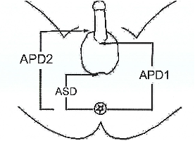
Using a digital caliper (Stainless steel electronic digital caliper, Original Equipment Manufacturer (OEM), Hong Kong) that reads in increments of 0.01mm, and with the patients in supine and frog-leg position, the AGD were measured. Three AGD parameters (
Figure 1) were measured viz: Anoscrotal Distance (ASD distance from the centre of the anus to the junction between the smooth perineal skin and the rugated skin of the scrotum), Anopenile Distance 1 (APD1; distance from the centre of the anus to the posterior border of the base of the penis), and Anopenile Distance 2 (APD2 distance from the centre of the anus to the anterior border of the base of the penis). Two researchers took the same measurements and the average for each measurement was calculated and used for analysis. Anogenital index (AGI) – AGD/patient’s weight (mm/kg) was calculated to control any effect of variation in body size on AGD.
A proforma was used for the data collection. Data collected include biodata, weight, height, birth weight, gestational age at birth, diagnosis, clinical and ultrasound location of the testis, AGD and AGI (AGD/patient’s weight).
2.3. Statistical Analysis
Data entry and analysis were done with the Statistical Package for Social Sciences (IBM; SPSS, Chicago, IL, USA) version 25 software. Descriptive statistics of socio-demographic and other background characteristics were done. Categorical variables were summarized with frequencies and proportions while the mean and standard deviation of continuous variables were presented.
The relationship between presence or absence of UDT (dependent variable) and AGD (independent variable) among the study subjects (test group and control group) was tested using the student t-test. Odds ratio with a 95% Confidence Interval was calculated. Significance level was set at 0.05.
3. Results
The mean age of the subjects with UDT was 5.43±3.97 years (0.5 to 13.0 years) while that of the participants with normal external genitalia was 6.16±3.59 years (0.5 to 13 years). There was no statistically significant difference in age between the 2 groups (p = 0.585). The mean height, weight and BMI for test group were 1.12±0.24m, 21.51±9.30Kg and 15.33±2.12Kg/m2 as against 1.15±.02m, 18.48±7.49Kg and 15.54±2.63Kg/m2 for the control group respectively. The mean GA for the test group was 38.77±1.34 weeks while that for the control group was 38.33±1.11weeks. The mean birth weight for the test group was 3.20±0.51Kg and 3.18±0.50Kg for the control group. There were no significant differences noted among the 2 groups in terms of the mean height, weight, body–mass index (BMI), gestational age and birth weight.
3.1. Comparison of Anogenital Distance Parameters Between the Two Groups
The mean anoscrotal distance (ASD) was 39.05±8.14 mm for the test group and 44.85±7.81 mm for the control group (p = 0.001). There were no significant differences in APD1 and APD2 between the 2 groups. The difference in anoscrotal index between the two groups was statistically significant (p = 0.022). There were no significant differences in anopenile index 1 and 2 between the test and the control groups (
Table 1).
3.2. Comparison of Anogenital Distance Parameters with Age Group of Participants in the Control Group
The mean ASD, APD1 and APD2 for the control group increased with increase in age across the 3 age groups of ≤ 3 years, 4 to 7 years and ≥ 8 years. The mean ASD for the three age groups were 40.63±4.64mm, 45.32±8.82mm and 48.33±7.81mm respectively. There was a significant difference in mean ASD between the age groups (p = 0.002). The mean APD1 were 71.89±.9.95mm for those ≤3 years, 84.67±6.38mm for those 4-7 years and 85.08±8.36mm for those ≥ 8 years (p = 0.000). The mean APD2 were 93.22±6.90mm, 98.08±19.322mm and 100.71±9.75mm. There was no significant difference in mean APD2 between the age group.
3.3. Comparison of Anogenital Distance Parameters with Age Group of Participants in the Test Group
The mean ASD, APD1 and APD2 for the test group showed no regular pattern across the 3 age groups of ≤ 3 years, 4 to 7 years and ≥ 8 years. The mean ASD for the 3 age groups were 36.97±10.56mm, 42.03±5.07mm and 38.96±5.93mm respectively. The mean APD1 were 69.39±14.07mm, 85.85±8.21mm and 79.98±8.84mm while the mean APD2 were 92.95±9.55mm, 99.48±4.74 and 97.53±8.82mm respectively. There were no significant differences in mean ASD, APD1 and APD2 between the 3 age groups in the test group.
3.4. Comparison of Anogenital Distance Parameters with Age Group of Participants Between the Test and Control Groups
The ASD, APD1 and APD2 were shorter in test groups when compared to the control groups across the 3 age groups. It was only the difference in mean ASD for those between the ages of 4 to 7 years in the test and control groups that was statistically significant with p-value of 0.002. The differences in mean of the other AGD parameters between the test and control across the 3 age groups were not statistically significant with p-values > 0.05 (
Table 2).
4. Discussion
This study showed that children with undescended testis (UDT) have shorter AGD parameters compared to those with normal external genitalia. The differences in AGD parameters persisted even after adjustment for weight (AGD/Wt). This was in accordance to findings made by other researchers
| [1] | Thankamony A, Lek N, Carroll D, Williams M, Dunger DB, Acerini CL, Ong KK, Hughes IA. Anogenital distance and penile length in infants with hypospadias or cryptorchidism: comparison with normative data. Environ Health Perspect. 2014; 122(2): 207-210. https://doi.org/10.1289/ehp.1307178 |
| [10] | Jain VG, Singal AK. Shorter anogenital distance correlates with undescended testis: a detailed genital anthropometric analysis in human newborns. Hum Reprod. 2013; 28(9): 2343-2349. https://doi.org/10.1093/humrep/det286 |
| [11] | Jiang DP, Geng HQ, Lin HW, Yu XN, Zhang XW, Yang SL et al. Relationship between anogenital distance and cryptorchidism in human newborns. Zhonghua Nan Ke Xue 2015; 21(5): 432-435. |
| [12] | Hua XG, Hu R, Hu CY, Li FL, Jiang W, Zhang XJ. Associations between hypospadias, cryptorchidism and anogenital distance: Systematic review and meta-analysis. Andrologia. 2018; 50(10): 1-8. https://doi.org/10.1111/and.13152 |
| [13] | Onyiriuka AN, Ikeany EM. Association between anogenital distance and external genital anomalies in Nigerian male newborn infants. NJOG. 2018; 12(2): 11-14. https://doi.org/10.3126/njog.v12i2.19942 |
| [14] | Hsieh MH, Breyer BN, Eisenberg ML, Baskin LS. Associations among hypospadias, cryptorchidism, anogenital distance, and endocrine disruption. Curr Urol Rep. 2008; 9(2): 137-142. https://doi.org/10.1007/s11934-008-0025-0 |
[1, 10-14]
. In our study, the mean ASD of children with UDT was significantly shorter than the mean ASD of children with normal external genitalia by 13.07% (P = 0.001). This is similar in pattern to the findings obtained by Jain et al
| [10] | Jain VG, Singal AK. Shorter anogenital distance correlates with undescended testis: a detailed genital anthropometric analysis in human newborns. Hum Reprod. 2013; 28(9): 2343-2349. https://doi.org/10.1093/humrep/det286 |
[10]
in India and Jaing et al
| [11] | Jiang DP, Geng HQ, Lin HW, Yu XN, Zhang XW, Yang SL et al. Relationship between anogenital distance and cryptorchidism in human newborns. Zhonghua Nan Ke Xue 2015; 21(5): 432-435. |
[11]
in China. The lower values of mean ASD seen in the studies by Jain et al and Jaing et al that used same measuring tool (calipers) when compared to the ASD values obtained in this study might be due to difference in age and weight of the participants. The mean age and weight of participants in this present study were 5.43 years and 21.51kg respectively while the participants in Asian studies were all newborn males with mean birth weight of 2.77kg. Longer perineal length in older children may be responsible for the longer ASD obtained in this study as anogenital distance has been reported to grow with the external genitalia from birth through puberty
| [10] | Jain VG, Singal AK. Shorter anogenital distance correlates with undescended testis: a detailed genital anthropometric analysis in human newborns. Hum Reprod. 2013; 28(9): 2343-2349. https://doi.org/10.1093/humrep/det286 |
| [15] | Eisenberg ML, Hsieh TC, Lipshultz LI. The relationship between anogenital distance and age. Int J Androl. 2013; 1(1): 90-93. https://doi.org/10.1111/j.2047-2927.2012.00019.x |
| [16] | Thankamony A, Ong KK, Dunger DB, Acerini CL, Hughes IA. Anogenital distance from birth to 2 years: a population study. Environ Health Perspect. 2009; 117(11): 1786-90. https://doi.org/10.1289/ehp.0900881 |
[10, 15, 16]
. This is alluded to by the progressive increase in AGD from those ≤ 3 years to those ≥ 8 years in the control group as noted in this study. The increase with age was not consistent in the test group probably due to variation in position (severity) of UDT among the age groups and the palpability or otherwise of the involved testis as well.
The AGD parameters were shorter in children with UDT across the 3 age groups when compared to those with NEG. However, it was only the difference in mean ASD in those with UDT versus those with NEG in the ages of 4 – 7 years that was statistically significant. This may be due to the fact that higher position of UDT is associated with shorter AGD and 58% of children with non-palpable testis in this study were in the age group of 4 – 7 years.
In another study by Hsieh et al
in the USA with similar age and weight distribution like in this present study, they demonstrated mean ASD of 43.80 mm for boys with UDT and 44.50 mm for those with NEG (P > 0.05). Although, measurements of AGD were performed in patients under anesthesia in their study, they used a different measuring tool (flexible surgical ruler) for measurement of AGD. Therefore, the higher values of ASD observed by Hsieh et al when compared to the present study may be due to difference in the instrument used in the measurement. The only published study on the relationship between AGD and UDT in Africa noted that the ASD was shorter in male neonates with UDT when compared to normal counterparts
| [13] | Onyiriuka AN, Ikeany EM. Association between anogenital distance and external genital anomalies in Nigerian male newborn infants. NJOG. 2018; 12(2): 11-14. https://doi.org/10.3126/njog.v12i2.19942 |
[13]
. The smaller values of mean AGD recorded in their study when compared to our work is most likely due to age and weight differences between participants. This difference in mean the ASD, however, did not reach statistical significance value. This may be as a result of small sample size of 11 male neonates with UDT versus 34 male neonates with normal external genitalia used in their study. Also, the use of a flexible inelastic tape as a tool for measurement of AGD in place of digital calipers in their study may have contributed to lack of statistically significant difference observed.
The mean APD1 of boys with UDT was shorter than the mean APD1 for those with normal external genitalia but the difference in mean APD1 was not statistically significant (P = 0.194). This is similar to the findings recorded by Hsieh et al
where the mean APD1 for boys with UDT (89.6mm) was shorter than the mean APD1 for those with normal external genitalia (93.20mm). This difference was also not statistically significant. For the boys with UDT, the mean APD2 was again shorter than what was obtained for those with normal external genitalia. The difference in the mean APD2 between the two groups was not statistically significant (p = 0.590). Amongst the published works reviewed, we could not find anyone relating the mean APD2 in boys with UDT and those with normal external genitalia for comparison.
Figure 1. Parameters of Male Angenital Distance.
Table 1. Comparison of anogenital distance and anogenital index.
AGD | Case | Control | |
Parameters | Mean±SD | Range | Mean±SD | Range | P-value |
ASD (mm) | 39.05±8.14 | 20.64-61.00 | 44.85±7.87 | 32.20-66.52 | 0.001* |
APD1 (mm) | 77.33±13.09 | 50.85-100.00 | 80.59±10.30 | 47.85-100.0 | 0.194 |
APD2 (mm) | 96.20±8.49 | 78.00-115.00 | 97.36±13.00 | 80.00-150.00 | 0.590 |
Anoscrotal Index (ASD/Wt) | 2.23±1.17 | 0.78-5.55 | 2.79±1.07 | 1.25-5.34 | 0.022* |
Anopenal Index 1 (APD1/Wt) | 4.30±1.83 | 1.68-8.33 | 5.05±1.94 | 2.37-8.88 | 0.067 |
Anopenal Index 2 (APD2/Wt) | 5.54±2.69 | 2.18-11.49 | 6.22±2.73 | 2.68-12.50 | 0.254 |
Table 2. Comparison of anogenital distance parameters with age group of participants between the test and control groups.
Age Group | AGD Parameter | Test Range | Mean±SD | Control Range | Mean±SD | P-value |
≤ 3 Yrs | ASD | 20.64 - 61.00 | 36.97±10.56 | 32.20 – 57.37 | 40.42±4.78 | 0.267 |
| APD1 | 50.85 - 100.00 | 69.39±14.07 | 47.85 -80.40 | 71.74±10.30 | 0.604 |
| APD2 | 78.00 - 115.00 | 92.87±7.47 | 80.00 – 100.00 | 92.95±9.55 | 0.978 |
4 – 7 Yrs | ASD | 30.00 – 48.00 | 42.03±5.07 | 36.00 – 65.00 | 45.32±8.82 | 0.254 |
| APD1 | 69.00 – 99.00 | 85.08±8.36 | 71.40 – 100.00 | 85.85±8.21 | 0.815 |
| APD2 | 89.04 – 106.14 | 98.08±19.32 | 80.00 – 150.00 | 99.48±4.74 | 0.801 |
≥ 8 Yrs | ASD | 26.99 – 48.00 | 38.96±5.93 | 39.00 – 66.52 | 48.33±7.75 | 0.002 |
| APD1 | 60.60 – 93.43 | 79.98±8.84 | 74.60 – 96.00 | 84.67±6.38 | 0.115 |
| APD2 | 78.40 – 110.00 | 97.53±8.82 | 80.40 – 120.00 | 100.71±9.75 | 0.382 |
5. Limitations of the Study
The present report is limited by the small sample size which did not provide adequate power to the study. Secondly, different authors used different instrument for measurement of AGD. This made comparison of data difficult.
6. Conclusion
This study has shown that boys with UDT had significantly shorter ASD compared with those with well descended testis. Further studies with larger sample size are needed to further substantiate the findings of this study.
Abbreviations
AEFUTHA: Alex Ekwueme Federal University Teaching Hospital Abakaliki (Formerly Known as FETHA - Federal Teaching Hospital Abakaliki)
AGD: Anogenital Distance
AGI: Anogenital Index
APD1: Anopenile Distance 1
APD1I: Anopenile Distance 1 Index
APD2: Anopenile Distance 2
APD2I: Anopenile Distance 2 Index
ASD: Anoscrotal Distance
ASDI: Anoscrotal Distance Index
HREC: Hospital Research and Ethics Committee
NEG: Normal External Genitalia
PSOP: Paediatric Surgery Outpatient
UDT: Undescended Testis
Acknowledgments
We acknowledge the assistance of all the Residents and nurses in Paediatric Surgery Unit that assisted us immensely during the period of data collection.
Ethics Approval and Consent to Participate
Written informed consent was obtained from the parents/guardians for their participation in the study, and for the publication of patient data and photographic images. Patients’ data and identification were anonymized prior to analysis. The study was approved by the Hospital Research and Ethics Committee (HREC) of AEFUTHA, with registration number FETHA/REC/VOL2/2019/145.
Author Contributions
OSE, ESO, EOA and OAA were the major contributors in conceptualization of the study, data collection, analysis and interpretation; and writing the manuscript. AIC was part of the surgical team and collaborated with data collection. AEN and UAO helped with the study design and revising the manuscript. All authors read, reviewed, and approved the final manuscript.
Funding
The authors received no financial support for this article’s research, authorship, and/or publication.
Data Availability Statement
The authors confirm that the data supporting the findings of this study are available from the corresponding author upon request.
Conflicts of Interest
The authors declare no conflicts of interests
References
| [1] |
Thankamony A, Lek N, Carroll D, Williams M, Dunger DB, Acerini CL, Ong KK, Hughes IA. Anogenital distance and penile length in infants with hypospadias or cryptorchidism: comparison with normative data. Environ Health Perspect. 2014; 122(2): 207-210.
https://doi.org/10.1289/ehp.1307178
|
| [2] |
Salazar-Martinez E, Romano-Riquer P, Yanez-Marquez E, Longnecker MP, Hernandez-Avila M. Anogenital distance in human male and female newborns: a descriptive, cross-sectional study. Environ Health. 2004; 3(1): 8-13.
https://doi.org/10.1186/1476-069X-3-8
|
| [3] |
Barthold JS. Undescended testis: Current theories of aetiology. Curr Opin Urol. 2008; 18(4): 395-399.
https://doi.org/10.1097/MOU.0b013e3283005869
|
| [4] |
Bowman CJ, Barlow NJ, Turner KJ, Wallace DG, Foster PM. Effects of in utero exposure to finasteride on androgen-dependent reproductive development in the male rat. Toxicol Sci. 2003; 74(2): 393-406.
https://doi.org/10.1093/toxsci/kfg128
|
| [5] |
Toppari J, Virtanen HE, Main KM, Skakkebaek NE. Cryptorchidism and hypospadias as a sign of testicular dysgenesis syndrome (TDS): environmental connection. Birth Defects Res A Clin Mol Teratol. 2010; 88(10): 910-917.
https://doi.org/10.1002/bdra.20707
|
| [6] |
Boisen KA, Chellakooty M, Schmidt IM, Kai CM, Damgaard IN, Suomi AM et al. Hypospadias in a cohort of 1072 Danish newborn boys: prevalence and relationship to placental weight, anthropometrical measurements at birth, and reproductive hormone levels at three months of age. J Clin Endocrinol Metab. 2005; 90(7): 4041-4046.
https://doi.org/10.1210/jc.2005-0302
|
| [7] |
Madden NP. Testis hydrocoele and varicocoele. In: Thomas DFM, Duffy PG, Rickwood AMK, eds. Essentials of Paediatric Urology. 2nd ed. London: Informa Healthcare; 2008. 247-256.
|
| [8] |
Hutson JM. Undescended testis, torsion, and varicocele. In: Grosfeld JL, O’Neill JA, Coran AG, Fonkalsrud EW, Caldamone AA, eds. Pediatric Surgery. 6th ed. Vol 1. Philadelphia: Mosby Elsevier; 2006. 1193-1205.
|
| [9] |
Lee JJ, Dairiki Shortliffe LM. Undescended testes and testicular tumors. In: Holcomb GW. Murphy JP, Ostlie DJ, St. Peter SD eds. Aschcraft’s Pediatric Surgery. 6th ed. New York: Elsevier Saunders; 2014. 689-699.
|
| [10] |
Jain VG, Singal AK. Shorter anogenital distance correlates with undescended testis: a detailed genital anthropometric analysis in human newborns. Hum Reprod. 2013; 28(9): 2343-2349.
https://doi.org/10.1093/humrep/det286
|
| [11] |
Jiang DP, Geng HQ, Lin HW, Yu XN, Zhang XW, Yang SL et al. Relationship between anogenital distance and cryptorchidism in human newborns. Zhonghua Nan Ke Xue 2015; 21(5): 432-435.
|
| [12] |
Hua XG, Hu R, Hu CY, Li FL, Jiang W, Zhang XJ. Associations between hypospadias, cryptorchidism and anogenital distance: Systematic review and meta-analysis. Andrologia. 2018; 50(10): 1-8.
https://doi.org/10.1111/and.13152
|
| [13] |
Onyiriuka AN, Ikeany EM. Association between anogenital distance and external genital anomalies in Nigerian male newborn infants. NJOG. 2018; 12(2): 11-14.
https://doi.org/10.3126/njog.v12i2.19942
|
| [14] |
Hsieh MH, Breyer BN, Eisenberg ML, Baskin LS. Associations among hypospadias, cryptorchidism, anogenital distance, and endocrine disruption. Curr Urol Rep. 2008; 9(2): 137-142.
https://doi.org/10.1007/s11934-008-0025-0
|
| [15] |
Eisenberg ML, Hsieh TC, Lipshultz LI. The relationship between anogenital distance and age. Int J Androl. 2013; 1(1): 90-93.
https://doi.org/10.1111/j.2047-2927.2012.00019.x
|
| [16] |
Thankamony A, Ong KK, Dunger DB, Acerini CL, Hughes IA. Anogenital distance from birth to 2 years: a population study. Environ Health Perspect. 2009; 117(11): 1786-90.
https://doi.org/10.1289/ehp.0900881
|
Cite This Article
-
APA Style
Egwu, O. S., Okwuchukwu, E. S., Abiodun, E. O., Aja, O. A., Nwali, A. E., et al. (2024). Anogenital Distance in Male Children with Normal External Genitelia and Those with Undescended Testis: A Comparative Study. Science Frontiers, 5(2), 96-101. https://doi.org/10.11648/j.sf.20240502.14
 Copy
|
Copy
|
 Download
ACS Style
Egwu, O. S.; Okwuchukwu, E. S.; Abiodun, E. O.; Aja, O. A.; Nwali, A. E., et al. Anogenital Distance in Male Children with Normal External Genitelia and Those with Undescended Testis: A Comparative Study. Sci. Front. 2024, 5(2), 96-101. doi: 10.11648/j.sf.20240502.14
Copy
|
Download
AMA Style
Egwu OS, Okwuchukwu ES, Abiodun EO, Aja OA, Nwali AE, et al. Anogenital Distance in Male Children with Normal External Genitelia and Those with Undescended Testis: A Comparative Study. Sci Front. 2024;5(2):96-101. doi: 10.11648/j.sf.20240502.14
Copy
|
Download
-
@article{10.11648/j.sf.20240502.14,
  author = {Omebe Sunday Egwu and Ekenze Sebastian Okwuchukwu and Elebute Olumide Abiodun and Obasi Akputa Aja and Afogu Emmanuel Nwali and Ulebe Augustine Obasi and Akpaette Iniofon Clement},
  title = {Anogenital Distance in Male Children with Normal External Genitelia and Those with Undescended Testis: A Comparative Study
},
  journal = {Science Frontiers},
  volume = {5},
  number = {2},
  pages = {96-101},
  doi = {10.11648/j.sf.20240502.14},
  url = {https://doi.org/10.11648/j.sf.20240502.14},
  eprint = {https://article.sciencepublishinggroup.com/pdf/10.11648.j.sf.20240502.14},
  abstract = {Background: Anogenital distance (AGD) is a hormone dependent anatomical landmark that serves as a measure of perineal growth. Previous studies have shown that it is shorter in children with undescended testis (UDT) when compared with those with normal external genitalia (NEG). In sub-Saharan Africa, however, there is paucity of information regarding the relationship between AGD and UDT, hence the need for this study. Objective: To determine whether AGD parameters are shorter in children with UDT when compared to those with normal external genitalia. Method: Three AGD parameters were measured using a digital caliper in 86 children (43 with UDT and 43 with normal external genitalia). These parameters include anoscrotal distance (ASD), anopenal distance 1 (APD1) and anopenal distance 2 (APD2). Data was collected over a 12-month period and analyzed using Statistical Package for Social Sciences (IBM SPSS) version 25 software. Results: Forty-three (43) boys with UDT were matched against the same number of boys with normal external genitalia in our study. There were no statistically significant differences among the two groups in the mean age, height, weight, body-mass index (BMI), gestational age and birth weight. The mean anoscrotal distance (ASD) and ASD index were 39.05±8.14 mm and 2.23±1.17mm/kg for the test group and 44.92±7.81mm and 2.79±1.07mm/kg for the control group respectively. The differences in the mean ASD and the mean ASD index between the test and the control groups were statistically significant with p < 0.05. The mean anopenile distance 1 (APD1) and anopenile distance 2 (APD2) were 77.33±13.09mm and 96.20±8.49mm for the test group and 80.63±10.15 mm and 97.48±12.86mm for the control group respectively. There were no statistically significant differences in the APD1 and APD2 between the 2 groups. Conclusion: The study showed that boys with UDT had consistently shorter AGD parameters than those with well descended testis.
},
 year = {2024}
}
Copy
|
Download
-
TY  - JOUR
T1  - Anogenital Distance in Male Children with Normal External Genitelia and Those with Undescended Testis: A Comparative Study

AU  - Omebe Sunday Egwu
AU  - Ekenze Sebastian Okwuchukwu
AU  - Elebute Olumide Abiodun
AU  - Obasi Akputa Aja
AU  - Afogu Emmanuel Nwali
AU  - Ulebe Augustine Obasi
AU  - Akpaette Iniofon Clement
Y1  - 2024/05/10
PY  - 2024
N1  - https://doi.org/10.11648/j.sf.20240502.14
DO  - 10.11648/j.sf.20240502.14
T2  - Science Frontiers
JF  - Science Frontiers
JO  - Science Frontiers
SP  - 96
EP  - 101
PB  - Science Publishing Group
SN  - 2994-7030
UR  - https://doi.org/10.11648/j.sf.20240502.14
AB  - Background: Anogenital distance (AGD) is a hormone dependent anatomical landmark that serves as a measure of perineal growth. Previous studies have shown that it is shorter in children with undescended testis (UDT) when compared with those with normal external genitalia (NEG). In sub-Saharan Africa, however, there is paucity of information regarding the relationship between AGD and UDT, hence the need for this study. Objective: To determine whether AGD parameters are shorter in children with UDT when compared to those with normal external genitalia. Method: Three AGD parameters were measured using a digital caliper in 86 children (43 with UDT and 43 with normal external genitalia). These parameters include anoscrotal distance (ASD), anopenal distance 1 (APD1) and anopenal distance 2 (APD2). Data was collected over a 12-month period and analyzed using Statistical Package for Social Sciences (IBM SPSS) version 25 software. Results: Forty-three (43) boys with UDT were matched against the same number of boys with normal external genitalia in our study. There were no statistically significant differences among the two groups in the mean age, height, weight, body-mass index (BMI), gestational age and birth weight. The mean anoscrotal distance (ASD) and ASD index were 39.05±8.14 mm and 2.23±1.17mm/kg for the test group and 44.92±7.81mm and 2.79±1.07mm/kg for the control group respectively. The differences in the mean ASD and the mean ASD index between the test and the control groups were statistically significant with p < 0.05. The mean anopenile distance 1 (APD1) and anopenile distance 2 (APD2) were 77.33±13.09mm and 96.20±8.49mm for the test group and 80.63±10.15 mm and 97.48±12.86mm for the control group respectively. There were no statistically significant differences in the APD1 and APD2 between the 2 groups. Conclusion: The study showed that boys with UDT had consistently shorter AGD parameters than those with well descended testis.

VL  - 5
IS  - 2
ER  -
Copy
|
Download