Research Article | | Peer-Reviewed

Optimizing CNN Kernel Sizes for Enhanced Melanoma Lesion Classification in Dermoscopy Images

Received: 15 June 2024     Accepted: 3 July 2024     Published: 15 July 2024
Views:       Downloads:
Abstract

Skin cancer, particularly melanoma, presents a significant global health challenge due to its increasing incidence and mortality rates. Current diagnostic methods relying on visual inspection and histopathological examination are subjective and time-consuming, often leading to delayed diagnoses. Recent advancements in machine and deep learning, particularly convolutional neural networks (CNNs), offer a promising avenue for transforming melanoma detection by automating precise classification of dermoscopy images. This study leverages a comprehensive dataset sourced from Kaggle, comprising 10,605 images categorized into benign and malignant classes. Methodologically, a custom CNN architecture is trained and evaluated using varying kernel sizes (3x3, 5x5, 7x7) to optimize melanoma lesion classification. Results demonstrate that smaller kernel sizes, notably 3x3, consistently yield superior accuracy of 93.00% and F1-scores of 96.00%, indicating their efficacy in distinguishing between benign and malignant lesions. The CNN model exhibits robust generalization capabilities with minimal overfitting, supported by high validation accuracy throughout training epochs. Comparative analysis with related studies highlights competitive performance, suggesting potential enhancements through advanced feature selection and optimization techniques. Despite these advancements, challenges such as dataset diversity and model optimization persist, particularly concerning underrepresented darker skin tones. The study underscores the transformative potential of CNNs in enhancing diagnostic accuracy and efficiency in dermatological practice, paving the way for improved patient outcomes through early detection and intervention strategies. Future research directions include refining segmentation techniques and expanding dataset evaluations to ensure the model's applicability across diverse clinical settings. Ultimately, this research contributes to advancing melanoma diagnosis by integrating cutting-edge deep learning methodologies with clinical practice, thereby addressing current limitations and driving forward innovations in dermatological image analysis.

Published in Machine Learning Research (Volume 9, Issue 2)
DOI 10.11648/j.mlr.20240902.11
Page(s) 26-38
Creative Commons

This is an Open Access article, distributed under the terms of the Creative Commons Attribution 4.0 International License (http://creativecommons.org/licenses/by/4.0/), which permits unrestricted use, distribution and reproduction in any medium or format, provided the original work is properly cited.

Copyright

Copyright © The Author(s), 2024. Published by Science Publishing Group

Keywords

Melanoma Lesion, Classification, Dermoscopy Image, Deep Leaning, CNN, Optimization, Kernel

1. Introduction
Skin cancers constitute one-third of all cancer diagnoses globally, and their prevalence is continuously increasing . Cancer initiates when healthy cells mutate and grow uncontrollably, forming a mass known as a tumor. Tumors can be either cancerous or benign, with cancerous tumors being malignant and capable of spreading to other parts of the body, while benign tumors grow without spreading . Skin cancer, particularly melanoma, poses a significant global health issue, with rising incidence and mortality rates worldwide . Melanoma is especially aggressive and prone to metastasizing, making early detection and treatment crucial for improving patient outcomes. A high occurrence of melanomas is found on the soles of the feet in people of African descent, with 81% of melanomas occurring in this location and 73.2% of cases being nodular .
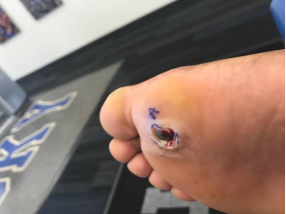
Figure 1. Melanoma found on Soles of the Feet.
Research in Nigeria has found that malignant melanoma accounts for 2.4% of all tumors, with 4.5% of skin lesions being malignant.
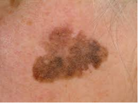
Figure 2. Lentigo Malignant Melanoma.
Traditional methods of diagnosing melanoma heavily depend on visual inspections and histopathological examinations, which can be subjective, time-consuming, and susceptible to variability among observers .
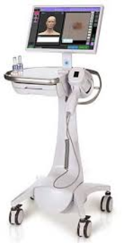
Figure 3. Visual Inspection Machine.
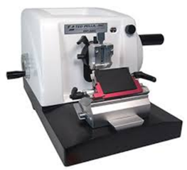
Figure 4. Histopathological Machine.
However, recent advancements in machine learning and deep learning have shown potential in transforming melanoma diagnosis by offering automated and precise classification of skin lesions based on dermoscopy images . Over the past few decades, the incidence of melanoma has been rising, particularly in areas with high ultraviolet (UV) radiation exposure . The disease, first described in the early 19th century, continues to be a significant concern due to its aggressive nature and the challenges associated with its early detection and diagnosis. The term “melanoma" was introduced by the Scottish surgeon Sir James Paget in 1857. Historically, clinical examination and basic visual inspection were the primary methods for detecting melanoma. However, these methods often lacked sensitivity and specificity, leading to delayed diagnoses and poor patient outcomes .
Dermoscopy, a non-invasive imaging technique, has enhanced the ability to visualize skin lesions with higher magnification and clarity than traditional clinical examination. Utilizing a handheld device called a dermatoscope, dermatologists can closely examine morphological features of skin lesions, including pigmentation patterns, vascular structures, architectural characteristics, asymmetry, irregular borders, multiple colors, and atypical structures. This enhanced visualization provided by dermoscopy aids in the early detection and differential diagnosis of melanoma and other skin conditions .
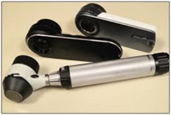
Figure 5. Dermatoscope.
Integrating dermoscopy images with deep learning techniques significantly enhances melanoma classification models, ultimately improving patient care. Deep learning, a subset of machine learning inspired by the brain's structure and function, has proven highly successful in various image recognition and classification tasks.
Convolutional Neural Networks (CNNs), a type of deep learning architecture, have achieved state-of-the-art performance in medical image analysis, including classifying skin lesions from dermoscopy images. CNNs automatically extract discriminative features from raw pixel data by learning hierarchical representations, making them effective in distinguishing different classes of skin lesions, including melanoma .
Despite the promise of integrating dermoscopy images with deep learning techniques, challenges such as dataset diversity scarcity, model optimization, and clinical integration hinder widespread adoption . Accurate melanoma lesion classification allows clinicians to differentiate between benign and malignant lesions, informing appropriate treatment strategies . These models exhibit high sensitivity and specificity in distinguishing melanoma from benign lesions, underscoring their potential to enhance diagnostic accuracy and efficiency. However, the scarcity of annotated, diverse skin datasets, particularly for dark skin tones, remains a significant challenge for deep learning model optimization . The proposed research aims to leverage deep learning techniques (CNN) to improve the accuracy, efficiency, and clinical utility of melanoma classification models. This effort seeks to develop an optimized model that enhances patient outcomes and reduces mortality rates associated with melanoma.
2. Related Works
This section reviews previous experimental research on melanoma lesion classification, focusing on diverse methodologies. Numerous studies have aimed to enhance the accuracy and efficiency of melanoma detection through various approaches, including traditional machine learning, advanced deep learning frameworks, and hybrid models.
Traditional machine learning methods, such as support vector machines (SVM), random forests, and decision trees, have been employed for melanoma classification. These techniques often utilize handcrafted features from dermoscopy images, like color, texture, and shape. Although effective to some extent, these methods sometimes struggle to capture the complex patterns inherent in skin lesions.
The advent of deep learning, particularly convolutional neural networks (CNNs), has significantly advanced melanoma lesion classification. CNNs excel at automatically learning features from images, leading to superior performance compared to traditional methods. Various CNN architectures, including ResNet, EfficientNet, and MobileNet, have been explored to optimize classification accuracy and computational efficiency. Hybrid models, which combine the strengths of multiple algorithms, have also been investigated to improve classification performance. For example, some studies integrate CNNs with feature selection techniques or utilize ensemble learning strategies to enhance robustness and accuracy. Additionally, optimization algorithms like genetic algorithms and particle swarm optimization have been applied to fine-tune model parameters and improve predictive outcomes.
Table 1. Summary of Previous Works.

Methods

Dataset Type/Size/Source

Result

Normal distribution-based segmentation and entropy-controlled features selection

NA

Achieved robust skin lesion detection and Classification

U-Net

Dermoscopy images / Not specified / Single source

Accuracy 95%

EfficientNetB0, Ant Colony Optimization, SVM

Custom dataset from ISIC images

Accuracy over 98% (CB-SVM)

MobileNetV3, DOLHGS feature selection algorithm

ISIC-2016, PH2 datasets

88.19%, 96.43%

Cat Swarm Optimization, Deep Learning, U-Net, MobileNet, GRU

ISIC2017, HAM10000 datasets

97.44%, 98.48%

U-Net, pre-trained models

N/A

AUC of 0.95, accuracy ~87%

Morphological filtering, Grab-cut, ResNet50, SVM

Real clinical skin lesions

99.87%

Intensity-based thresholding, PET/CT scans

53 PET/CT images

Median F1-score of 0.7500

Region growing, DBSCAN clustering

Two datasets

Sensitivity 0.91-0.92

Delaunay Triangulation, binary masks

PH2 database

Sensitivity 93.5%, specificity 87.1%

K-means, GOA, SURF, CNN

ISIC-2017, ISIC-2018, PH-2 datasets

98.42%

Improved K-means, LANM, LVP, GLCM, DCNN

N/A

0.86379 accuracy

Modified Probabilistic Neural Network (MPNN), SMO

N/A

0.98% accuracy

3. Materials and Methods
In this section, we describe the methodology employed in this research, providing a detailed account of the processes involved in the choice of dataset source, data preparation, and the use of CNNs for classifying melanoma lesions in dermoscopy images.
3.1. Dataset
This research utilizes the Melanoma Skin Cancer dataset from Kaggle, specifically sourced from (https://www.kaggle.com/datasets/hasnainjaved/melanoma-skin-cancer-dataset-of-10000-images/code). The dataset, with a total size of 104MB, comprises 10,605 images divided into two classes: benign and malignant. The training set includes 5,000 benign and 4,605 malignant images, while the test set contains 500 benign and 500 malignant images (See Figures 6 and 7). To ensure a comprehensive evaluation of the base CNN model's classification performance, we initially trained the model without cleaning the dataset. For this purpose, 80% of the dataset was used for training, 10% for validation, and the remaining 10% for testing. This approach allowed us to assess the model's effectiveness and robustness in classifying melanoma lesions from dermoscopy images, using the dataset in its raw form. By doing so, we could establish a baseline performance metric before applying any data preprocessing or cleaning techniques. This methodology helps to provide a clear understanding of the model's capabilities and limitations in handling real-world, unprocessed data.
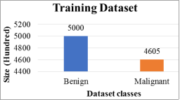
Figure 6. Un-preprocessed training dataset.
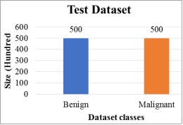
Figure 7. Un-preprocessed test dataset.
3.2. Proposed Framework
The diagram in Figure 8 shows a comprehensive workflow for melanoma lesion classification using Convolutional Neural Networks (CNN) with varying kernel sizes.
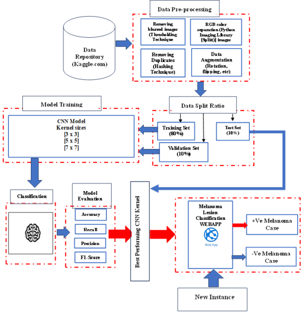
Figure 8. Proposed Framework.
The process begins with sourcing the dataset from Kaggle, ensuring a robust foundation for the project. In the Data Pre-processing phase, several key steps are taken to enhance the quality of the dataset. Noisy images are removed using techniques like Thresholding and Filtering, while images are standardized by converting them to RGB or grayscale formats. Image enhancement techniques such as Histogram Equalization and Gamma Correction are applied to improve the visual quality. Additionally, data augmentation methods are employed to increase the diversity and size of the dataset, thereby improving the model's ability to generalize.
The dataset is then split into three parts: Training Set (80%), Validation Set (10%), and Test Set (10%). The majority of the data is used to train the CNN model, while a portion is reserved for validating the model's performance during training, and the rest is used for final testing after the model is trained.
Model Training involves experimenting with CNN kernel sizes of [3x3], [5x5], and [7x7] to determine the optimal configuration. The trained model is then used for the Classification phase, where it distinguishes between positive (+Ve) and negative (-Ve) melanoma cases.
Model Evaluation is conducted using metrics such as Accuracy, Precision, Recall, and F1-Score to assess the performance of the model with different kernel sizes. Based on these evaluation metrics, the best-performing CNN kernel size is selected. Finally, the chosen model, featuring the optimal kernel size, is employed to classify new instances of melanoma, ensuring high accuracy and reliability in real-world applications. This structured approach ensures a thorough evaluation and optimization process, leading to an effective melanoma lesion classification system.
3.3. Data Preprocessing
We processed the dataset by following a detailed preprocessing workflow, starting with the initial download of 9605 training images from Kaggle.com. During this phase, we identified and removed 1246 blurred images, resulting in a directory of 8359 clean images. Further refinement involved eliminating 11 duplicate images, leaving us with a total of 8348 clean images. Next, we split all 8348 images into their respective Red, Green, and Blue (RGB) channels, creating separate directories for each channel. These were then combined, resulting in a folder containing 25044 RGB images. Additionally, by combining these RGB images with the cleaned image directory, the total increased to 33392 images.
To enhance the dataset's diversity, data augmentation was applied to the cleaned set of images. Starting with 9,605 images from Kaggle, 1,246 blurred images and 11 duplicates were removed, leaving a clean dataset of 8,358 images. Techniques such as rotation (up to 40 degrees), shifting (20% width and height), shearing (20 degrees), zooming (20%), and horizontal flipping were utilized. The ImageDataGenerator method was employed to generate augmented images, creating five variations of each original image, resulting in 17,400 augmented images. This augmentation process aimed to diversify the dataset, improving model generalization by introducing variations in orientation, perspective, and appearance. This systematic approach enhances the dataset's richness and robustness for melanoma lesion classification.
By combining the augmented images with the RGB, cleaned, and primary source images, we achieved a comprehensive dataset of 50834 images.
This meticulous preprocessing workflow ensured that we had a robust and diverse dataset, essential for training a reliable and accurate CNN model for melanoma lesion classification.
Table 2. Summary statistics of the preprocessed dataset.

Description

Number of Images

Initial Training Images Downloaded from Kaggle.com

9605

Blurred Images Removed

1246

Clean Images Directory (after removing blurred images)

8359

Duplicate Images Removed

11

Total Clean Images Directory (after removing duplicates)

8348

Red Channel Images

8348

Green Channel Images

8348

Blue Channel Images

8348

Combined Folder (Addition of RGB images)

25044

Combined Folder (RGB images and cleaned images)

33392

Images Collected from Primary Source

42

Augmented Images Directory

17400

Combined Folder (Augmented + RGB + Cleaned + Primary)

50834

3.4. Model Training
We conducted a series of comprehensive classification experiments by utilizing various CNN kernel sizes to classify melanoma lesions. The fundamental design of the CNN and the training parameters are detailed in Table 3.
Table 3. CNN Model Design and Training Parameters.

Aspect

Custom CNN

Number of Epochs

20

Batch Size

32

Learning Rate

0.001

Early Stopping

Patience: 7

Model Architecture

Custom CNN

Input Image Size

(224, 224, 3)

Convolutional Blocks

4

Neurons in Dense Layer

512

Dropout Rate

0.5

Loss Function

Binary Crossentropy

Optimization Algorithm

Adam

4. Results
This section describes the results obtained from using a CNN model for melanoma lesion classification. It includes a detailed account of the experimental setup, training and testing outcomes, and comparative evaluations. The performance of the models is assessed using metrics such as accuracy, precision, recall, and F1-score.
4.1. Experimental Setup
For our experiments, we utilized a high-performance personal computer with an Intel Core i7-8700 processor (3.2-4.6GHz) and an NVIDIA GeForce GTX 1080Ti GPU (16GB GDDR5X). This configuration, complemented by 64GB DDR4 RAM and 1TB of storage, ensured efficient processing and handling of intensive tasks. The system ran on Windows 10, providing a stable environment for development. Anaconda3 was used as the development platform, with Python 3.9 as the primary programming language. The project employed TensorFlow 10.0 and Keras for building and training the CNN models, with Django supporting web development tasks. This setup facilitated the efficient management of dependencies, streamlined development, and allowed us to focus on optimizing our CNN models for melanoma lesion classification.
4.2. Training Results
This section graphically illustrates the various training results obtained.
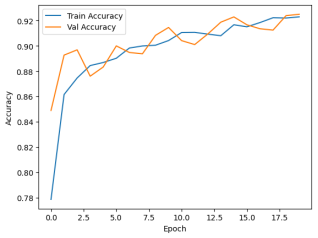
Figure 9. Training/Validation Accuracy Graph for Uncleaned Dataset.
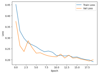
Figure 10. Training/Validation Loss Graph for Uncleaned Dataset.
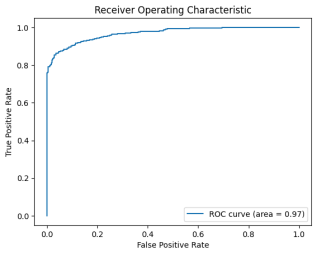
Figure 11. ROC/AUC Graph for Uncleaned Dataset.
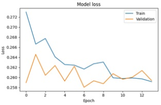
Figure 12. Training/Validation Loss Graph for Preprocessed Dataset.
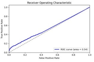
Figure 13. ROC/AUC Graph for Preprocessed Dataset.
4.3. Test Results
This section presents the various outcomes obtained from the experiments conducted.
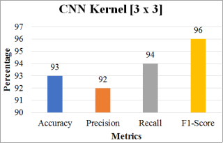
Figure 14. CNN Kernel [3 x 3] Test Results.
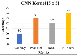
Figure 15. CNN Kernel [5 x 5] Test Results.
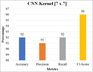
Figure 16. CNN Kernel [7 x 7] Test Results.
4.4. Kernel Filters Comparison Evaluation
This section presents a comparative performance evaluation of various CNN kernel filters.
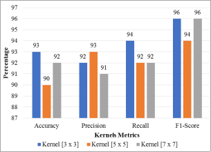
Figure 17. CNN Kernel Sizes Comparison Results.
Table 4. Comparison Evaluation of CNN Kernels using Preprocessed Training Set.

CNN Model

Training Epochs

80% Training Set (Pre-processed)

Training Accuracy

Training Loss

Validation Accu

Validation Loss

Kernel [3 x 3]

20

40666 images

92.64%

0.2581

92.69%

0.2581

Kernel [5 x 5]

20

40666 images

91.42%

0.2599

92.76%

0.2636

Kernel [7 x 7]

20

40666 images

92.69%

0.2607

92.68%

0.2603

Table 5. Comparison of CNN Kernels using Preprocessed Test Set.

CNN Model

10% Test Set (Pre-processed)

Accuracy

Precision

Recall

F1-Score

Kernel [3 x 3]

5086 images

93.00

92.00

94.00

96.00

Kernel [5 x 5]

5086 images

90.00

93.00

92.00

94.00

Kernel [7 x 7]

5086 images

92.00

91.00

92.00

96.00

Table 6. Learning Rate and Optimizer Comparison.

Learning Rate

Optimizer

Accuracy (%)

Loss

Comments

0.01

Adam

88.6

0.34

Too high, leads to overshooting

0.001

Adam

92.5

0.23

Chosen for optimal balance

0.0001

Adam

90.3

0.26

Too low, slower convergence

5. Discussion
This section analyzes and explains the findings from the research.
The presented CNN model demonstrates substantial promise in the field of dermatological image classification, specifically for melanoma detection. The training and validation accuracy graph (Figure 9) shows a steady increase in both accuracies over 20 epochs, starting from 77.86% and 84.90% respectively, and culminating at 92.3% and 92.5%. This progression indicates the model's strong learning capability and effective generalization to unseen data, evidenced by minimal overfitting. The consistent tracking of validation accuracy with training accuracy throughout the epochs highlights the robustness of the model. This performance is further corroborated by the training and validation loss graph (Figure 10). The steady decline in loss values suggests improved model accuracy over time, with the final epoch showing convergence of training and validation losses at low values. This indicates that the model has successfully minimized errors and achieved a reliable performance level in classifying dermoscopy images.
Figure 11 illustrates a high AUC of 0.97 on the uncleaned dataset, which signifies strong discrimination between melanoma and non-melanoma cases. However, the analysis notes the importance of evaluating precision and recall for a comprehensive performance assessment. This high AUC suggests that while the model is proficient in classification, further tests on diverse datasets are necessary to confirm its practical utility and rule out dataset-specific biases.
On a preprocessed dataset, Figures 12 and 13 present a slightly different picture. While the model achieves a stable convergence in training and validation loss at approximately 0.258 (Figure 12), the ROC curve and AUC value of 0.54 (Figure 13) indicate less-than-ideal performance. This discrepancy underscores the need for further optimization and refinement to improve the model’s ability to distinguish between positive and negative cases.
Comparing kernel sizes, Table 4 and Table 5 provide insights into the model's performance with different configurations. The CNN with a [3 x 3] kernel size achieves the highest accuracy and F1-score, demonstrating its effectiveness in both training (92.64% accuracy) and test sets (93.00% accuracy). The [5 x 5] and [7 x 7] kernels also perform well but show slightly lower accuracies and higher losses, indicating that smaller kernel sizes might be more efficient for this specific task. Table 6 compares the performance of different learning rates (0.01, 0.001, 0.0001) under the Adam optimizer in the context of melanoma lesion classification. Accuracy (%) assesses how effectively the model classifies the validation set, while loss reflects the binary cross-entropy post-training. Insights into each rate's performance are detailed in the comments column.
The results highlight that a learning rate of 0.001 with Adam achieved a significant 92.5% accuracy and a low 0.23 loss, demonstrating superior performance in accuracy and convergence speed. In contrast, a 0.01 rate initially progressed faster but tended to overshoot, whereas a 0.0001 rate converged more slowly with slightly reduced accuracy compared to 0.001. This analysis provides crucial insights into the impact of learning rates on training outcomes, offering valuable guidance for optimizing deep learning configurations to enhance the efficiency of melanoma lesion classification models.
When compared with related works, our model exhibits competitive performance. For instance, the research work done by , achieved over 98% accuracy by combining EfficientNetB0 with Ant Colony Optimization. This suggests that incorporating nature-inspired optimization techniques could further enhance our model's accuracy and computational efficiency. Similarly, another researcher utilized MobileNetV3 and a novel feature selection algorithm to achieve up to 96.43% accuracy, highlighting the potential benefits of advanced feature selection methods in boosting model performance .
Again, remarkable accuracies of 97.44% and 98.48% was achieved using a combination of Cat Swarm Optimization and Deep Learning techniques . This indicates that hybrid approaches combining optimization algorithms with deep learning could be highly effective. Similarly, another researcher also achieved significant improvements by integrating mole probability into image inputs . This suggests that, incorporating additional contextual information could enhance model performance.
The morphological filtering and ABCD rule-based image processing, achieved nearly perfect accuracy as demonstrated by . This indicates that preprocessing techniques and rule-based methods can significantly enhance the effectiveness of deep learning models in dermatological image classification.
The importance of robust segmentation techniques showed promising sensitivity and precision metrics and were emphasized by . These studies suggest that further improvements in our model could involve enhancing segmentation accuracy.
Future research should focus on incorporating advanced feature selection and optimization techniques, as seen in the works of . Additionally, integrating contextual information and advanced preprocessing methods could further enhance model performance . Expanding the model's evaluation to more diverse and larger datasets will be crucial for ensuring its robustness and generalizability in real-world clinical settings .
Finally, while the current proposed CNN model demonstrates robust performance and strong generalization capabilities; integrating advanced techniques and broadening the scope of evaluation could further enhance its efficacy in melanoma detection and other dermatological classification tasks.
6. Conclusions
The research into CNN models for melanoma lesion classification marks a notable advancement in dermatological image analysis. It underscores the efficacy of deep learning techniques, particularly convolutional neural networks (CNNs), in enhancing the accuracy and efficiency of diagnosing melanoma from dermoscopy images. Through extensive experimentation with various CNN kernel sizes, the study finds that smaller kernels, such as [3 x 3], consistently outperform larger ones like [5 x 5] and [7 x 7], achieving superior accuracy and F1-scores. This suggests that finer granularity in kernel size contributes significantly to the model's capability to differentiate between benign and malignant lesions.
The evaluation metrics, including accuracy, precision, recall, and F1-score, indicate robust performance across both training and test datasets post preprocessing. The model demonstrates strong generalization abilities, showing minimal overfitting and maintaining consistent validation accuracy with training accuracy over epochs. However, challenges persist, particularly in optimizing performance across diverse datasets, including those representing darker skin tones where data scarcity remains an issue.
The comparative analysis with related studies highlights the competitive nature of the proposed CNN model. It achieves comparable accuracy levels to state-of-the-art methods while identifying opportunities for enhancement through the incorporation of advanced feature selection algorithms and optimization techniques inspired by natural processes, such as ant colony optimization and cat swarm optimization.
Looking ahead, future research should prioritize refining segmentation techniques and integrating additional contextual information to further bolster the model's diagnostic accuracy using advanced deep learning techniques such as Autoencoder, Generative Adversarial Network (GAN), Transfer Learning (VGG, ResNet, Inception, EfficientNet), Recurrent Neural Metwork (RNN), Attention Mechanism and Transformer Network. Moreover, expanding the scope of evaluation to encompass larger and more varied datasets will be pivotal in validating the model's robustness and applicability in real-world clinical scenarios.
This investigation underscores the transformative potential of CNNs in redefining melanoma diagnosis through automated and precise classification of skin lesions. By addressing current limitations and building upon these findings, the study aims to significantly advance patient outcomes by facilitating early detection and intervention strategies for melanoma and other dermatological conditions.
Abbreviations

AI

Artificial Intelligence

ML

Machine Learning

DL

Deep Learning

CNN

Convolutional Neural Network

UV

Ultraviolet

RGB

Red, Green, Blue

SVM

Support Vector Machines

ROC

Receiver Operating Characteristic

AUC

Area Under the Curve

HAM10000

Human Against Machine with 10000 Training Images

ISIC

International Skin Imaging Collaboration

GA

Genetic Algorithm

GRU

Gated Recurrent Unit

SMO

Sequential Minimal Optimization

MPNN

Modified Probabilistic Neural Network

DOLHGS

Density Over-Lap Histogram Global Similarity

GLCM

Gray-Level Co-occurrence Matrix

DBSCAN

Density-Based Spatial Clustering of Applications with Noise

LANM

Local Active Neighborhood Model

LVP

Local Variance Patterns

GLCM

Gray-Level Co-occurrence Matrix

Acknowledgments
We extend our sincere gratitude to C. N. John, a Machine Learning Engineer, for his dedicated efforts in data preprocessing and model training for this study. We also wish to thank Mrs. Lovelyn Chukwunyere, Secretary of the Ethical Committee Board at the Federal Medical Centre (FMC) Owerri, Imo State, Nigeria, for her invaluable assistance in clarifying the concept of melanoma lesions during our visits to obtain primary datasets.
Author Contributions
Adetokunbo Macgregor John-Otumu: Conceptualization, Data curation, Investigation, Methodology, Visualization, Supervision, Resources, Writing, Review and Editing
Rebecca Ogechi Ekemonye: Conceptualization, Data curation, Investigation, Methodology, Visualization, Resources, Writing
Toochi Chima Ewunonu: Investigation, Visualization, Resources, Review
Victor Onyekachi Aniugo: Investigation, Visualization, Resources, Review
Ogadimma Thaddeus Okonkwo: Investigation, Visualization, Resources, Review
Data Availability Statement
The data that support the findings of this study can be found at: https://www.kaggle.com/datasets/hasnainjaved/melanoma-skin-cancer-dataset-of-10000-images/code (a publicly available repository url)
Conflicts of Interest
The authors declare no conflicts of interest.
References
[1] Chaturvedi, S. S.; Gupta, K.; Prasad, P. S. Skin lesion analyser: An efficient seven-way multi-class skin cancer classification using MobileNet. In Advances in Intelligent Systems and Computing; Springer: Singapore, 2020, 165–176.
[2] Bi, L., Kim, J., Ahn, E., Kumar, A., Feng, D., Fulham, M. Step-wise integration of deep class-specific learning for dermoscopic image segmentation. Pattern Recognit. 2019, 85, 78–89.
[3] Sonia, R. (2016). Melanoma Image Classification System by NSCT Features and Bayes Classification. 2(2), 27–33.
[4] Rehman, A.; Khan, M. A.; Mehmood, Z.; Saba, T.; Sardaraz, M.; Rashid, M. Microscopic melanoma detection and classification: A framework of pixel-based fusion and multilevel features reduction. Microsc. Res. Tech. 2020, 83, 410–423.
[5] Jain, S., Pise, N. Computer aided Melanoma skin cancer detection using Image Processing. Procedia - Procedia Computer Science. 2015, 48(ICCC), 735–740.
[6] Seeja, R. D., Suresh, A. Deep Learning Based Skin Lesion Segmentation and Classification of Melanoma Using Support Vector Machine. 2019a, 20, 1555–1561.
[7] Xie, Y., Zhang, J., Xia, Y., Shen, C. A Mutual Bootstrapping Model for Automated Skin Lesion Segmentation and Classification. 2019. 3, 1–12.
[8] Banjan, N., Dalvi, P., Athavale, N. Melanoma Skin Cancer Detection by Segmentation and Feature Extraction using combination of OTSU and STOLZ Algorithm Technique. 2017, 4(4), 21–25.
[9] Fornaciali, M., Avila, S., Carvalho, M., Valle, E. Statistical Learning Approach for Robust Melanoma Screening. In: 27th Conference on Graphics, Patterns and Images (SIBGRAPI), 2014, p. 319-326.
[10] Alsahafi, Y. S., Kassem, M. A., Hosny, K. M. Skin Net: A novel deep residual network for skin lesions classification using multilevel feature extraction and cross channel correlation with detection of outlier. Journal of Big Data. 2023.
[11] Dandu, R., Murthy, M. V., Kumar, Y. B. R. Transfer learning for segmentation with hybrid classification to Detect Melanoma Skin Cancer. Heliyon, 2023, 9(4), e15416.
[12] Sumithra, R., Suhil, M., Guru, D. S. Segmentation and Classification of Skin Lesions for Disease Diagnosis. - Procedia Computer Science, 2015, 45, 76–85.
[13] Khan, M. A., Akram, T., Sharif, M., Shahzad, A., Alhussein, M., Haider, S. I., Altamrah, A. An implementation of normal distribution-based segmentation and entropy-controlled features selection for skin lesion detection and classification. 2018, 1–20.
[14] Palivela, L. H., Athanesious, J., Deepika, V., & Vignesh, M. Segmentation and Classification of Skin Lesions from Dermoscopic Images. Journal of Scientific & Industrial Research, 2021.80(4), 328–335.
[15] Imran, T., Alghamdi, A. S., & Alkatheiri, M. S. Enhanced Skin Cancer Classification using Deep Learning and Nature-based Feature Optimization. Engineering, Technology and Applied Science Research, 2024, 14(1), 12702–12710.
[16] Dahou, A., Aseeri, A. O., Mabrouk, A., Ibrahim, R. A., Al-Betar, M. A., Elaziz, M. A. Optimal Skin Cancer Detection Model Using Transfer Learning and Dynamic-Opposite Hunger Games Search. Diagnostics, 2023, 13(9), 1–20.
[17] Rajendran, V. A., Shanmugam, S. Automated Skin Cancer Detection and Classification using Cat Swarm Optimization with a Deep Learning Model. Engineering, Technology & Applied Science Research. 2024. 14(1), 12734–12739.
[18] Verma, P. Deep Learning Techniques for Skin Lesion Segmentation and Classification. Diagnostics, 2021, 11(5): 811.
[19] Li, C., Miao, F., Gao, G. A Novel Progressive Image Classification Method Based on Hierarchical Convolutional Neural Networks. 2021, 16, 1–1726.
[20] Salma, W., Eltrass, A. S. Automated deep learning approach for classification of malignant melanoma and benign skin lesions. 2022, 32643–32660.
[21] Ine, K. M., Neyns, B., Vandemeulebroucke, J. Vrije Universiteit Brussel Computer-aided detection and segmentation of malignant melanoma lesions on whole-body 18F-FDG PET / CT using an interpretable deep learning approach Publication date: License: Computer Methods and Programs in Biomedicine Compu. Computer Methods and Programs, 2022.
[22] Owka, S. U. R. Segmentation of the melanoma lesion and its border. 2022, 32(4), 683–699.
[23] Pennisi, A., Bloisi, D. D., Nardi, D., Giampetruzzi, A. R., Mondino, C., Facchiano, A. Skin lesion image segmentation using Delaunay Triangulation for melanoma detection. Computerized Medical Imaging and Graphics, 2016, 52, 89–103.
[24] Rakhra, M., Cazzato, G., Hossain, M. S. Retracted: A Novel Hybrid Deep Learning Approach for Skin. 2023.
[25] Chang, W., Huang, A., Chen, Y., Lin, C., Tsai, J. The feasibility of using manual segmentation in a multifeature computer-aidediagnosis system for classification of skin lesions: A Retrospective comparative study. 2015, 1–8.
Cite This Article
  • APA Style

    John-Otumu, A. M., Ekemonye, R. O., Ewunonu, T. C., Aniugo, V. O., Okonkwo, O. T. (2024). Optimizing CNN Kernel Sizes for Enhanced Melanoma Lesion Classification in Dermoscopy Images. Machine Learning Research, 9(2), 26-38. https://doi.org/10.11648/j.mlr.20240902.11

    Copy | Download

    ACS Style

    John-Otumu, A. M.; Ekemonye, R. O.; Ewunonu, T. C.; Aniugo, V. O.; Okonkwo, O. T. Optimizing CNN Kernel Sizes for Enhanced Melanoma Lesion Classification in Dermoscopy Images. Mach. Learn. Res. 2024, 9(2), 26-38. doi: 10.11648/j.mlr.20240902.11

    Copy | Download

    AMA Style

    John-Otumu AM, Ekemonye RO, Ewunonu TC, Aniugo VO, Okonkwo OT. Optimizing CNN Kernel Sizes for Enhanced Melanoma Lesion Classification in Dermoscopy Images. Mach Learn Res. 2024;9(2):26-38. doi: 10.11648/j.mlr.20240902.11

    Copy | Download

  • @article{10.11648/j.mlr.20240902.11,
      author = {Adetokunbo Macgregor John-Otumu and Rebecca Ogechi Ekemonye and Toochi Chima Ewunonu and Victor Onyekachi Aniugo and Ogadimma Thaddeus Okonkwo},
      title = {Optimizing CNN Kernel Sizes for Enhanced Melanoma Lesion Classification in Dermoscopy Images
    },
      journal = {Machine Learning Research},
      volume = {9},
      number = {2},
      pages = {26-38},
      doi = {10.11648/j.mlr.20240902.11},
      url = {https://doi.org/10.11648/j.mlr.20240902.11},
      eprint = {https://article.sciencepublishinggroup.com/pdf/10.11648.j.mlr.20240902.11},
      abstract = {Skin cancer, particularly melanoma, presents a significant global health challenge due to its increasing incidence and mortality rates. Current diagnostic methods relying on visual inspection and histopathological examination are subjective and time-consuming, often leading to delayed diagnoses. Recent advancements in machine and deep learning, particularly convolutional neural networks (CNNs), offer a promising avenue for transforming melanoma detection by automating precise classification of dermoscopy images. This study leverages a comprehensive dataset sourced from Kaggle, comprising 10,605 images categorized into benign and malignant classes. Methodologically, a custom CNN architecture is trained and evaluated using varying kernel sizes (3x3, 5x5, 7x7) to optimize melanoma lesion classification. Results demonstrate that smaller kernel sizes, notably 3x3, consistently yield superior accuracy of 93.00% and F1-scores of 96.00%, indicating their efficacy in distinguishing between benign and malignant lesions. The CNN model exhibits robust generalization capabilities with minimal overfitting, supported by high validation accuracy throughout training epochs. Comparative analysis with related studies highlights competitive performance, suggesting potential enhancements through advanced feature selection and optimization techniques. Despite these advancements, challenges such as dataset diversity and model optimization persist, particularly concerning underrepresented darker skin tones. The study underscores the transformative potential of CNNs in enhancing diagnostic accuracy and efficiency in dermatological practice, paving the way for improved patient outcomes through early detection and intervention strategies. Future research directions include refining segmentation techniques and expanding dataset evaluations to ensure the model's applicability across diverse clinical settings. Ultimately, this research contributes to advancing melanoma diagnosis by integrating cutting-edge deep learning methodologies with clinical practice, thereby addressing current limitations and driving forward innovations in dermatological image analysis.
    },
     year = {2024}
    }
    

    Copy | Download

  • TY  - JOUR
    T1  - Optimizing CNN Kernel Sizes for Enhanced Melanoma Lesion Classification in Dermoscopy Images
    
    AU  - Adetokunbo Macgregor John-Otumu
    AU  - Rebecca Ogechi Ekemonye
    AU  - Toochi Chima Ewunonu
    AU  - Victor Onyekachi Aniugo
    AU  - Ogadimma Thaddeus Okonkwo
    Y1  - 2024/07/15
    PY  - 2024
    N1  - https://doi.org/10.11648/j.mlr.20240902.11
    DO  - 10.11648/j.mlr.20240902.11
    T2  - Machine Learning Research
    JF  - Machine Learning Research
    JO  - Machine Learning Research
    SP  - 26
    EP  - 38
    PB  - Science Publishing Group
    SN  - 2637-5680
    UR  - https://doi.org/10.11648/j.mlr.20240902.11
    AB  - Skin cancer, particularly melanoma, presents a significant global health challenge due to its increasing incidence and mortality rates. Current diagnostic methods relying on visual inspection and histopathological examination are subjective and time-consuming, often leading to delayed diagnoses. Recent advancements in machine and deep learning, particularly convolutional neural networks (CNNs), offer a promising avenue for transforming melanoma detection by automating precise classification of dermoscopy images. This study leverages a comprehensive dataset sourced from Kaggle, comprising 10,605 images categorized into benign and malignant classes. Methodologically, a custom CNN architecture is trained and evaluated using varying kernel sizes (3x3, 5x5, 7x7) to optimize melanoma lesion classification. Results demonstrate that smaller kernel sizes, notably 3x3, consistently yield superior accuracy of 93.00% and F1-scores of 96.00%, indicating their efficacy in distinguishing between benign and malignant lesions. The CNN model exhibits robust generalization capabilities with minimal overfitting, supported by high validation accuracy throughout training epochs. Comparative analysis with related studies highlights competitive performance, suggesting potential enhancements through advanced feature selection and optimization techniques. Despite these advancements, challenges such as dataset diversity and model optimization persist, particularly concerning underrepresented darker skin tones. The study underscores the transformative potential of CNNs in enhancing diagnostic accuracy and efficiency in dermatological practice, paving the way for improved patient outcomes through early detection and intervention strategies. Future research directions include refining segmentation techniques and expanding dataset evaluations to ensure the model's applicability across diverse clinical settings. Ultimately, this research contributes to advancing melanoma diagnosis by integrating cutting-edge deep learning methodologies with clinical practice, thereby addressing current limitations and driving forward innovations in dermatological image analysis.
    
    VL  - 9
    IS  - 2
    ER  - 

    Copy | Download

Author Information
  • Department of Information Technology, Federal University of Technology, Owerri, Nigeria

    Biography: Adetokunbo MacGregor John-Otumu is a lecturer and AI researcher in the Department of Information Technology, Federal University of Technology, Owerri, Nigeria. He served as a Post Doctoral Research Fellow at Morgan State University, Baltimore, MD, USA, in 2021. Dr. John-Otumu earned his PhD in Computer Science, specializing in Artificial Intelligence, from Ebonyi State University, Nigeria. He also holds a Master of Science in Computer Science from Ambrose Alli University, Ekpoma, and another Master of Science in Information Technology from the National Open University of Nigeria. As a prolific contributor to the field, Dr. John-Otumu is affiliated with numerous professional organizations including IEEE, ACM, IAENG, IACSIT, NCS, CPN, AAAI, and ISOC. He serves on various journal review boards, such as IJACSA, and is recognized as an IBM Certified AI Analyst. His extensive involvement in international research collaborations highlights his commitment to advancing knowledge in Machine Learning, Deep Learning, Multi-Agent Systems, Computer Vision, and Health Informatics with numerous publications and invitations as a Keynote Speaker at AI and Computer Science Summits.

    Research Fields: Machine Learning, Deep Learning, Computer Vision, MAS, Health Informatics

  • Department of Information Technology, Federal University of Technology, Owerri, Nigeria

    Biography: Rebecca Ogechi Ekemonye is is currently working towards a Master's degree in Information Management Technology, focusing on Data Science, in the Department of Information Technology at the Federal University of Technology, Owerri, Nigeria. She completed her Bachelor of Technology (BTech) in Information Management Technology at the same university in 2019. Additionally, she holds a National Diploma (ND) in Library Science from Federal Polytechnic Nekede, Owerri, Imo State, obtained in 2013. Her research interests mainly lie in Machine Learning and Deep Learning.

    Research Fields: Machine Learning, Deep Learning, Computer Vision

  • Department of Cybersecurity, Federal University of Technology, Owerri, Nigeria

    Biography: Toochi Chima Ewunonu is a lecturer and researcher in the Department of Cybersecurity at the Federal University of Technology, Owerri. He holds a PhD in Communication Engineering from Nnamdi Azikiwe University, an M.Eng in Communication Engineering from FUTO, and a B.Eng in Electrical Engineering from the University of Nigeria, Nsukka. Additionally, he has a Professional Diploma in Education from Alvan Ikoku University of Education and is a certified teacher with TRCN. Dr. Chima is a registered engineer with COREN and a member of NSE and IEEE. He is proficient in ICT, VSAT, wireless technology, and electrical power systems. His research interests include Communication Engineering, Embedded Systems, AI, Cyber Security, Wireless Sensor Networks, and IoT. He has an extensive publication record across various media.

    Research Fields: Communication Engineering, Embedded Systems, AI, Cyber Security, Wireless Sensor Networks, and IoT

  • Department of Mechatronics Engineering, Federal University of Technology, Owerri, Nigeria

    Biography: Victor Onyekachi Aniugo is a lecturer in the Department of Mechatronics Engineering at the Federal University of Technology, Owerri, Imo State, Nigeria. He serves as a student advisor and the Departmental SIWES Coordinator. He holds a Ph.D. in Control & Instrumentation Engineering from the Department of Electrical & Electronic Engineering at Enugu State University of Science and Technology (ESUT), where he also earned his M.Eng and B.Eng degrees in the same field. Dr. Aniugo is registered with the Council for the Regulation of Engineering in Nigeria and is a corporate member of the Nigerian Society of Engineers. His research interests include microcontroller and microprocessor systems, robotics, and process automation management. Dr. Aniugo is dedicated to advancing engineering education and research.

    Research Fields: Microcontroller and Microprocessor Systems, Robotics, and Process Automation Management

  • Department of Information Technology, Federal University of Technology, Owerri, Nigeria

    Biography: Ogadimma Thaddeus Okonkwo is a lecturer in the Department of Information Technology, at the Federal University of Technology, Owerri. He holds a PhD in Computer Science from Imo State University, Owerri, Nigeria He also holds a Master of Science in Computer Science from same university and another Master of Science in Information Technology from the National Open University of Nigeria. Additionally, he is a registered member of Nigeria Computer Society (NCS). His research interests include Articifical Intelligence / Machine Learning.

    Research Fields: Artificial Intelligence, Mchine Deep Learning and Deep Learning