Blunt abdominal trauma (BAT) is a significant cause of morbidity in the pediatric population, with injuries to solid organs occurring far more commonly than injuries to the hollow viscera. Small bowel perforation resulting from low-energy blunt abdominal trauma is rare and frequently presents with delayed or subtle clinical manifestations, posing a diagnostic challenge. Timely recognition, accurate diagnosis, and early surgical intervention are essential to minimize morbidity and improve clinical outcomes. We report the case of a 13-year-old boy presented with severe abdominal pain one day following a fall while drinking water, resulting in blunt abdominal trauma and a lacerated wound over the right iliac fossa. The wound had been sutured at a peripheral hospital. He subsequently developed progressively worsening diffuse abdominal pain associated with fever and hematuria. Contrast-enhanced computed tomography (CECT) revealed disruption of the anterior abdominal wall musculature, multiple distal ileal perforations, pneumoperitoneum, hemoperitoneum, and traumatic peritonitis without solid organ injury. Emergency exploratory laparotomy demonstrated seven perforations involving a 15-20 cm segment of distal ileum approximately 20-25 cm proximal to the ileocecal junction, associated with a large mesenteric tear. Segmental resection of approximately 20 cm distal ileum and Primary end-to-end ileoileal anastomosis was done. The postoperative recovery was uneventful. Persistent abdominal pain after even trivial blunt abdominal trauma should prompt evaluation for hollow viscus injury. This case emphasises that timely CT imaging and early surgical intervention are essential for preventing life-threatening complications and improving patient outcomes.
This is an Open Access article, distributed under the terms of the Creative Commons Attribution 4.0 International License (http://creativecommons.org/licenses/by/4.0/), which permits unrestricted use, distribution and reproduction in any medium or format, provided the original work is properly cited.
Trauma remains one of the leading causes of mortality and disability among children worldwide. Blunt abdominal trauma accounts for nearly 90% of pediatric abdominal injuries, with motor vehicle accidents, bicycle injuries, falls, sports injuries, and direct blows being common mechanisms. Although injuries to the liver and spleen predominate, hollow viscus injuries occur in less than 5% of pediatric blunt abdominal trauma cases but carry significantly higher morbidity because diagnosis is frequently delayed
[2]
Bège T, Brunet C, Berdah SV. Hollow viscus injury due to blunt trauma: A review. J Visc Surg. 2016 Aug; 153, S61-S68.
The small intestine is the most commonly injured hollow organ, particularly the jejunum near the ligament of Treitz and the distal ileum near the ileocecal junction due to their relatively fixed anatomical positions. Mechanisms of bowel injury include crushing of the bowel against the vertebral column, rapid increase in intraluminal pressure, and mesenteric shearing forces. Delay in diagnosis beyond 8-12 hours has been associated with increased septic complications, prolonged hospitalization, and mortality.
[3]
Richards J. R., Knopf N. A., Wang L., McGahan J. P. Blunt abdominal trauma in children: Evaluation with emergency US. Radiology. 2002; 222(3): 749-754.
We report an unusual case of multiple distal ileal perforations following an apparently trivial fall in a child that required emergency bowel resection and primary anastomosis. Written appropriate consent was obtained from patient’s parents. They provided permission for the use of clinical information and images for academic and publication purposes. This case reinforces the critical role of early surgical intervention to prevent lifetime complications.
2. Case Report
A 13-year-old male presented to the Emergency Department of Saraswathi Institute of Medical Sciences (SIMS), Hapur, with complaints of severe abdominal pain for one day following blunt abdominal trauma.
According to the history, the child had accidentally fallen while drinking water and sustained an injury over the right lower abdomen. Initial treatment was received at a nearby hospital, where a lacerated wound measuring approximately 3 × 0.5 cm over the right iliac fossa was sutured.
Following discharge, the patient developed progressively worsening abdominal pain. The pain was sudden in onset, diffuse, gradually progressive, sharp, and stabbing in nature. It was associated with fever and passage of blood in urine. There was no history of vomiting, loss of consciousness, or respiratory complaints.
There was no significant past medical or surgical history. Family history was non-contributory. Personal history revealed decreased appetite, disturbed sleep, and altered bowel and bladder habits. Clinical Examination on admission, the patient appeared ill and uncomfortable due to pain with mild dehydration and tachycardia. Local examination revealed a sutured lacerated wound measuring approximately 3 × 0.5 cm over the right iliac fossa. Abdominal examination demonstrated Generalized tenderness, Guarding, Board-like rigidity, Rebound tenderness and Absent bowel sounds. These findings were suggestive of generalized peritonitis secondary to hollow viscus perforation.
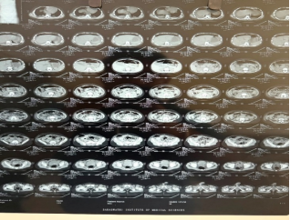
Chest radiography showed no evidence of pneumothorax but mild bibasal atelectatic changes. Contrast-enhanced CT scan of the abdomen and pelvis showed Large penetrating soft tissue injury involving the right iliac fossa, Extensive subcutaneous emphysema extending to the right hypochondrium, Disruption of the anterior abdominal wall musculature, Circumferential thickening and hyperenhancement of distal ileal loops, Multiple traumatic mural defects in the distal ileum, the largest measuring approximately 8.4 mm, Large pneumoperitoneum, Moderate hemoperitoneum with pelvic blood clot, Diffuse traumatic peritonitis, Reactive mesenteric lymphadenopathy, Mild bilateral pleural effusion with adjacent bibasal compressive atelectasis The radiological findings were highly suggestive of multiple traumatic small bowel perforations requiring emergency surgery (Figure 1).
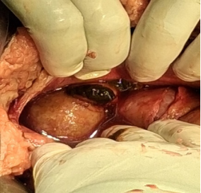
The patient underwent Emergency exploratory laparotomy of duration approximately 3.5 hours, with estimated blood loss of approximately 20-50 mL. Emergency exploratory laparotomy through a midline incision showed Large hemoperitoneum, Large intraperitoneal blood clot, Diffuse fecal contamination, Seven perforations involving approximately 15-20 cm of distal ileum, Injured bowel located approximately 20-25 cm proximal to the ileocecal junction, Large mesenteric tear near the ileocecal junction, Edematous distal ileum (Figure 2).
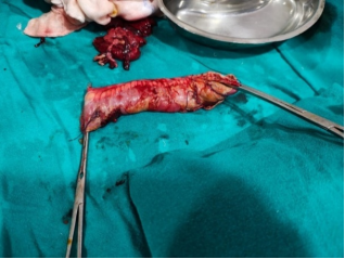
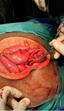
Segmental ileal resection with primary end-to-end anastomosis, repair of the mesenteric tear, evacuation of hematoma, extensive peritoneal lavage, and drain placement were performed successfully (Figures 3 & 4). Postoperative recovery was uneventful, and the patient was discharged in a stable condition with satisfactory wound healing.
Figure 4. Operative photograph following bowel resection and primary end-to-end anastomosis.
3. Discussion
Traumatic perforation of the small intestine following blunt abdominal trauma remains uncommon in children. Due to the elasticity of the pediatric abdominal wall, severe intra-abdominal injuries may occur despite minimal external signs. Clinical diagnosis is challenging because symptoms often evolve gradually over several hours
[1]
Staab V, Naganathan S, McGuire M, Pinto JM, Pall H. Gastrointestinal Perforation with Blunt Abdominal Trauma in Children. Children (Basel). 2024; 11(6): 612.
The distal ileum is particularly vulnerable because of its relative fixation near the ileocecal junction. Three principal mechanisms explain bowel injury after blunt trauma: compression of the bowel against the vertebral column, rapid increase in intraluminal pressure leading to rupture, and shearing forces causing mesenteric tears and bowel ischemia
[2]
Bège T, Brunet C, Berdah SV. Hollow viscus injury due to blunt trauma: A review. J Visc Surg. 2016 Aug; 153, S61-S68.
Computed tomography remains the investigation of choice in hemodynamically stable patients. Important CT findings include pneumoperitoneum, bowel wall thickening, mural discontinuity, mesenteric fat stranding, extraluminal air, and free intraperitoneal fluid. Nevertheless, CT may occasionally underestimate bowel injury, and repeated clinical examination remains essential
[3]
Richards J. R., Knopf N. A., Wang L., McGahan J. P. Blunt abdominal trauma in children: Evaluation with emergency US. Radiology. 2002; 222(3): 749-754.
Hasan Madenci, Cüneyt Uğur, Taha Demirci. A rare small intestine injury without free gas image on radiological imaging after blunt abdominal trauma in a child: ileal perforation. Journal of Emergency Medicine Case Reports. 2022 Mar. 1; 13(1): 31-3.
. Even in pediatric abdominal trauma, imaging findings may initially be normal despite clinically significant bowel perforation. Darlington and Anitha reported an infant with multiple abdominal stab injuries in whom abdominal radiography and ultrasonography failed to demonstrate free air or free fluid, yet emergency laparotomy revealed gastric perforation, mesenteric tear, and ileal laceration. The authors emphasized that persistent clinical suspicion and timely surgical exploration are crucial when bowel injury is suspected despite inconclusive imaging
[4]
Darlington DC, Anitha FSG. Multiple stab injuries in an infant with silent bowel perforations: a case report. Int Surg J. 2018; 5(10): 3407-3409.
. Similarly, Madenci et al. described a 4-year-old boy with blunt abdominal trauma in whom both standing abdominal radiography and the initial CT scan showed no free intraperitoneal air despite an ileal perforation. The child developed worsening abdominal tenderness and vomiting during observation, and repeat imaging demonstrated only bowel wall edema and mesenteric fat contamination without pneumoperitoneum. Exploratory laparotomy performed 56 hours after injury revealed a terminal ileal perforation 15–20 cm proximal to the ileocecal valve, which was successfully repaired primarily. The authors emphasized that the absence of free intraperitoneal gas does not exclude hollow viscus injury and advocated close clinical monitoring with repeated physical examination in all children with blunt abdominal trauma
[5]
Hasan Madenci, Cüneyt Uğur, Taha Demirci. A rare small intestine injury without free gas image on radiological imaging after blunt abdominal trauma in a child: ileal perforation. Journal of Emergency Medicine Case Reports. 2022 Mar. 1; 13(1): 31-3.
Similarly, another reported case involved a 4-year-old girl initially believed to have sustained accidental blunt abdominal trauma following a fall. She presented with delayed abdominal pain, multiple bruises, and free intraperitoneal fluid, while CT findings suggested perforated appendicitis. Diagnostic laparoscopy, however, revealed mesenteric hematomas, a jejunal serosal injury, and a terminal ileal perforation near the ileocecal valve, requiring surgical repair. Subsequent investigation confirmed non-accidental injury. Although the patient initially recovered well and was discharged on postoperative day 11, she was readmitted two days later with abdominal pain and vomiting due to adhesive intestinal obstruction. Re-laparotomy with adhesiolysis was successfully performed, and she recovered uneventfully, being discharged five days later. This case illustrates that pediatric bowel perforation may mimic other intra-abdominal conditions on imaging and highlights the importance of maintaining a high index of suspicion, correlating radiological findings with serial clinical assessment, and ensuring close postoperative follow-up because adhesive bowel obstruction may complicate recovery after surgical repair
[6]
Meyer J, Schäfer FM, Stehr M, Burgmeier C. A case of non-accidental injury with ileal perforation. Klinische Pädiatrie. 2025 May 6.
The presence of seven perforations involving a short segment of distal ileum in our patient represents an unusual injury pattern. Multiple adjacent perforations are best managed by segmental bowel resection with primary anastomosis rather than individual closure because this approach removes devitalized tissue, minimizes the risk of leakage, and provides better long-term outcomes. Darlington et al. similarly highlighted that the choice between primary repair and bowel resection should be individualized according to the extent of bowel injury, degree of contamination, and tissue viability
[7]
Öztürk H, Önen A, Otçu S, Dokucu Aİ, Yağmur Y, Kaya M, Yücesan S. Diagnostic delay increases morbidity in children with gastrointestinal perforation from blunt abdominal trauma. Surgery today. 2003 Mar; 33(3): 178-82.
Recent evidence by Bašković et al. further emphasizes that successful management of pediatric blunt abdominal trauma depends on early recognition, systematic clinical assessment, appropriate use of imaging, and timely surgical intervention when hollow viscus injury is suspected. Their review highlights that delayed diagnosis remains the most important predictor of morbidity and advocates serial abdominal examinations and multidisciplinary management in children with equivocal imaging findings or persistent abdominal symptoms.
[12]
Bašković M, Keretić D, Lacković M, Borić Krakar M, Pogorelić Z. The Diagnosis and Management of Pediatric Blunt Abdominal Trauma-A Comprehensive Review. Diagnostics (Basel). 2024 Oct 10; 14(20): 2257.
. A retrospective descriptive study by Schacherer and Miller et al involving 105 patients admitted with blunt abdominal trauma (BAT) over an 8-year period found that BAT in children predominantly affects boys younger than 10 years, with road traffic accidents (RTAs) being the most common cause. Clinical examination combined with appropriate imaging, particularly ultrasonography, plays a crucial role in determining the management strategy
[13]
Schacherer, N.; Miller, J.; Petronis, K. Pediatric blunt abdominal trauma: Recognition and management in the emergency department. Pediatr.Emerg.Med.Pract. 2020, 17, 1–59.
. Singh et al concluded that CECT findings such as free intraperitoneal air, solid organ injury with hemodynamically instability but also cases with clinical examination and minor mesenteric injuries should be subjected to either laparoscopy or exploratory laparotomy without any delay
[15]
Singh AK, Bais PS, Chand K, Dey S. Mesenteric Injury in Blunt Abdominal Trauma in Children: Is Early Surgical Intervention Need of the Hour? J Indian Assoc Pediatr Surg. 2022 Jul-Aug; 27(4): 381-386.
The absence of associated solid organ injury despite extensive bowel trauma further underscores the rarity of our case. Early diagnosis, prompt exploratory laparotomy, resection of the injured bowel segment with primary anastomosis, and meticulous postoperative care resulted in an excellent clinical outcome, reinforcing the importance of maintaining a high index of suspicion for bowel injury even after apparently low-energy blunt abdominal trauma in children
[8]
Albanese CT, Meza MP, Gardner MJ, Smith SD, Rowe MI, Lynch JM. Is computed tomography a useful adjunct to the clinical examination for the diagnosis of pediatric gastrointestinal perforation from blunt abdominal trauma in children?. Journal of Trauma and Acute Care Surgery. 1996 Mar 1; 40(3): 417-21.
Small bowel injuries including jejunal and ileal perforations, mesenteric tears, and transections; most cases involved a single perforation
Exploratory laparotomy with primary repair or segmental bowel resection and primary anastomosis according to the extent of injury
Good outcomes with early diagnosis and surgery.
Madenci et al.
[5]
Hasan Madenci, Cüneyt Uğur, Taha Demirci. A rare small intestine injury without free gas image on radiological imaging after blunt abdominal trauma in a child: ileal perforation. Journal of Emergency Medicine Case Reports. 2022 Mar. 1; 13(1): 31-3.
Memeti S, Sulejmani H, Kamiloski M, Selmani R, Rushiti Q, Cokleska N, et al. Blunt polytrauma in two pediatric patients: surgical vs. conservative management–a two-case study. Kos J Surg. 2026; 10(2): 14–18.
[10]
7 year old/girl
Blunt Abdominal Trauma
Splenic rupture, bowel perforation
Emergency laparotomy (splenectomy, bowel resection and primary repair)
Rapid postoperative clinical and laboratory improvement
Katwal et al.
[11]
Katwal S, Ghimire A, Joshi N. Uncommon presentation: isolated small bowel perforation after blunt abdominal trauma in a pediatric patient; a case report. Ann Med Surg (Lond). 2023 Nov 1; 85(12): 6222-6226.
Emergency exploratory laparotomy, resection of affected ileal segment with primary end-to-end ileo-ileal anastomosis and peritoneal lavage
Uneventful recovery; discharged in stable condition on postoperative day 10
4. Conclusion
This case emphasizes that apparently trivial blunt abdominal trauma in children can conceal severe hollow viscus injuries. Persistent abdominal pain, fever, and signs of peritonitis following trauma should immediately raise suspicion for bowel perforation. Early CT imaging, repeated clinical assessment, and timely exploratory laparotomy remain the cornerstones of successful management.
Diwakar Sharma: Data curation, Formal Analysis, Investigation, Visualization, Writing – original draft, Writing – review & editing
Conflicts of Interest
The authors declare no conflicts of interest.
References
[1]
Staab V, Naganathan S, McGuire M, Pinto JM, Pall H. Gastrointestinal Perforation with Blunt Abdominal Trauma in Children. Children (Basel). 2024; 11(6): 612.
Richards J. R., Knopf N. A., Wang L., McGahan J. P. Blunt abdominal trauma in children: Evaluation with emergency US. Radiology. 2002; 222(3): 749-754.
Hasan Madenci, Cüneyt Uğur, Taha Demirci. A rare small intestine injury without free gas image on radiological imaging after blunt abdominal trauma in a child: ileal perforation. Journal of Emergency Medicine Case Reports. 2022 Mar. 1; 13(1): 31-3.
Öztürk H, Önen A, Otçu S, Dokucu Aİ, Yağmur Y, Kaya M, Yücesan S. Diagnostic delay increases morbidity in children with gastrointestinal perforation from blunt abdominal trauma. Surgery today. 2003 Mar; 33(3): 178-82.
Albanese CT, Meza MP, Gardner MJ, Smith SD, Rowe MI, Lynch JM. Is computed tomography a useful adjunct to the clinical examination for the diagnosis of pediatric gastrointestinal perforation from blunt abdominal trauma in children?. Journal of Trauma and Acute Care Surgery. 1996 Mar 1; 40(3): 417-21.
Memeti S, Sulejmani H, Kamiloski M, Selmani R, Rushiti Q, Cokleska N, et al. Blunt polytrauma in two pediatric patients: surgical vs. conservative management–a two-case study. Kos J Surg. 2026; 10(2): 14–18.
[11]
Katwal S, Ghimire A, Joshi N. Uncommon presentation: isolated small bowel perforation after blunt abdominal trauma in a pediatric patient; a case report. Ann Med Surg (Lond). 2023 Nov 1; 85(12): 6222-6226.
Bašković M, Keretić D, Lacković M, Borić Krakar M, Pogorelić Z. The Diagnosis and Management of Pediatric Blunt Abdominal Trauma-A Comprehensive Review. Diagnostics (Basel). 2024 Oct 10; 14(20): 2257.
Singh AK, Bais PS, Chand K, Dey S. Mesenteric Injury in Blunt Abdominal Trauma in Children: Is Early Surgical Intervention Need of the Hour? J Indian Assoc Pediatr Surg. 2022 Jul-Aug; 27(4): 381-386.
Majumdar, R. K., Sharma, D. (2026). Multiple Distal Ileal Perforations Following Blunt Abdominal Trauma in a Child: A Rare Case Report. Journal of Surgery, 14(4), 96-100. https://doi.org/10.11648/j.js.20261404.15
Majumdar, R. K.; Sharma, D. Multiple Distal Ileal Perforations Following Blunt Abdominal Trauma in a Child: A Rare Case Report. J. Surg.2026, 14(4), 96-100. doi: 10.11648/j.js.20261404.15
Majumdar RK, Sharma D. Multiple Distal Ileal Perforations Following Blunt Abdominal Trauma in a Child: A Rare Case Report. J Surg. 2026;14(4):96-100. doi: 10.11648/j.js.20261404.15
@article{10.11648/j.js.20261404.15,
  author = {Rajib Kumar Majumdar and Diwakar Sharma},
  title = {Multiple Distal Ileal Perforations Following Blunt Abdominal Trauma in a Child: A Rare Case Report},
  journal = {Journal of Surgery},
  volume = {14},
  number = {4},
  pages = {96-100},
  doi = {10.11648/j.js.20261404.15},
  url = {https://doi.org/10.11648/j.js.20261404.15},
  eprint = {https://article.sciencepublishinggroup.com/pdf/10.11648.j.js.20261404.15},
  abstract = {Blunt abdominal trauma (BAT) is a significant cause of morbidity in the pediatric population, with injuries to solid organs occurring far more commonly than injuries to the hollow viscera. Small bowel perforation resulting from low-energy blunt abdominal trauma is rare and frequently presents with delayed or subtle clinical manifestations, posing a diagnostic challenge. Timely recognition, accurate diagnosis, and early surgical intervention are essential to minimize morbidity and improve clinical outcomes. We report the case of a 13-year-old boy presented with severe abdominal pain one day following a fall while drinking water, resulting in blunt abdominal trauma and a lacerated wound over the right iliac fossa. The wound had been sutured at a peripheral hospital. He subsequently developed progressively worsening diffuse abdominal pain associated with fever and hematuria. Contrast-enhanced computed tomography (CECT) revealed disruption of the anterior abdominal wall musculature, multiple distal ileal perforations, pneumoperitoneum, hemoperitoneum, and traumatic peritonitis without solid organ injury. Emergency exploratory laparotomy demonstrated seven perforations involving a 15-20 cm segment of distal ileum approximately 20-25 cm proximal to the ileocecal junction, associated with a large mesenteric tear. Segmental resection of approximately 20 cm distal ileum and Primary end-to-end ileoileal anastomosis was done. The postoperative recovery was uneventful. Persistent abdominal pain after even trivial blunt abdominal trauma should prompt evaluation for hollow viscus injury. This case emphasises that timely CT imaging and early surgical intervention are essential for preventing life-threatening complications and improving patient outcomes.},
 year = {2026}
}
TY  - JOUR
T1  - Multiple Distal Ileal Perforations Following Blunt Abdominal Trauma in a Child: A Rare Case Report
AU  - Rajib Kumar Majumdar
AU  - Diwakar Sharma
Y1  - 2026/08/24
PY  - 2026
N1  - https://doi.org/10.11648/j.js.20261404.15
DO  - 10.11648/j.js.20261404.15
T2  - Journal of Surgery
JF  - Journal of Surgery
JO  - Journal of Surgery
SP  - 96
EP  - 100
PB  - Science Publishing Group
SN  - 2330-0930
UR  - https://doi.org/10.11648/j.js.20261404.15
AB  - Blunt abdominal trauma (BAT) is a significant cause of morbidity in the pediatric population, with injuries to solid organs occurring far more commonly than injuries to the hollow viscera. Small bowel perforation resulting from low-energy blunt abdominal trauma is rare and frequently presents with delayed or subtle clinical manifestations, posing a diagnostic challenge. Timely recognition, accurate diagnosis, and early surgical intervention are essential to minimize morbidity and improve clinical outcomes. We report the case of a 13-year-old boy presented with severe abdominal pain one day following a fall while drinking water, resulting in blunt abdominal trauma and a lacerated wound over the right iliac fossa. The wound had been sutured at a peripheral hospital. He subsequently developed progressively worsening diffuse abdominal pain associated with fever and hematuria. Contrast-enhanced computed tomography (CECT) revealed disruption of the anterior abdominal wall musculature, multiple distal ileal perforations, pneumoperitoneum, hemoperitoneum, and traumatic peritonitis without solid organ injury. Emergency exploratory laparotomy demonstrated seven perforations involving a 15-20 cm segment of distal ileum approximately 20-25 cm proximal to the ileocecal junction, associated with a large mesenteric tear. Segmental resection of approximately 20 cm distal ileum and Primary end-to-end ileoileal anastomosis was done. The postoperative recovery was uneventful. Persistent abdominal pain after even trivial blunt abdominal trauma should prompt evaluation for hollow viscus injury. This case emphasises that timely CT imaging and early surgical intervention are essential for preventing life-threatening complications and improving patient outcomes.
VL  - 14
IS  - 4
ER  -
Majumdar, R. K., Sharma, D. (2026). Multiple Distal Ileal Perforations Following Blunt Abdominal Trauma in a Child: A Rare Case Report. Journal of Surgery, 14(4), 96-100. https://doi.org/10.11648/j.js.20261404.15
Majumdar, R. K.; Sharma, D. Multiple Distal Ileal Perforations Following Blunt Abdominal Trauma in a Child: A Rare Case Report. J. Surg.2026, 14(4), 96-100. doi: 10.11648/j.js.20261404.15
Majumdar RK, Sharma D. Multiple Distal Ileal Perforations Following Blunt Abdominal Trauma in a Child: A Rare Case Report. J Surg. 2026;14(4):96-100. doi: 10.11648/j.js.20261404.15
@article{10.11648/j.js.20261404.15,
  author = {Rajib Kumar Majumdar and Diwakar Sharma},
  title = {Multiple Distal Ileal Perforations Following Blunt Abdominal Trauma in a Child: A Rare Case Report},
  journal = {Journal of Surgery},
  volume = {14},
  number = {4},
  pages = {96-100},
  doi = {10.11648/j.js.20261404.15},
  url = {https://doi.org/10.11648/j.js.20261404.15},
  eprint = {https://article.sciencepublishinggroup.com/pdf/10.11648.j.js.20261404.15},
  abstract = {Blunt abdominal trauma (BAT) is a significant cause of morbidity in the pediatric population, with injuries to solid organs occurring far more commonly than injuries to the hollow viscera. Small bowel perforation resulting from low-energy blunt abdominal trauma is rare and frequently presents with delayed or subtle clinical manifestations, posing a diagnostic challenge. Timely recognition, accurate diagnosis, and early surgical intervention are essential to minimize morbidity and improve clinical outcomes. We report the case of a 13-year-old boy presented with severe abdominal pain one day following a fall while drinking water, resulting in blunt abdominal trauma and a lacerated wound over the right iliac fossa. The wound had been sutured at a peripheral hospital. He subsequently developed progressively worsening diffuse abdominal pain associated with fever and hematuria. Contrast-enhanced computed tomography (CECT) revealed disruption of the anterior abdominal wall musculature, multiple distal ileal perforations, pneumoperitoneum, hemoperitoneum, and traumatic peritonitis without solid organ injury. Emergency exploratory laparotomy demonstrated seven perforations involving a 15-20 cm segment of distal ileum approximately 20-25 cm proximal to the ileocecal junction, associated with a large mesenteric tear. Segmental resection of approximately 20 cm distal ileum and Primary end-to-end ileoileal anastomosis was done. The postoperative recovery was uneventful. Persistent abdominal pain after even trivial blunt abdominal trauma should prompt evaluation for hollow viscus injury. This case emphasises that timely CT imaging and early surgical intervention are essential for preventing life-threatening complications and improving patient outcomes.},
 year = {2026}
}
TY  - JOUR
T1  - Multiple Distal Ileal Perforations Following Blunt Abdominal Trauma in a Child: A Rare Case Report
AU  - Rajib Kumar Majumdar
AU  - Diwakar Sharma
Y1  - 2026/08/24
PY  - 2026
N1  - https://doi.org/10.11648/j.js.20261404.15
DO  - 10.11648/j.js.20261404.15
T2  - Journal of Surgery
JF  - Journal of Surgery
JO  - Journal of Surgery
SP  - 96
EP  - 100
PB  - Science Publishing Group
SN  - 2330-0930
UR  - https://doi.org/10.11648/j.js.20261404.15
AB  - Blunt abdominal trauma (BAT) is a significant cause of morbidity in the pediatric population, with injuries to solid organs occurring far more commonly than injuries to the hollow viscera. Small bowel perforation resulting from low-energy blunt abdominal trauma is rare and frequently presents with delayed or subtle clinical manifestations, posing a diagnostic challenge. Timely recognition, accurate diagnosis, and early surgical intervention are essential to minimize morbidity and improve clinical outcomes. We report the case of a 13-year-old boy presented with severe abdominal pain one day following a fall while drinking water, resulting in blunt abdominal trauma and a lacerated wound over the right iliac fossa. The wound had been sutured at a peripheral hospital. He subsequently developed progressively worsening diffuse abdominal pain associated with fever and hematuria. Contrast-enhanced computed tomography (CECT) revealed disruption of the anterior abdominal wall musculature, multiple distal ileal perforations, pneumoperitoneum, hemoperitoneum, and traumatic peritonitis without solid organ injury. Emergency exploratory laparotomy demonstrated seven perforations involving a 15-20 cm segment of distal ileum approximately 20-25 cm proximal to the ileocecal junction, associated with a large mesenteric tear. Segmental resection of approximately 20 cm distal ileum and Primary end-to-end ileoileal anastomosis was done. The postoperative recovery was uneventful. Persistent abdominal pain after even trivial blunt abdominal trauma should prompt evaluation for hollow viscus injury. This case emphasises that timely CT imaging and early surgical intervention are essential for preventing life-threatening complications and improving patient outcomes.
VL  - 14
IS  - 4
ER  -





 Share This Article
 Twitter
 Linked In
 Facebook
 Copy
Copy Download