Pregnancy induces a range of hormonal and physiological changes and also affect the brain. Yet the specific cerebral morphometric markers and their associated molecular profiles throughout pregnancy remain poorly understood. In this study, we investigated the cerebral morphometric changes in 23 pregnant women using T1-weighted MRI scans, with pregnancy progression quantified by post-menstrual age (PMA). We performed a whole-brain regression analysis to examine how gray matter volume (GMV) was influenced by PMA, and further explored the molecular profiles of these changes by integrating GMV findings with the JuSpace toolbox. Our analysis revealed that with PMA increased, there was a significant reduction in the left medial frontal gyrus (MFG) GMV, suggesting structural brain changes associated with pregnancy progression. Spatial correlation analyses did not reveal any significant associations between neurotransmitter distribution and the observed GMV changes. Gene enrichment analysis pointed to an important molecular shift: protein binding was the most significantly enriched term during pregnancy. This suggests that molecular mechanisms related to protein binding may play a crucial role in the neurobiological adaptations observed during pregnancy. In conclusion, our findings provide new insights into how pregnancy is associated with alterations in both brain structure and molecular profiles. The decreased GMV in the left MFG and the changes in molecular functions contribute to our understanding of the neural and biological mechanisms underlying pregnancy. These findings offer a foundation for future research into maternal brain health and the long-term effects of pregnancy on brain structure and function.
| Published in | International Journal of Psychological and Brain Sciences (Volume 10, Issue 1) |
| DOI | 10.11648/j.ijpbs.20251001.13 |
| Page(s) | 29-36 |
| Creative Commons |
This is an Open Access article, distributed under the terms of the Creative Commons Attribution 4.0 International License (http://creativecommons.org/licenses/by/4.0/), which permits unrestricted use, distribution and reproduction in any medium or format, provided the original work is properly cited. |
| Copyright |
Copyright © The Author(s), 2025. Published by Science Publishing Group |
Pregnancy, Left Medial Frontal Gyrus, Neurotransmitter, Gene Enrichment
Participant ID | Age | Scan PMA (weeks) | Scan status | TIV (mm3) |
|---|---|---|---|---|
sub001 | 35 | 33.86 | 2 days after Postpartum | 1439.6 |
sub002 | 31 | 38.43 | Prenatal | 1471.7 |
sub003 | 36 | 29.29 | Prenatal | 1317.8 |
sub004 | 24 | 37.71 | 2 days after Postpartum | 1304.5 |
sub005 | 30 | 34.00 | Prenatal | 1406.5 |
sub006 | 30 | 26.29 | Prenatal | 1281.3 |
sub007 | 27 | 27.57 | Prenatal | 1328.2 |
sub008 | 29 | 35.29 | Prenatal | 1305.3 |
sub009 | 31 | 32.14 | Prenatal | 1551.0 |
sub010 | 23 | 37.00 | 1 day after Cesarean section | 1418.5 |
sub011 | 23 | 29.14 | 2 days after Cesarean section | 1217.7 |
sub012 | 35 | 26.57 | Prenatal | 1107.5 |
sub013 | 31 | 39.00 | Prenatal | 1481.2 |
sub014 | 33 | 15.43 | Prenatal | 1307.0 |
sub015 | 33 | 14.71 | Prenatal | 1183.2 |
sub016 | 41 | 34.43 | Prenatal | 1492.5 |
sub017 | 30 | 31.00 | Prenatal | 1202.4 |
sub018 | 30 | 35.86 | 4 days after Cesarean section | 1176.0 |
sub019 | 34 | 30.71 | 1 day after Cesarean section | 1332.2 |
sub020 | 23 | 28.71 | Prenatal | 1445.1 |
sub021 | 27 | 39.29 | 1 day after Postpartum | 1502.6 |
sub022 | 33 | 33.71 | 2 days after Postpartum | 1408.0 |
sub023 | 34 | 36.43 | 3 days after Postpartum | 1391.4 |
Characteristic | Correlation | |
|---|---|---|
5-HT1a_1 | r | -0.04 |
p | 0.717 | |
5-HT1a_2 | r | -0.03 |
p | 0.830 | |
5-HT1b_1 | r | -0.10 |
p | 0.447 | |
5-HT1b_2 | r | 0.02 |
p | 0.780 | |
5-HT2a_1 | r | 0.06 |
p | 0.486 | |
5-HT2a_2 | r | 0.08 |
p | 0.529 | |
5-HT4 | r | 0.30 |
p | 0.741 | |
CB1 | r | -0.05 |
p | 0.775 | |
D1 | r | -0.02 |
p | 0.829 | |
D2_1 | r | -0.07 |
p | 0.440 | |
D2_2 | r | -0.05 |
p | 0.586 | |
DAT | r | -0.06 |
p | 0.553 | |
FDOPA | r | -0.06 |
p | 0.506 | |
GABAa_1 | r | 0.07 |
p | 0.433 | |
GABAa_2 | r | 0.09 |
p | 0.521 | |
MU_1 | r | -0.05 |
p | 0.807 | |
MU_2 | r | -0.06 |
p | 0.819 | |
NAT | r | 0.02 |
p | 0.798 | |
SERT_1 | r | -0.06 |
p | 0.508 | |
SERT_2 | r | -0.04 |
p | 0.661 | |
SERT_3 | r | 0.08 |
p | 0.373 | |
VAChT_1 | r | -0.07 |
p | 0.432 | |
VAChT_2 | r | -0.00 |
p | 0.990 | |
VAChT_3 | r | 0.01 |
p | 0.943 | |
mGluR5_1 | r | -0.02 |
p | 0.918 | |
mGluR5_2 | r | -0.01 |
p | 0.964 | |
mGluR5_3 | r | -0.12 |
p | 0.406 |
PMA | Post-menstrual Age |
GMV | Gray Matter Volume |
MFG | Medial Frontal Gyrus |
TIV | Total Intracranial Volume |
FOV | Field of View |
5-HT | 5-hydroxytryptamine (Serotonin) |
CB1 | Cannabinoid Type 1 |
D | Dopamine Receptor |
DAT | Dopamine Transporter |
FDOPA | Fluorodopa |
GABAa | Gamma-Aminobutyric Acid a |
MOR | Mu Opioid Receptor |
NAT | Noradrenaline Transporter |
SERT | Serotonin Transporter |
VAChT | Vesicular Ace-tylcholine Transporter |
mGluR5 | Metabotropic Glutamate Type 5 |
BP | Biological Process |
CC | Cellular Component |
MF | Molecular Function |
| [1] | P. Duarte-Guterman, B. Leuner, and L. A. M. Galea, "The long and short term effects of motherhood on the brain", Frontiers in Neuroendocrinology, vol. 53, p. 100740, Apr 2019. |
| [2] | E. F. Cárdenas, A. Kujawa, and K. L. Humphreys, "Neurobiological changes during the peripartum period: implications for health and behavior", Social Cognitive and Affective Neuroscience, vol. 15, no. 10, pp. 1097-1110, Nov 10 2020. |
| [3] | H. Luo et al., "Effects of normal pregnancy on maternal EEG, TCD, and cerebral cortical volume", Brain and Cognition, vol. 140, p. 105526, Apr 2020. |
| [4] | H. L. Cao et al., "Interactions between overweight/obesity and alcohol dependence impact human brain white matter microstructure: evidence from DTI", European Archives of Psychiatry and Clinical Neuroscience, 2024. |
| [5] | E. Hoekzema et al., "Pregnancy leads to long-lasting changes in human brain structure", Nature Neuroscience, vol. 20, no. 2, pp. 287-296, Feb 2017. |
| [6] | N. Chechko, J. Dukart, S. Tchaikovski, C. Enzensberger, I. Neuner, and S. Stickel, "The expectant brain-pregnancy leads to changes in brain morphology in the early postpartum period", Cerebral Cortex, vol. 32, no. 18, pp. 4025-4038, Sep 4 2022. |
| [7] | Z. Zhang et al., "Neural mechanisms underlying the processing of emotional stimuli in individuals with depression: An ALE meta-analysis study", Psychiatry Research, vol. 313, p. 114598, Jul 2022. |
| [8] | E. Zhou et al., "Prediction of anxious depression using multimodal neuroimaging and machine learning", Neuroimage, vol. 285, p. 120499, Jan 2024. |
| [9] | P. J. Brunton and J. A. Russell, "The expectant brain: adapting for motherhood", Nature Reviews Neuroscience, vol. 9, no. 1, pp. 11-25, Jan 2008. |
| [10] | P. Dayan and Q. J. Huys, "Serotonin, inhibition, and negative mood", PLOS Computational Biology, vol. 4, no. 2, p. e4, Feb 2008. |
| [11] | B. M. Ruf and Z. Bhagwagar, "The 5-HT1B receptor: a novel target for the pathophysiology of depression", Current Drug Targets, vol. 10, no. 11, pp. 1118-38, Nov 2009. |
| [12] | X. Li et al., "mGluR5 in hippocampal CA1 pyramidal neurons mediates stress-induced anxiety-like behavior", Neuropsychopharmacology, vol. 48, no. 8, pp. 1164-1174, Jul 2023. |
| [13] | P. H. Desan, W. W. Woodmansee, S. M. Ryan, T. K. Smock, and S. F. Maier, "Monoamine neurotransmitters and metabolites during the estrous cycle, pregnancy, and the postpartum period", Pharmacology Biochemistry and Behavior, vol. 30, no. 3, pp. 563-8, Jul 1988. |
| [14] | M. L. Wright et al., "Pregnancy-associated systemic gene expression compared to a pre-pregnancy baseline, among healthy women with term pregnancies", Frontiers in Immunology, vol. 14, p. 1161084, 2023. |
| [15] | P. J. Brunton and J. A. Russell, "Endocrine induced changes in brain function during pregnancy", Brain Research, vol. 1364, pp. 198-215, Dec 10 2010. |
| [16] | L. M. Glynn, E. P. Davis, C. A. Sandman, and W. A. Goldberg, "Gestational hormone profiles predict human maternal behavior at 1-year postpartum", Hormones and Behavior, vol. 85, pp. 19-25, Sep 2016. |
| [17] | A. B. Janssen et al., "A Role for the Placenta in Programming Maternal Mood and Childhood Behavioural Disorders", Journal of Neuroendocrinology, vol. 28, no. 8, p. n/a, Aug 2016. |
| [18] | G. Li et al., "Perceived stress, self-efficacy, and the cerebral morphometric markers in binge-drinking young adults", Neuroimage Clinical, vol. 32, p. 102866, 2021. |
| [19] | J. Dukart et al., "JuSpace: A tool for spatial correlation analyses of magnetic resonance imaging data with nuclear imaging derived neurotransmitter maps", Human Brain Mapping, vol. 42, no. 3, pp. 555-566, Feb 15 2021. |
| [20] | Y. Li et al., "Functional Networks of Reward and Punishment Processing and Their Molecular Profiles Predicting the Severity of Young Adult Drinking", Brain Sciences, vol. 14, no. 6, Jun 18 2024. |
| [21] | M. Dricu and S. Frühholz, "Perceiving emotional expressions in others: Activation likelihood estimation meta-analyses of explicit evaluation, passive perception and incidental perception of emotions", Neuroscience & Biobehavioral Reviews, vol. 71, pp. 810-828, Dec 2016. |
| [22] | Y. B. Ahmed et al., "Limbic and cortical regions as functional biomarkers associated with emotion regulation in bipolar disorder: A meta-analysis of neuroimaging studies", Journal of Affective Disorders, vol. 323, pp. 506-513, Feb 15 2023. |
| [23] | X. Y. Gou et al., "The conscious processing of emotion in depression disorder: a meta-analysis of neuroimaging studies", Frontiers in Psychiatry, vol. 14, p. 1099426, 2023. |
| [24] | T. M. Guimaraes, J. P. Machado-de-Sousa, J. A. S. Crippa, M. R. C. Guimaraes, and J. E. C. Hallak, "Arterial spin labeling in patients with schizophrenia: a systematic review", Archives of Clinical Psychiatry, vol. 43, no. 6, pp. 151-156, Nov-Dec 2016. |
| [25] | E. Barba-Müller, S. Craddock, S. Carmona, and E. Hoekzema, "Brain plasticity in pregnancy and the postpartum period: links to maternal caregiving and mental health", Archives of Women's Mental Health, vol. 22, no. 2, pp. 289-299, Apr 2019. |
| [26] | A. Rolls, H. Schori, A. London, and M. Schwartz, "Decrease in hippocampal neurogenesis during pregnancy: a link to immunity", Molecular Psychiatry, vol. 13, no. 5, pp. 468-9, May 2008. |
| [27] | M. S. Clark and J. F. Neumaier, "The 5-HT1B receptor: behavioral implications", Psychopharmacology bulletin, vol. 35, no. 4, pp. 170-85, 2001 2001. |
| [28] | A. Deschwanden et al., "Reduced metabotropic glutamate receptor 5 density in major depression determined by [(11)C]ABP688 PET and postmortem study", American Journal of Psychiatry, vol. 168, no. 7, pp. 727-34, Jul 2011. |
| [29] | Z. Chaker et al., "Pregnancy-responsive pools of adult neural stem cells for transient neurogenesis in mothers", Science, vol. 382, no. 6673, pp. 958-963, Nov 24 2023. |
| [30] | K. I. Matsuda, T. Takahashi, S. Morishita, and M. Tanaka, "Histological analysis of neuronal changes in the olfactory cortex during pregnancy", Heliyon, vol. 10, no. 5, p. e26780, Mar 15 2024. |
| [31] | M. J. Wiegman, L. V. Bullinger, M. M. Kohlmeyer, T. C. Hunter, and M. J. Cipolla, "Regional expression of aquaporin 1, 4, and 9 in the brain during pregnancy", Reproductive Sciences, vol. 15, no. 5, pp. 506-16, May 2008. |
| [32] | Z. Khant Aung, D. R. Grattan, and S. R. Ladyman, "Pregnancy-induced adaptation of central sensitivity to leptin and insulin", Molecular and Cellular Endocrinology, vol. 516, p. 110933, Oct 1 2020. |
| [33] | R. Haddad-Tóvolli and M. Claret, "Metabolic and feeding adjustments during pregnancy", Nature Reviews Endocrinology, vol. 19, no. 10, pp. 564-580, Oct 2023. |
| [34] | A. González-Arenas, A. G. Piña-Medina, O. González-Flores, A. Galván-Rosas, G.-A. Porfirio, and I. Camacho-Arroyo, "Sex hormones and expression pattern of cytoskeletal proteins in the rat brain throughout pregnancy", The Journal of Steroid Biochemistry and Molecular Biology, vol. 139, pp. 154-8, Jan 2014. |
APA Style
Su, Y., Ren, X., Sun, Z., Li, S., Li, G. (2025). Cerebral Morphometric Markers and Molecular Profiles in Pregnant Women: A Cross-Sectional Study. International Journal of Psychological and Brain Sciences, 10(1), 29-36. https://doi.org/10.11648/j.ijpbs.20251001.13
ACS Style
Su, Y.; Ren, X.; Sun, Z.; Li, S.; Li, G. Cerebral Morphometric Markers and Molecular Profiles in Pregnant Women: A Cross-Sectional Study. Int. J. Psychol. Brain Sci. 2025, 10(1), 29-36. doi: 10.11648/j.ijpbs.20251001.13
@article{10.11648/j.ijpbs.20251001.13,
  author = {Yanan Su and Xiaohang Ren and Ziyan Sun and Shufang Li and Guangfei Li},
  title = {Cerebral Morphometric Markers and Molecular Profiles in Pregnant Women: A Cross-Sectional Study
},
  journal = {International Journal of Psychological and Brain Sciences},
  volume = {10},
  number = {1},
  pages = {29-36},
  doi = {10.11648/j.ijpbs.20251001.13},
  url = {https://doi.org/10.11648/j.ijpbs.20251001.13},
  eprint = {https://article.sciencepublishinggroup.com/pdf/10.11648.j.ijpbs.20251001.13},
  abstract = {Pregnancy induces a range of hormonal and physiological changes and also affect the brain. Yet the specific cerebral morphometric markers and their associated molecular profiles throughout pregnancy remain poorly understood. In this study, we investigated the cerebral morphometric changes in 23 pregnant women using T1-weighted MRI scans, with pregnancy progression quantified by post-menstrual age (PMA). We performed a whole-brain regression analysis to examine how gray matter volume (GMV) was influenced by PMA, and further explored the molecular profiles of these changes by integrating GMV findings with the JuSpace toolbox. Our analysis revealed that with PMA increased, there was a significant reduction in the left medial frontal gyrus (MFG) GMV, suggesting structural brain changes associated with pregnancy progression. Spatial correlation analyses did not reveal any significant associations between neurotransmitter distribution and the observed GMV changes. Gene enrichment analysis pointed to an important molecular shift: protein binding was the most significantly enriched term during pregnancy. This suggests that molecular mechanisms related to protein binding may play a crucial role in the neurobiological adaptations observed during pregnancy. In conclusion, our findings provide new insights into how pregnancy is associated with alterations in both brain structure and molecular profiles. The decreased GMV in the left MFG and the changes in molecular functions contribute to our understanding of the neural and biological mechanisms underlying pregnancy. These findings offer a foundation for future research into maternal brain health and the long-term effects of pregnancy on brain structure and function.
},
 year = {2025}
}
TY  - JOUR
T1  - Cerebral Morphometric Markers and Molecular Profiles in Pregnant Women: A Cross-Sectional Study

AU  - Yanan Su
AU  - Xiaohang Ren
AU  - Ziyan Sun
AU  - Shufang Li
AU  - Guangfei Li
Y1  - 2025/02/21
PY  - 2025
N1  - https://doi.org/10.11648/j.ijpbs.20251001.13
DO  - 10.11648/j.ijpbs.20251001.13
T2  - International Journal of Psychological and Brain Sciences
JF  - International Journal of Psychological and Brain Sciences
JO  - International Journal of Psychological and Brain Sciences
SP  - 29
EP  - 36
PB  - Science Publishing Group
SN  - 2575-1573
UR  - https://doi.org/10.11648/j.ijpbs.20251001.13
AB  - Pregnancy induces a range of hormonal and physiological changes and also affect the brain. Yet the specific cerebral morphometric markers and their associated molecular profiles throughout pregnancy remain poorly understood. In this study, we investigated the cerebral morphometric changes in 23 pregnant women using T1-weighted MRI scans, with pregnancy progression quantified by post-menstrual age (PMA). We performed a whole-brain regression analysis to examine how gray matter volume (GMV) was influenced by PMA, and further explored the molecular profiles of these changes by integrating GMV findings with the JuSpace toolbox. Our analysis revealed that with PMA increased, there was a significant reduction in the left medial frontal gyrus (MFG) GMV, suggesting structural brain changes associated with pregnancy progression. Spatial correlation analyses did not reveal any significant associations between neurotransmitter distribution and the observed GMV changes. Gene enrichment analysis pointed to an important molecular shift: protein binding was the most significantly enriched term during pregnancy. This suggests that molecular mechanisms related to protein binding may play a crucial role in the neurobiological adaptations observed during pregnancy. In conclusion, our findings provide new insights into how pregnancy is associated with alterations in both brain structure and molecular profiles. The decreased GMV in the left MFG and the changes in molecular functions contribute to our understanding of the neural and biological mechanisms underlying pregnancy. These findings offer a foundation for future research into maternal brain health and the long-term effects of pregnancy on brain structure and function.

VL  - 10
IS  - 1
ER  -
Department of Biomedical Engineering, College of Chemistry and Life Science, Beijing University of Technology, Beijing, China; Beijing International Science and Technology Cooperation Base for Intelligent Physiological Measurement and Clinical Transformation, Beijing, China
Department of Biomedical Engineering, College of Chemistry and Life Science, Beijing University of Technology, Beijing, China; Beijing International Science and Technology Cooperation Base for Intelligent Physiological Measurement and Clinical Transformation, Beijing, China
Department of Radiology, Tongji Hospital, Tongji Medical College, Huazhong University of Science and Technology, Wuhan, China
National Clinical Research Center for Obstetrics and Gynecology, Tongji Hospital, Tongji Medical College, Huazhong University of Science and Technology, Wuhan, China
Department of Biomedical Engineering, College of Chemistry and Life Science, Beijing University of Technology, Beijing, China; Beijing International Science and Technology Cooperation Base for Intelligent Physiological Measurement and Clinical Transformation, Beijing, China
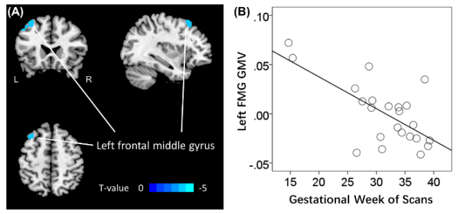
Figure 1. Brain region showing GMV in correlation with PMA. (A) Left frontal middle gyrus (FMG) GMV showing negative correlation with PMA; (B) Scatter plot of correlation between PMA and the left MFG GMV. PMA: post-menstrual age, TIV: total intracranial volume, GMV: gray matter volume. Note that the residuals are plotted here with age and TIV accounted for in the regression.
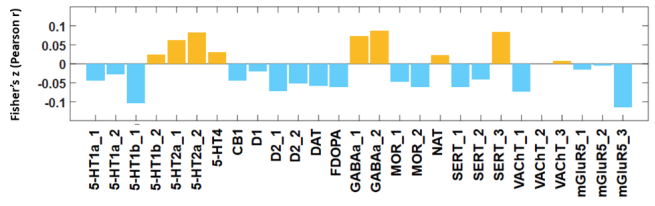
Figure 2. Correlations between T values of whole-brain regression of the GMV against PMA and neurotransmitter distribution maps, orange/blue each represents positive/negative Pearson's. Abbreviations: 5-HT,5-hydroxytryptamine (serotonin); CB1, cannabinoid type 1; D, dopamine receptor; DAT, dopamine transporter; FDOPA, fluorodopa, an analog of L-DOPA to assess the nigrostriatal dopamine system; GABAa, gamma-aminobutyric acid a; MOR, mu opioid receptor; NAT, noradrenaline transporter; SERT, serotonin transporter; VAChT, vesicular acetylcholine transporter; mGluR5, metabotropic glutamate type 5. PMA: post-menstrual age, GMV: gray matter volume.
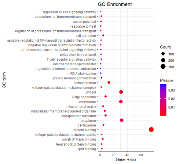
Figure 3. Bubble map of gene enrichment. The horizontal coordinate is the ratio of the number of genes associated with the Go term to the total number of target genes in the target gene set, %. The ordinate lists Go terms for significant gene enrichment, with the first 15 terms representing a specific Biological Process (BP), and the 16th to 25th terms being Cellular Component (CC). Terms 26-30 stand for Molecular Function (MF).
Information

