The large volume of toxic acid mine drainage wastewater generated from the pyritic oxidation of coal and gold mine result in serious environmental pollution because of the problem of waste disposal. The aim of this study is to use iron-rich raw acid mine drainage (RAMD) as a substitute to commercial reagent grade iron salt to synthesize iron nanoparticles. Chemical reduction method was employed to synthesize iron nanoparticles using sodium borohydride as reductant. The synthesized iron nanoparticles from RAMD and reagent grade iron salt solutions were quantified and characterized using analytical techniques such as ion chromatography (IC), Inductively coupled plasma-optical-emission spectroscopy (ICP-OES), X-ray diffraction (XRD), high resolution scanning electron microscopy (HRSEM), High resolution transmission electron microscopy-Selected area electron diffraction (HRTEM-SAED), X-ray fluorescence (XRF), Brunauer-Emmett-Teller (BET), Fourier Transform infrared (FTIR) spectroscopy, atomic force microscopy (AFM), and Thermogravimetric analysis (TGA). The ICP-OES result revealed high iron concentration (4784.13 mg/L) and IC sulphate concentration (27, 204. 72 mg/L that iron sulphate salt was present in the RAMD solution. XRD results identified magnetic pure iron mineral phase for both samples and the SEM results revealed spherical crystal particle morphology as long interwoven strand with beads. The HRTEM results revealed a bead-like necklace structure with average particle size of 28.48 ± 4.2 nm and 24.23 ± 2.17 nm for iron nanoparticles synthesized from RAMD (A) and ferric chloride (B) respectively. The XRF elemental composition of the synthesized nanoparticles revealed A (97.4%) and B (99.9%) iron (Fe). BET surface area results for A is 89 ± 3.13 m2/g and B is 93 ± 3.16 m2/g, FTIR results revealed O-H, CO2, Fe and FeO absorption peaks and the AFM results revealed more agglomeration in sample A than in B. The TGA of both synthesized iron nanoparticles were thermally stable. In conclusion, the iron-rich RAMD wastewater was found to be a good substitute for reagent grade iron salt use for making quality iron nanoparticles.
| Published in | International Journal of Materials Science and Applications (Volume 14, Issue 5) |
| DOI | 10.11648/j.ijmsa.20251405.14 |
| Page(s) | 212-223 |
| Creative Commons |
This is an Open Access article, distributed under the terms of the Creative Commons Attribution 4.0 International License (http://creativecommons.org/licenses/by/4.0/), which permits unrestricted use, distribution and reproduction in any medium or format, provided the original work is properly cited. |
| Copyright |
Copyright © The Author(s), 2025. Published by Science Publishing Group |
Raw Acid Mine Drainage, Chemical Reduction, Reductant, Iron Nanoparticles, Characterization
ELEMENTS | BAI (% atomic wt) | BFCI (% atomic wt) |
|---|---|---|
Fe | 70.08 ± 5.85 | 75.8 ± 3.96 |
O | 20.04 ± 0.6 | 24.11 ± 1.38 |
S | 8.26 ± 0.03 | ND |
Al | 1.66 ± 0.01 | ND |
Sample | Fe2O3 | SiO2 | Al2O3 | CaO |
|---|---|---|---|---|
BAI | 97.4 ± 0.01 | 0.07 ± 0.01 | 0.68 ± 0.01 | 0.15 ± 0.01 |
BFCI | 99.6 ± 0.03 | ND | ND | ND |
RAMD | Raw Acid Mine Drainage |
IC | Ion Chromatography |
ICP-OES | Inductively Coupled Plasma Optical Emission Spectroscopy |
XRD | X-ray Diffraction |
XRF | X-ray Fluorescence |
HRSEM | High Resolution Scanning Electron Microscopy |
HRTEM | High Resolution Transmission Electron Microscopy |
BET | Brunauer-Emmett-Teller |
FTIR | Fourier Transform Infra-red Spectroscopy |
AFM | Atomic Force Microscopy |
TGA | Thermogravimetric Analysis |
AMD | Acid Mine Drainage |
PTE | Potentially Toxic Elements |
PAHs | Polycyclic Aromatic Hydrocarbons |
PHCs | Polyhydrocarbons |
NaBH4 | Sodium Borohydride |
ATR | Attenuated Total Reflectance |
BAI | Borohydride AMD Iron Nanoparticles |
BFCI | Borohydride Ferric Chloride Iron Nanoparticles |
SAED | Selective Area Electron Diffraction |
EDS | Energy Dispersive Spectroscopy |
EDX | Energy Dispersive X-ray |
DSC | Defferential Scanning Calorimetry |
| [1] | Retka, J., Rzepa, G., Bajda, T., & Drewniak, L. (2020). The use of mining waste materials for the treatment of acid and alkaline mine wastewater. Minerals, 10(12), 1061, 2020. |
| [2] | Alegbe, M. J., Ayanda, O. S., Ndungu, P., Nechaev, A., Fatoba, O. O., & Petrik, L. F. (2019). Physicochemical characteristics of acid mine drainage, simultaneous remediation and use as feedstock for value added products. Journal of Environmental Chemical Engineering, 7(3), 103097, 2019. |
| [3] | Hatar, H., Rahim, S. A., Razi, W. M., & Sahrani, F. K. November). (2013). Heavy metals content in acid mine drainage at abandoned and active mining area. In AIP conference proceedings (Vol. 1571, No. 1, pp. 641-646). American Institute of Physics, 2013. |
| [4] | McCarthy, T. S. (2011). The impact of acid mine drainage in South Africa. South African journal of science, 107, 5-6, 2011. |
| [5] | Cheng, H., Hu, Y., Luo, J., Xu, B., & Zhao, J. (2009). Geochemical processes controlling fate and transport of arsenic in acid mine drainage (AMD) and natural systems. Journal of hazardous materials, 165(1-3), 13-26, 2009. |
| [6] | Aghaei, E., Wang, Z., Tadesse, B., Tabelin, C. B., Quadir, Z., & Alorro, R. D. (2021). Performance evaluation of Fe-Al bimetallic particles for the removal of potentially toxic elements from combined acid mine drainage-effluents from refractory gold ore processing. Minerals, 11(6), 590, 2021. |
| [7] | Kefeni, K. K., Msagati, T. A., & Mamba, B. B. (2017). Acid mine drainage: Prevention, treatment options, and resource recovery: A review. Journal of cleaner production, 151, 475-493, 2017. |
| [8] | Naidu, G., Ryu, S., Thiruvenkatachari, R., Choi, Y., Jeong, S., & Vigneswaran, S. A (2019). Critical review on remediation, reuse, and resource recovery from acid mine drainage. Environmental pollution, 247, 1110-1124, 2019. |
| [9] | Sánchez, M., Sabio, L., Gálvez, N., Capdevila, M., & Dominguez‐Vera, J. M. (2017). Iron chemistry at the service of life. IUBMB life, 69(6), 382-388, 2017. |
| [10] | Pasinszki, T., & Krebsz, M. (2020). Synthesis and application of zero-valent iron nanoparticles in water treatment, environmental remediation, catalysis, and their biological effects. Nanomaterials, 10(5), 917, 2020. |
| [11] | Stoia, M., Tamaş, A., Rusu, G., & Moroşanu, J. (2016). Synthesis of Magnetic Iron Oxides from Ferrous Sulfate and Substitutes Amines. Studia Universitatis Babes-Bolyai, Chemia, 61(4), 2016. |
| [12] | Javed, R., Zia, M., Naz, S., Aisida, S. O., Ain, N. U., & Ao, Q. (2020). Role of capping agents in the application of nanoparticles in biomedicine and environmental remediation: recent trends and future prospects. Journal of Nanobiotechnology, 18, 1-15, 2020. |
| [13] | Szczyglewska, P., Feliczak-Guzik, A., & Nowak, I. (2023). Nanotechnology-general aspects: A chemical reduction approach to the synthesis of nanoparticles. Molecules, 28(13), 4932, 2023. |
| [14] | Ijaz, I., Gilani, E., Nazir, A., & Bukhari, A. (2020). Detail review on chemical, physical and green synthesis, classification, characterizations and applications of nanoparticles. Green Chemistry Letters and Reviews, 13(3), 223-245, 2020. |
| [15] | Shavel, A., Rodríguez‐González, B., Spasova, M., Farle, M., & Liz‐Marzán, L. M. (2007). Synthesis and characterization of iron/iron oxide core/shell nanocubes. Advanced functional materials, 17(18), 3870-3876, 2007. |
| [16] | Bhaumik, A., Samanta, S., & Mal, N. K. (2005). Iron oxide nanoparticles stabilized inside highly ordered mesoporous silica. Pramana, 65(5), 855-862, 2005. |
| [17] | Ghorbani, H. R., Safekordi, A. A., Attar, H., Rezayat Sorkhabadi, S. M. (2011). Biological and non-biological methods for silver nanoparticles synthesis. Chem. Biochem. Eng. Q. 25 (3), 317-326, 2011. |
| [18] | Crane, R., & Scott, T. (2012). Nanoscale zero-valent iron: Future prospects for an emerging water treatment technology. Journal of Hazardous Materials, 211, 112-125, 2012. |
| [19] | Sun, Y. P., Li, X. Q., Cao, J., Zhang, W. X., & Wang, H. P. (2006). Characterization of zero-valent iron nanoparticles. Advances in colloid and interface science, 120(1-3), 47-56, 2006. |
| [20] | Mukherjee, J., & Gupta, M. N. (2016). Lipase coated clusters of iron oxide nanoparticles for biodiesel synthesis in a solvent free medium. Bioresource technology, 209, 166-171, 2016. |
| [21] | Katiyar, J. K., Sharma, A. K., & Pandey, B. (2018). Synthesis of iron-copper alloy using electrical discharge machining. Materials and Manufacturing Processes, 33(14), 1531-1538, 2018. |
| [22] | Niculescu, A. G., Chircov, C., & Grumezescu, A. M. (2022). Magnetite nanoparticles: Synthesis methods-A comparative review. Methods, 199, 16-27, 2022. |
| [23] | Darvina, Y., Yulfriska, N., Rifai, H., Dwiridal, L., & Ramli, R. April). (2019). Synthesis of magnetite nanoparticles from iron sand by ball-milling. In Journal of Physics: Conference Series (Vol. 1185, No. 1, p. 012-017). IOP Publishing, 2019. |
| [24] | Zhang, Z., & Wen, G. (2020). Synthesis and characterization of carbon-encapsulated magnetite, martensite and iron nanoparticles by high-energy ball milling method. Materials Characterization, 167, 110502,.2020. |
| [25] | Alegbe, M. J., Moronkola, B. A., Osundiya, M. O., Adekolurejo, E., Ajewole, B. S., & Petrik, L. F. (2022). Synthesis of iron nanoparticles from acid mine drainage using hydrazine as reductant. Am. J. Chem, 12, 23-31, 2022. |
| [26] | Chekli, L., Bayatsarmadi, B., Sekine, R., Sarkar, B., Shen, A. M., Scheckel, K. G.,... & Donner, E. (2016). Analytical characterisation of nanoscale zero-valent iron: A methodological review. Analytica chimica acta, 903, 13-35, 2016. |
| [27] | Madhavi, V., Prasad, T. N. V. K. V., & Madhavi, G. (2013). Synthesis and spectral characterization of iron based micro and nanoparticles. Iranica Journal of Energy & Environment, 4(4), 2013. |
| [28] | Mitar, I., Guć, L., Soldin, Ž., Vrankić, M., Paut, A., Prkić, A., & Krehula, S. (2021). Rapid microwave method for synthesis of iron oxide particles under specific conditions. Crystals, 11(4), 383, 2021. |
| [29] | Henam, S. D., Ahmad, F., Shah, M. A., Parveen, S., & Wani, A. H. (2019). Microwave synthesis of nanoparticles and their antifungal activities. Spectrochimica Acta Part A: Molecular and Biomolecular Spectroscopy, 213, 337-341, 2019. |
| [30] | Aivazoglou, E., Metaxa, E., & Hristoforou, E. (2018). Microwave-assisted synthesis of iron oxide nanoparticles in biocompatible organic environment. AIP Advances, 8(4), 2018. |
| [31] | Lu, L., Ai, Z., Li, J., Zheng, Z., Li, Q., and Zhang, L. (2007). Synthesis and Characterization of Fe-Fe2O3 Core-Shell Nanowires and Nanonecklaces. crystal growth and design, 7(2), 459-464, 2007. |
| [32] | Kandpal, N. D., Sah, N., Loshali, R., Joshi, R., & Prasad, J. (2014). Co-precipitation method of synthesis and characterization of iron oxide nanoparticles, 2014. |
| [33] | Khalil, M. I. (2015). Co-precipitation in aqueous solution synthesis of magnetite nanoparticles using iron (III) salts as precursors. Arabian Journal of Chemistry, 8(2), 279-284, 2015. |
| [34] | Rahmawati, R., Taufiq, A., Sunaryono, S., Fuad, A., Yuliarto, B., Suyatman, S., & Kurniadi, D. (2018). Synthesis of magnetite (Fe3O4) nanoparticles from iron sands by coprecipitation-ultrasonic irradiation methods. J. Mater. Environ. Sci, 9(3), 155-160, 2018. |
| [35] | Takai, Z. I., Mustafa, M. K., Asman, S., & Sekak, K. A. (2019). Preparation and characterization of magnetite (Fe3O4) nanoparticles by sol-gel method. Int. J. Nanoelectron. Mater, 12, 37-46, 2019. |
| [36] | Khan, I., Morishita, S., Higashinaka, R., Matsuda, T. D., Aoki, Y., Kuzmann, E.,... & Kubuki, S. (2021). Synthesis, characterization and magnetic properties of ε-Fe2O3 nanoparticles prepared by sol-gel method. Journal of magnetism and magnetic materials, 538, 168264, 2021. |
| [37] | Taha, A. B., Essa, M. S., & Chiad, B. T. (2022). Spectroscopic study of iron oxide nanoparticles synthesized via hydrothermal method. Chem. Methodol, 6(12), 977-984, 2022. |
| [38] | Long, L. Q., Hue, T. T. B., Hoan, N. X., Cuong, L. V., Thang, P. D., Hoang, T., & Truc, T. A.(2016). Growth mechanism and stability of magnetite nanoparticles synthesized by the hydrothermal method. Journal of Nanoscience and Nanotechnology, 16(7), 7373-7379, 2016. |
| [39] | Yadav, V. K., Ali, D., Khan, S. H., Gnanamoorthy, G., Choudhary, N., Yadav, K. K.,... & Manhrdas, S. (2020). Synthesis and characterization of amorphous iron oxide nanoparticles by the sonochemical method and their application for the remediation of heavy metals from wastewater. Nanomaterials, 10(8), 1551, 2020. |
| [40] | Yadav, V. K., Ali, D., Khan, S. H., Gnanamoorthy, G., Choudhary, N., Yadav, K. K., ... & Manhrdas, S. (2020). Synthesis and characterization of amorphous iron oxide nanoparticles by the sonochemical method and their application for the remediation of heavy metals from wastewater. Nanomaterials. 10(8), 1551. |
| [41] | Shin, N., Saravanakumar, K., & Wang, M. H. Sonochemical mediated synthesis of iron oxide (Fe 3 O 4 and Fe 2 O 3) nanoparticles and their characterization, cytotoxicity and antibacterial properties. Journal of Cluster Science, 30, 669-675, 2019. |
| [42] | Wang, Y., Nkurikiyimfura, I., & Pan, Z. (2015). Sonochemical synthesis of magnetic nanoparticles. Chemical Engineering Communications, 202(5), 616-621, 2015. |
| [43] | Glasgow, W., Fellows, B., Qi, B., Darroudi, T., Kitchens, C., Ye, L.,... & Mefford, O. T. (2016). Continuous synthesis of iron oxide (Fe3O4) nanoparticles via thermal decomposition. Particuology, 26, 47-53, 2016. |
| [44] | Unni, M., Uhl, A. M., Savliwala, S., Savitzky, B. H., Dhavalikar, R., Garraud, N.,... & Rinaldi, C. (2017). Thermal decomposition synthesis of iron oxide nanoparticles with diminished magnetic dead layer by controlled addition of oxygen. ACS nano, 11(2), 2284-2303, 2017. |
| [45] | Wei & Viadero, Jr. (2007). Synthesis of magnetite nanoparticles with ferric iron recovered from acid mine drainage: Implications for environmental engineering. Colloids and surfaces A: physicochem. Eng. Aspects, 294, 280-282, 2007. |
| [46] | Wang, X. Y., Narita, A., & Müllen, K. (2017). Precision synthesis versus bulk-scale fabrication of graphenes. Nature Reviews Chemistry, 2(1), 0100., 2017. |
| [47] | Minnich, A., Dresselhaus, M. S., Ren, Z. F., & Chen, G. (2009). Bulk nanostructured thermoelectric materials: current research and future prospects. Energy & Environmental Science, 2(5), 466-479, 2009. |
| [48] | Li, X. Q., & Zhang, W. X. (2006a). Iron nanoparticles: The core− shell structure and unique properties for Ni (II) sequestration. Langmuir, 22(10), 4638-4642, 2006a. |
| [49] | Lin, K.-S., Chang, N.-B., and Chuang, T.-D. (2008). Fine structure characterization of zero-valent iron nanoparticles for decontamination of nitrites and nitrates in wastewater and groundwater. Science and Technology of Advanced Materials, 9(2), 025015, 2008. |
| [50] | Yuvakkumar, R., Elango, V., Rajendran, V., and Kannan, N. (2011). Preparation and characterization of zero valent iron nanoparticles. Digest Journal of Nanomaterials and Biostructures, 6(4, October-December), 1771-1776, 2011. |
| [51] | Nurmi, J. T., Tratnyek, P. G., Sarathy, V., Baer, D. R., amonette, J. E., Specher, K., Wang, C., Linenhan, J. C., Matson, D. W., Leepenn, R., and Driessen, M. D. (2005). Characterization and properties of metallic iron nanoparticles: Spectroscopy, electrochemistry and kinetics. Environ. Sci. Technol, 39, 1221-1230, 2005. |
| [52] | Ghosh Chaudhuri, R., & Paria, S. (2012). Core/shell nanoparticles: classes, properties, synthesis mechanisms, characterization, and applications. Chemical reviews, 112(4), 2373-2433, 2012. |
| [53] | Li, X.-q., Elliott, D. W., & Zhang, W.-x. (2006b). Zero-valent iron nanoparticles for abatement of environmental pollutants: materials and engineering aspects. Critical Reviews in Solid State and Materials Sciences, 31(4), 111-122, 2006b. |
| [54] | Wang, C. B., & Zhang, W. X. (1997). Synthesizing nanoscale iron particles for rapid and complete Dechlorination of TCE and PCBS. Environmental science and technology, 31(7), 2154-2156, 1997. |
| [55] | Cheng, J., Ma, R., Chen, X., Shi, D., Liu, F., & Zhang, X. (2011). Effect of ferric ions on the morphology and size of magnetite nanocrystals synthesized by ultrasonic irradiation. Crystal Research and Technology, 46(7), 723-730, 2011. |
| [56] | Menezes, J. C. S. S., Silva, R. A., Arce, I. S., & Schneider, I. A. H. (2010). Production of a poly-alumino-iron sulphate coagulant by chemical precipitation of a coal mining acid drainage. Mineral engineering, 23, 249-251, 2010. |
| [57] | Kanel, S., Greneche, J., & Choi, H. (2006). Arsenic (V) removal from groundwater using nanoscale zero valent ironas a colloidal reactive barrier material. Environ. Sci. Technol., 40, 2045-2050, 2006. |
| [58] | Kazemzadeh, H., Ataie, A., & Rashchi, F. (2012). Synthesis of magnetite nano-particles by reverse co-precipitation. In International Journal of Modern Physics: Conference Series (Vol. 5, pp. 160-167). World Scientific Publishing Company, 2012. |
| [59] | Celebi, O., Üzüm, Ç., Shahwan, T., & Erten, H. N. (2007). A radiotracer study of the adsorption behavior of aqueous Ba2+ ions on nanoparticles of zero-valent iron. Journal of Hazardous Materials, 148(3), 761-767, 2007. |
| [60] | Giraldo, L., & Moreno-Piraján, J. C. Synthesis of magnetite nanoparticles and exploring their application in the removal of Pt2+ and Au3+ ions from aqueous solutions. European Chemical Bulletin, 2(7), 445-452, 2013. |
| [61] | Karatapanis, A. E., Petrakis, D. E., & Stalikas, C. D. A layered magnetic iron/iron oxide nanoscavenger for the analytical enrichment of ng-L− 1 concentration levels of heavy metals from water. Analytica Chimica Acta, 726, 22-27, 2012. |
| [62] | Wang, Q., Kanel, S. R., Park, H., Ryu, A., & Choi, H. Controllable synthesis, characterization, and magnetic properties of nanoscale zerovalent iron with specific high Brunauer-Emmett-Teller surface area Received:. J Nanopart Res. 11, 749-755, 2009. |
| [63] | Li, Y., & Zhang, F. S. Catalytic oxidation of Methyl Orange by an amorphous FeOOH catalyst developed from a high iron-containing fly ash. Chemical Engineering Journal, 158(2), 148-153, 2010. |
| [64] | Chmielewská, E., Tylus, W., Drábik, M., Majzlan, J., Kravčak, J., Williams, C., & Čaplovič, Ľ. Structure investigation of nano-FeO (OH) modified clinoptilolite tuff for antimony removal. Microporous and Mesoporous Materials, 248, 222-233, 2017. |
| [65] | Gotić, M., Dražić, G., & Musić, S. Hydrothermal synthesis of α-Fe2O3 nanorings with the help of divalent metal cations, Mn2+, Cu2+, Zn2+ and Ni2+. Journal of Molecular Structure, 993(1-3), 167-176, 2011. |
| [66] | Roonasi, P. (2007). Adsorption and surface reaction properties of synthesized magnetite nano-particles (Doctoral dissertation, Luleå tekniska universitet), 2007. |
| [67] | Sundrarajan, M., & Ramalakshmi, M. Novel cubic magnetite nanoparticle synthesis using room temperature ionic liquid. Journal of Chemistry, 9, 1070-1076, 2012. |
| [68] | Cai, W., & Wan, J. Facile synthesis of superparamagnetic magnetite nanoparticles in liquid polyols. Journal of Colloid and Interface Science, 305(2), 366-370, 2007. |
| [69] | Duhan, S., & Devi, S. Synthesis and structural characterization of iron oxide-silica nanocomposites prepared by the sol gel method. International Journal of Electronics Engineering, 2(1), 89-92, 2010. |
| [70] | Ghosh, M. K., Poinern, G. E. J., Issa, T. B., & Singh, P. Arsenic adsorption on goethite nanoparticles produced through hydrazine sulfate assisted synthesis method. Korean Journal of Chemical Engineering, 29, 95-102, 2012. |
| [71] | Panwar, V., Kumar, P., Bansal, A., Ray, S. S., & Jain, S. L. PEGylated magnetic nanoparticles (PEG@ Fe3O4) as cost effective alternative for oxidative cyanation of tertiary amines via CH activation. Applied catalysis A: general, 498, 25-31, 2015. |
APA Style
John, A. M., Adekemi, M. B., Ojo, F. O., Felicia, P. L. (2025). Synthesis and Characterization of Iron Nanoparticles from Acid Mine Drainage Using Sodium Borohydride as Reductant. International Journal of Materials Science and Applications, 14(5), 212-223. https://doi.org/10.11648/j.ijmsa.20251405.14
ACS Style
John, A. M.; Adekemi, M. B.; Ojo, F. O.; Felicia, P. L. Synthesis and Characterization of Iron Nanoparticles from Acid Mine Drainage Using Sodium Borohydride as Reductant. Int. J. Mater. Sci. Appl. 2025, 14(5), 212-223. doi: 10.11648/j.ijmsa.20251405.14
AMA Style
John AM, Adekemi MB, Ojo FO, Felicia PL. Synthesis and Characterization of Iron Nanoparticles from Acid Mine Drainage Using Sodium Borohydride as Reductant. Int J Mater Sci Appl. 2025;14(5):212-223. doi: 10.11648/j.ijmsa.20251405.14
@article{10.11648/j.ijmsa.20251405.14,
  author = {Alegbe Monday John and Moronkola Bridget Adekemi and Fatoba Olarenwaju Ojo and Petrik Leslie Felicia},
  title = {Synthesis and Characterization of Iron Nanoparticles from Acid Mine Drainage Using Sodium Borohydride as Reductant
},
  journal = {International Journal of Materials Science and Applications},
  volume = {14},
  number = {5},
  pages = {212-223},
  doi = {10.11648/j.ijmsa.20251405.14},
  url = {https://doi.org/10.11648/j.ijmsa.20251405.14},
  eprint = {https://article.sciencepublishinggroup.com/pdf/10.11648.j.ijmsa.20251405.14},
  abstract = {The large volume of toxic acid mine drainage wastewater generated from the pyritic oxidation of coal and gold mine result in serious environmental pollution because of the problem of waste disposal. The aim of this study is to use iron-rich raw acid mine drainage (RAMD) as a substitute to commercial reagent grade iron salt to synthesize iron nanoparticles. Chemical reduction method was employed to synthesize iron nanoparticles using sodium borohydride as reductant. The synthesized iron nanoparticles from RAMD and reagent grade iron salt solutions were quantified and characterized using analytical techniques such as ion chromatography (IC), Inductively coupled plasma-optical-emission spectroscopy (ICP-OES), X-ray diffraction (XRD), high resolution scanning electron microscopy (HRSEM), High resolution transmission electron microscopy-Selected area electron diffraction (HRTEM-SAED), X-ray fluorescence (XRF), Brunauer-Emmett-Teller (BET), Fourier Transform infrared (FTIR) spectroscopy, atomic force microscopy (AFM), and Thermogravimetric analysis (TGA). The ICP-OES result revealed high iron concentration (4784.13 mg/L) and IC sulphate concentration (27, 204. 72 mg/L that iron sulphate salt was present in the RAMD solution. XRD results identified magnetic pure iron mineral phase for both samples and the SEM results revealed spherical crystal particle morphology as long interwoven strand with beads. The HRTEM results revealed a bead-like necklace structure with average particle size of 28.48 ± 4.2 nm and 24.23 ± 2.17 nm for iron nanoparticles synthesized from RAMD (A) and ferric chloride (B) respectively. The XRF elemental composition of the synthesized nanoparticles revealed A (97.4%) and B (99.9%) iron (Fe). BET surface area results for A is 89 ± 3.13 m2/g and B is 93 ± 3.16 m2/g, FTIR results revealed O-H, CO2, Fe and FeO absorption peaks and the AFM results revealed more agglomeration in sample A than in B. The TGA of both synthesized iron nanoparticles were thermally stable. In conclusion, the iron-rich RAMD wastewater was found to be a good substitute for reagent grade iron salt use for making quality iron nanoparticles.
},
 year = {2025}
}
TY  - JOUR
T1  - Synthesis and Characterization of Iron Nanoparticles from Acid Mine Drainage Using Sodium Borohydride as Reductant

AU  - Alegbe Monday John
AU  - Moronkola Bridget Adekemi
AU  - Fatoba Olarenwaju Ojo
AU  - Petrik Leslie Felicia
Y1  - 2025/09/23
PY  - 2025
N1  - https://doi.org/10.11648/j.ijmsa.20251405.14
DO  - 10.11648/j.ijmsa.20251405.14
T2  - International Journal of Materials Science and Applications
JF  - International Journal of Materials Science and Applications
JO  - International Journal of Materials Science and Applications
SP  - 212
EP  - 223
PB  - Science Publishing Group
SN  - 2327-2643
UR  - https://doi.org/10.11648/j.ijmsa.20251405.14
AB  - The large volume of toxic acid mine drainage wastewater generated from the pyritic oxidation of coal and gold mine result in serious environmental pollution because of the problem of waste disposal. The aim of this study is to use iron-rich raw acid mine drainage (RAMD) as a substitute to commercial reagent grade iron salt to synthesize iron nanoparticles. Chemical reduction method was employed to synthesize iron nanoparticles using sodium borohydride as reductant. The synthesized iron nanoparticles from RAMD and reagent grade iron salt solutions were quantified and characterized using analytical techniques such as ion chromatography (IC), Inductively coupled plasma-optical-emission spectroscopy (ICP-OES), X-ray diffraction (XRD), high resolution scanning electron microscopy (HRSEM), High resolution transmission electron microscopy-Selected area electron diffraction (HRTEM-SAED), X-ray fluorescence (XRF), Brunauer-Emmett-Teller (BET), Fourier Transform infrared (FTIR) spectroscopy, atomic force microscopy (AFM), and Thermogravimetric analysis (TGA). The ICP-OES result revealed high iron concentration (4784.13 mg/L) and IC sulphate concentration (27, 204. 72 mg/L that iron sulphate salt was present in the RAMD solution. XRD results identified magnetic pure iron mineral phase for both samples and the SEM results revealed spherical crystal particle morphology as long interwoven strand with beads. The HRTEM results revealed a bead-like necklace structure with average particle size of 28.48 ± 4.2 nm and 24.23 ± 2.17 nm for iron nanoparticles synthesized from RAMD (A) and ferric chloride (B) respectively. The XRF elemental composition of the synthesized nanoparticles revealed A (97.4%) and B (99.9%) iron (Fe). BET surface area results for A is 89 ± 3.13 m2/g and B is 93 ± 3.16 m2/g, FTIR results revealed O-H, CO2, Fe and FeO absorption peaks and the AFM results revealed more agglomeration in sample A than in B. The TGA of both synthesized iron nanoparticles were thermally stable. In conclusion, the iron-rich RAMD wastewater was found to be a good substitute for reagent grade iron salt use for making quality iron nanoparticles.

VL  - 14
IS  - 5
ER  -
Department of Chemistry, Lagos State University, Lagos, Nigeria; Department of Chemistry, University of the Western Cape, Bellville, South Africa
Department of Chemistry, Lagos State University, Lagos, Nigeria
Department of Chemistry, University of the Western Cape, Bellville, South Africa
Department of Chemistry, University of the Western Cape, Bellville, South Africa
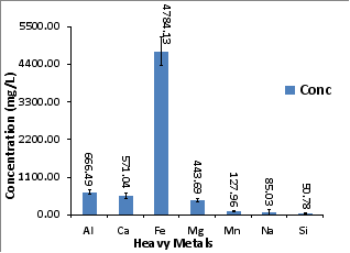
Figure 1. ICP concentration (mg/g) of major species in RAMD. Experimental conditions: concentration of RAMD = 4784.13 mg/L, pH = 2.06, EC = 7.92 and TDS = 6/14, n = 3.
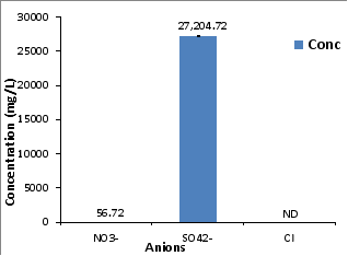
Figure 2. IC concentration (mg/g) of major anions in RAMD. Experimental conditions: concentration of RAMD SO42- anions = 27, 204.72 mg/L, pH = 2.06, EC = 7.92 and TDS = 6.14, n = 3.
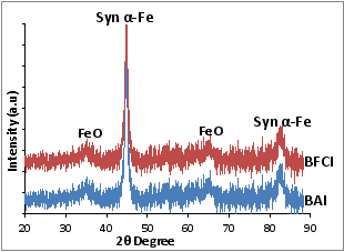
Figure 3. Powder X-Ray Diffraction pattern of synthesized iron nanoparticles BAI and BFCI. Experimental conditions: Experimental conditions: concentration of RAMD SO42- anions = 27, 204.72 mg/L, pH = 2.06, EC = 7.92 and TDS = 6.14, n = 3.
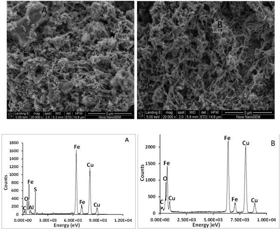
Figure 4. SEM image and EDS spectral analysis of BAI (A) and BFCI (B) nanoparticles. Experimental conditions: concentration of RAMD SO42- anions = 27, 204.72 mg/L, pH = 2.06, EC = 7.92 and TDS = 6.14, n = 3.
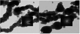
Figure 5. HRTEM-SAED morphology of BAI (A) and BFCI (B) nanoparticles. Experimental conditions: concentration of RAMD SO42- anions = 27, 204.72 mg/L, pH = 2.06, EC = 7.92 and TDS = 6.14, n = 3.
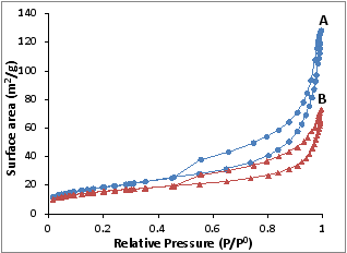
Figure 6. BET surface area N2 adsorption-desorption hysteresis loop of BAI and BFCI nanoparticles. Experimental conditions: concentration of RAMD SO42- anions = 27, 204.72 mg/L, pH = 2.06, EC = 7.92 and TDS = 6.14, n = 3.
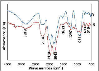
Figure 7. FTIR spectral analysis synthesized magnetic iron nanoparticles BAI (A) and BFCI (B). Experimental conditions: concentration of RAMD SO42- anions = 27, 204.72 mg/L, pH = 2.06, EC = 7.92 and TDS = 6.14, n = 3.
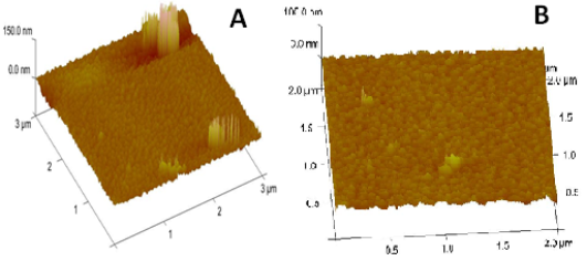
Figure 8. AFM analysis of synthesized magnetic iron nanoparticles BAI (A) and BFCI (B). Experimental conditions: concentration of RAMD SO42- anions = 27, 204.72 mg/L, pH = 2.06, EC = 7.92 and TDS = 6.14, n = 3.
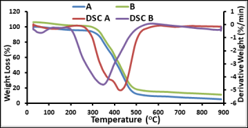
Figure 9. TGA analysis of synthesized magnetic iron nanoparticles BAI (A) and BFCI (B). Experimental conditions: concentration of RAMD SO42- anions = 27, 204.72 mg/L, pH = 2.06, EC = 7.92 and TDS = 6.14, n = 3.
Information

