Background: Measurements of the normal portal venous dimension in a species population is so crucial. portal vein can be measured by several methods for assessing different parameters, Computed topography (CT) and sonographic imaging are common examples. The diagnosis of portal hypertension depends on the transverse portal vein diameter (normal diameter from 6-15 mm). The aim of this study is to assess the portal vein diameter and correlate the diameter with age, sex, weight, height and BMI in Sudanese populations. Methodology: This was a descriptive cross-sectional study that included a sample of Sudanese adults who were requested to have routine abdominal sonographic scanning due to reasons not related to liver or portal vein problems. The study was carried out at the Radiology Department of Qatar Alnada Hospital, Umbadda, Khartoum, Sudan 2022. Results: In this study, 376 participants were included of which more than half were females (56.1%, n=211), while 165 (43.9%) were males. The mean age of the participants was 38.12 + 15.57 years. The mean portal vein diameter was 10.79 + 1.27 millimeters. From this study the portal vein diameter varied with age (p value = 0.000), weight (p value = 0.002), height (p value = 0.000) and sex (p value = 0.000). It is not related to BMI (p value = 0.3). Conclusion: This study revealed that the portal vein diameter has positive correlation with age, sex, height, and weight of the person and has no correlation with the BMI.
This is an Open Access article, distributed under the terms of the Creative Commons Attribution 4.0 International License (http://creativecommons.org/licenses/by/4.0/), which permits unrestricted use, distribution and reproduction in any medium or format, provided the original work is properly cited.
The splenic vein and superior mesenteric vein meet beneath the pancreas neck to produce the portal vein. In front of the inferior vena cava, it is situated behind the pancreas and the first section of the duodenum. The smaller omentum, which is situated below the hepatic artery and the bile duct, creates the anterior boundary of the epiploic foramen and extends virtually vertically upward in the free edge until it reaches the porta hepatis. From this point, the right and left branches emerge and enter the respective halves of the liver. 75% of the liver's blood supply is located there
[1]
Keith L. Moore, Arthur F. Dalley II, Anne M. R. Agur. Clinical Oriented Anatomy. 7th edition. Philadelphia: Lippincott Williams &Wilkins; 2014. p262, 268.
[2]
Richard S. Snell. Clinical Anatomy by Regions. 9th edition. Philadelphia: Lippincott Williams &Wilkins; 2012. p 194, 196.
[1, 2]
.
The right and left gastric veins, as well as the superior pancreaticoduodenal vein, are drained by the portal vein
[3]
Sinnatamby CS, Last RJ, editors. Last’s anatomy: regional and applied. 12th ed. Edinburgh; New York: Churchill Livingstone/Elsevier; 2011. 548 p266, 267.
[3]
. The portal vein has an 8 cm length and an 8 mm width. The five locations for portosystemic anastomosis are the periumbilical region, the liver's bare area, the upper end of the anal canal, and the retroperitoneal sections
[3]
Sinnatamby CS, Last RJ, editors. Last’s anatomy: regional and applied. 12th ed. Edinburgh; New York: Churchill Livingstone/Elsevier; 2011. 548 p266, 267.
[3]
.
The diameter of the vein and the thickness of the portal vein wall both increase when the portal vein pressure rises
[4]
Abdel-Wahab MF, Esmat G, Farrag A, El-Boraey YA, Strickland GT. Grading of Hepatic Schistosomiasis by the Use of Ultrasonography. Am J Trop Med Hyg. 1992 Apr 1; 46(4): 403–8.
. The transverse portal vein diameter, which is normally between 6 and 15 millimeters in size, is what determines the diagnosis of portal hypertension
[5]
Weinreb J, Kumari S, Phillips G, Pochaczevsky R. Portal vein measurements by real-time sonography. Am J Roentgenol. 1982; 139(3): 497–499.
[5]
. If the diameter exceeds 15 millimeters, the diagnosis is suggested
[6]
Lafortune M, Marleau D, Breton G, Viallet A, Lavoie P, Huet PM. Portal venous system measurements in portal hypertension. Radiology. 1984; 151(1): 27–30.
According to research done in 2013 by Jayanta Bhattacharya et al. in India, the average portal vein diameter in males was 10.37 + 1.03 mm while in females, it was 9.82 + 1.01 mm. They stated that when both sexs were combined, the PVD was 10.02 + 0.89mm
[7]
Bhattacharya J, Das A, Bhowmik A. Sonographical assessment of portal vein diameter in Northern part of West Bengal, India. Octa Journal of Environmental Research. 2013 Sep 1; 1(3).
[7]
.
In 2015, a study was undertaken in Nigeria by Usman et al. The mean age of the participants in the study was 43.78 12.97 years, with 187 (74.8%) males and 63 (25.2%) females between the ages of 19 and 77. The primary PV's mean diameter was 10.87 mm plus or minus 0.81. Age and respiratory phases were linked with the PV diameter (P 0.05). PV diameters in males and females were statistically different (P 0.05), with female values being higher
[8]
Usman AU, Ibinaiye P, Ahidjo A, Tahir A, Sa'ad ST, Mustapha Z, Tahir N, Garko S. Determination of normal portal vein diameter on ultrasound scan among adults in northeastern Nigeria. Archives of International Surgery. 2015 Jul 1; 5(3): 143.
[8]
.
Geleto et al in Ethiopia in 2016 came to the conclusion that there were 195 people there. The median age of the participants was 35 years, and 121 of them (62.1%) were men. With a respirophasic variance of 25.6% and a normal mean portal vein width of 10.6 mm 1.8 SD, the study's findings are clear. Age and sex appeared to have a substantial impact on the normal mean portal vein diameter
[9]
Geleto G, Getnet W, Tewelde T. Mean normal portal vein diameter using sonography among clients coming to radiology department of Jimma University Hospital, Southwest Ethiopia. Ethiopian journal of health sciences. 2016; 26(3): 237-42.
Luntsi et al in 2017 reported that for both sexes, the mean portal vein diameter in Nigeria was 9.60 1.41 mm. Males had a mean value of 9.71 1.42 mm while females had a mean value of 9.35 1.46 mm. The PV diameter and the anthropometric measurements showed a positive connection
[10]
Luntsi G, Sani M, Zira JD, Ivor NC, Garba SH. Sonographic assessment of the portal vein diameter in apparently healthy adults in a Northern Nigerian population. African Health Sciences. 2016; 16(4): 1163-8.
In 2017, Gareeballah et al used ultrasonography (US) to determine the portal vein diameter (PVD) in Sudanese patients. According to the survey, the average PVD in the Sudanese population is 10.7 1.47 mm. Males have a somewhat larger mean PVD than females have. The study discovered that there was a strong positive link between portal vein diameter and height and weight but no significant correlation between portal vein diameter and participant age or body mass index
[11]
Gareeballah A, Hassan IA, Elzaki M, Ibraheem SS, Abelwahab B, Siddig A. Measurement of normal portal vein using ultrasound in Sudanese. Glob Adv Res J Med Med Sci. 2017; 6: 336-40.
[11]
.
According to Jha et al in Nepal in 2023, the mean portal vein width was 9.57 0.66mm for both sexes. The PV diameter and age, height, weight, and body mass index all showed positive correlations (P 0.05)
[12]
Jha AK, Shah SS, Sah D, Akthar MK, Mahato RK, Rimal B, Chaurasiya C, Jha RK, Thakur AK, Adhikari T. Measurement of Normal Portal Vein Diameter by Ultrasound. Journal of National Medical College. 2023 Jul 30; 8(1): 17-20.
For the reason of comparison of the results obtained by the studies mentioned. (table 1)
Table 1. Summarizes the PVD in different populations.
Study
Country
Mean of PVD
Comments
Bhattacharya et al
[7]
Bhattacharya J, Das A, Bhowmik A. Sonographical assessment of portal vein diameter in Northern part of West Bengal, India. Octa Journal of Environmental Research. 2013 Sep 1; 1(3).
[7]
India
10.02 mm + 0.89
The PV values higher in males
Usman et al
[8]
Usman AU, Ibinaiye P, Ahidjo A, Tahir A, Sa'ad ST, Mustapha Z, Tahir N, Garko S. Determination of normal portal vein diameter on ultrasound scan among adults in northeastern Nigeria. Archives of International Surgery. 2015 Jul 1; 5(3): 143.
[8]
Nigeria
10.87 mm ± 0.81
Positive correlation with age. The PV diameter higher in female.
Geleto et al
[9]
Geleto G, Getnet W, Tewelde T. Mean normal portal vein diameter using sonography among clients coming to radiology department of Jimma University Hospital, Southwest Ethiopia. Ethiopian journal of health sciences. 2016; 26(3): 237-42.
The normal mean portal vein diameter seemed to have varied significantly by age and sex.
Luntsi et al
[10]
Luntsi G, Sani M, Zira JD, Ivor NC, Garba SH. Sonographic assessment of the portal vein diameter in apparently healthy adults in a Northern Nigerian population. African Health Sciences. 2016; 16(4): 1163-8.
There was a positive correlation between the PV diameter and the anthropometric variables.
Gareeballah et al
[11]
Gareeballah A, Hassan IA, Elzaki M, Ibraheem SS, Abelwahab B, Siddig A. Measurement of normal portal vein using ultrasound in Sudanese. Glob Adv Res J Med Med Sci. 2017; 6: 336-40.
[11]
Sudan
10.7 mm ±1.47
The mean PV diameter in male is higher. There was no significant correlation between PV diameter and the age, BMI and there was significant positive correlation between PV diameter, height and weight.
Jha et al
[12]
Jha AK, Shah SS, Sah D, Akthar MK, Mahato RK, Rimal B, Chaurasiya C, Jha RK, Thakur AK, Adhikari T. Measurement of Normal Portal Vein Diameter by Ultrasound. Journal of National Medical College. 2023 Jul 30; 8(1): 17-20.
There was a positive correlation between the PV diameter and age, height, weight and body mass index.
3. Methods
This was a descriptive cross-sectional study was done at the Radiology Department, Qatar Alnada Hospital, Umbadda, Khartoum, Sudan in the period from November 2021 to October 2022 for Sudanese adults who were requested to undergo sonographic scanning for reasons other than liver or portal vein problems. Data was collected through a total coverage of Sudanese adult referred to the radiology department of the Qatar Alnada hospital. Participants with on the following were excluded from the study; non-Sudanese patients, patients with history or symptoms or sings suggesting the presence of hepato-biliary disease, patients who had any abdominal surgery, pregnant ladies or children <18 years.
Interview of the participants was done individually after obtaining his informed consent and explanation of the advantages of the research. Confidentiality in handling patient information was reassured.
Anthropometric measurements were recorded first after reporting the sex and age. Height of the participant was measured with the patient standing on his feet looking forward to identify the highest point of the head. While the weight was measured with weighing scale. BMI was calculated and divided into six groups according to WHO classification.
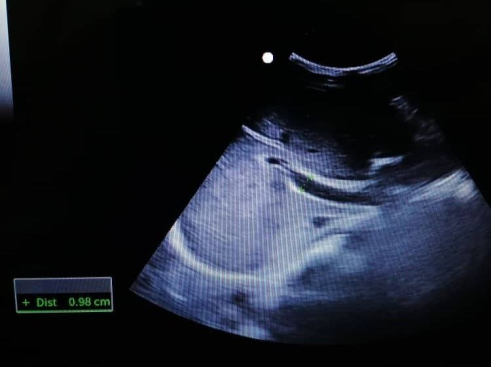
The ultrasonography device used was Mindray® model DC-N3 (China). The measurements were done by the primary investigator after having training to measure the portal vein diameter by a certified sonographer. The transverse diameter of the portal vein was measured from the outer wall of the vein to the other outer wall and the point of measurement was 1 cm from the point of entrance into the hepatic hilum. (Figure 1)
Data were analyzed using Statistical Package for Social Sciences –SPSS version 20 [SPSS. IBM®].
Descriptive and comparative tests were used and results were displayed in tables and figures.
Ethical clearance obtained from the Ethical Review Board of the Hospital.
Informed consent was obtained from the patients who t the inclusion criteria, patients were been met individually to explain the nature and the justification of the study.
All the information of the participants was handled with confidentiality, voluntary non rewarded contribution.
4. Results
376 people were enrolled in this study. Table 2 displays the fundamental characteristics of the population under investigation.
Table 2. Frequency of the basic characteristics.
Mean
Median
Mode
St. deviation
Sex
-
-
Female
-
Age
38.12
34
30
15.577
Weight in kg
64.8
64
65
13.94
Height in cm
163.3
163
165
8.62
PV diameter in mm
10.79
10.95
11
1.27
BMI
24.35
23.9
19.6
5.31
More than half of the participants were females (56.1%, n=211), while 165 (43.9%) were males. The ranged from 18 years to 71 years while the mean age of the distribution of the participants was 38.12 ± 15.57 years. Regarding the weight of the participants, the mean was 64.88 ± 13.94 kilograms. While for the height, the mean was 163.38 ± 8.62 centimeters. The mean BMI was 24.36 ± 5.32. The mean portal vein diameter was 10.79 ± 1.27 millimeters.
Independent t-test was applied to assess the difference of mean Portal vein in males and females, and found a statistically significant difference as the p-value = 0.000.
The portal vein diameter had positive correlation with age (p value = 0.000), weight (p value = 0.002), height (p value = 0.000) and sex (p value = 0.000). Portal vein diameter is not related to BMI (p value = 0.3). (table 3)
Table 3. Correlations between PVD and age, weight and height PVD.
Pearson Correlation
P-value
Age
0.192 **
0.000
Weight
0.161 **
0.002
Height
0.190 **
0.000
BMI
0.069
0.0183
** mean significant correlation
5. Discussion
The goal of this study was to evaluate the portal vein diameter and how it relates to factors including age, sex, weight, height, and body mass index. Patients who had no complaints about the portal vein or liver had ultrasound imaging for the measurement.
Given that Gareeballah et al study had a smaller sample size, the findings of this study are comparable to those of that study. Both studies came to the same conclusion: the portal vein fluctuates with height and weight but is unrelated to BMI
[11]
Gareeballah A, Hassan IA, Elzaki M, Ibraheem SS, Abelwahab B, Siddig A. Measurement of normal portal vein using ultrasound in Sudanese. Glob Adv Res J Med Med Sci. 2017; 6: 336-40.
[11]
. The similarity in the reported results could be the consequence of these studies' similar methodologies.
Similar findings were revealed by studies carried out in Ethiopia and Nigeria, two other regional nations. For Geleto et al. in Ethiopians and Luntsi et al. in Nigerians, the typical portal vein diameters (PVD) were 10.8 1.8 mm and 10.5 1.6 mm, respectively
[9]
Geleto G, Getnet W, Tewelde T. Mean normal portal vein diameter using sonography among clients coming to radiology department of Jimma University Hospital, Southwest Ethiopia. Ethiopian journal of health sciences. 2016; 26(3): 237-42.
Luntsi G, Sani M, Zira JD, Ivor NC, Garba SH. Sonographic assessment of the portal vein diameter in apparently healthy adults in a Northern Nigerian population. African Health Sciences. 2016; 16(4): 1163-8.
. Due to the prevalence of shared tribes between Sudan and these nations, this similarity may be genetic in nature or the result of migration. The same assessment techniques were used in both of these investigations, which may also account for the identical outcomes. Additionally, the findings of these investigations showed that PVD varied with age, sex, and BMI
[9]
Geleto G, Getnet W, Tewelde T. Mean normal portal vein diameter using sonography among clients coming to radiology department of Jimma University Hospital, Southwest Ethiopia. Ethiopian journal of health sciences. 2016; 26(3): 237-42.
Luntsi G, Sani M, Zira JD, Ivor NC, Garba SH. Sonographic assessment of the portal vein diameter in apparently healthy adults in a Northern Nigerian population. African Health Sciences. 2016; 16(4): 1163-8.
The portal vein diameter was found to be 9.57 + 0.66 mm in other international studies, which is consistent with the results of our investigation
[12]
Jha AK, Shah SS, Sah D, Akthar MK, Mahato RK, Rimal B, Chaurasiya C, Jha RK, Thakur AK, Adhikari T. Measurement of Normal Portal Vein Diameter by Ultrasound. Journal of National Medical College. 2023 Jul 30; 8(1): 17-20.
Aqeel S, Amjad S, Tauqir J, Fatima M. Sonographic correlation of portal vein diameter with gender in population of Lahore. The Professional Medical Journal. 2022 Apr 30; 29(05): 607-12.
. The BMI exhibited a positive correlation, which is the opposite of what this study found
[12]
Jha AK, Shah SS, Sah D, Akthar MK, Mahato RK, Rimal B, Chaurasiya C, Jha RK, Thakur AK, Adhikari T. Measurement of Normal Portal Vein Diameter by Ultrasound. Journal of National Medical College. 2023 Jul 30; 8(1): 17-20.
Aqeel S, Amjad S, Tauqir J, Fatima M. Sonographic correlation of portal vein diameter with gender in population of Lahore. The Professional Medical Journal. 2022 Apr 30; 29(05): 607-12.
, but the PV diameter and age, height, and weight all showed positive correlations
[12]
Jha AK, Shah SS, Sah D, Akthar MK, Mahato RK, Rimal B, Chaurasiya C, Jha RK, Thakur AK, Adhikari T. Measurement of Normal Portal Vein Diameter by Ultrasound. Journal of National Medical College. 2023 Jul 30; 8(1): 17-20.
According to this study, the portal vein diameter positively correlates with a person's age, sex, height, and weight but not with their BMI. Clinicians and anatomists alike should pay attention to the study's findings. They must consider how sex, age, height, and weight can affect the portal vein.
Abbreviations
CT
Computed Topography
US
Ultrasonography
PV
Portal Vein
PVD
Portal Vein Diameter
BMI
Body Mass Index
SPSS. IBM®
Statistical Package for Social Sciences
Data Availability
Data available on request from authors.
Conflicts of Interest
The authors declare no conflicts of interest.
References
[1]
Keith L. Moore, Arthur F. Dalley II, Anne M. R. Agur. Clinical Oriented Anatomy. 7th edition. Philadelphia: Lippincott Williams &Wilkins; 2014. p262, 268.
[2]
Richard S. Snell. Clinical Anatomy by Regions. 9th edition. Philadelphia: Lippincott Williams &Wilkins; 2012. p 194, 196.
[3]
Sinnatamby CS, Last RJ, editors. Last’s anatomy: regional and applied. 12th ed. Edinburgh; New York: Churchill Livingstone/Elsevier; 2011. 548 p266, 267.
[4]
Abdel-Wahab MF, Esmat G, Farrag A, El-Boraey YA, Strickland GT. Grading of Hepatic Schistosomiasis by the Use of Ultrasonography. Am J Trop Med Hyg. 1992 Apr 1; 46(4): 403–8.
Weinreb J, Kumari S, Phillips G, Pochaczevsky R. Portal vein measurements by real-time sonography. Am J Roentgenol. 1982; 139(3): 497–499.
[6]
Lafortune M, Marleau D, Breton G, Viallet A, Lavoie P, Huet PM. Portal venous system measurements in portal hypertension. Radiology. 1984; 151(1): 27–30.
Bhattacharya J, Das A, Bhowmik A. Sonographical assessment of portal vein diameter in Northern part of West Bengal, India. Octa Journal of Environmental Research. 2013 Sep 1; 1(3).
[8]
Usman AU, Ibinaiye P, Ahidjo A, Tahir A, Sa'ad ST, Mustapha Z, Tahir N, Garko S. Determination of normal portal vein diameter on ultrasound scan among adults in northeastern Nigeria. Archives of International Surgery. 2015 Jul 1; 5(3): 143.
[9]
Geleto G, Getnet W, Tewelde T. Mean normal portal vein diameter using sonography among clients coming to radiology department of Jimma University Hospital, Southwest Ethiopia. Ethiopian journal of health sciences. 2016; 26(3): 237-42.
Luntsi G, Sani M, Zira JD, Ivor NC, Garba SH. Sonographic assessment of the portal vein diameter in apparently healthy adults in a Northern Nigerian population. African Health Sciences. 2016; 16(4): 1163-8.
Gareeballah A, Hassan IA, Elzaki M, Ibraheem SS, Abelwahab B, Siddig A. Measurement of normal portal vein using ultrasound in Sudanese. Glob Adv Res J Med Med Sci. 2017; 6: 336-40.
[12]
Jha AK, Shah SS, Sah D, Akthar MK, Mahato RK, Rimal B, Chaurasiya C, Jha RK, Thakur AK, Adhikari T. Measurement of Normal Portal Vein Diameter by Ultrasound. Journal of National Medical College. 2023 Jul 30; 8(1): 17-20.
Aqeel S, Amjad S, Tauqir J, Fatima M. Sonographic correlation of portal vein diameter with gender in population of Lahore. The Professional Medical Journal. 2022 Apr 30; 29(05): 607-12.
Mustafa, M. A., Awad, K., Koko, A. O. M. (2024). Sonographic Measurement and Sex Variability of Portal Vein in a Sample of Sudanese Adults. International Journal of Medical Imaging, 12(3), 68-72. https://doi.org/10.11648/j.ijmi.20241203.11
Mustafa, M. A.; Awad, K.; Koko, A. O. M. Sonographic Measurement and Sex Variability of Portal Vein in a Sample of Sudanese Adults. Int. J. Med. Imaging2024, 12(3), 68-72. doi: 10.11648/j.ijmi.20241203.11
Mustafa MA, Awad K, Koko AOM. Sonographic Measurement and Sex Variability of Portal Vein in a Sample of Sudanese Adults. Int J Med Imaging. 2024;12(3):68-72. doi: 10.11648/j.ijmi.20241203.11
@article{10.11648/j.ijmi.20241203.11,
  author = {Mohammedelghazali Abuelgassim Mustafa and Khalid Awad and Alaa Osman Mohamed Koko},
  title = {Sonographic Measurement and Sex Variability of Portal Vein in a Sample of Sudanese Adults
},
  journal = {International Journal of Medical Imaging},
  volume = {12},
  number = {3},
  pages = {68-72},
  doi = {10.11648/j.ijmi.20241203.11},
  url = {https://doi.org/10.11648/j.ijmi.20241203.11},
  eprint = {https://article.sciencepublishinggroup.com/pdf/10.11648.j.ijmi.20241203.11},
  abstract = {Background: Measurements of the normal portal venous dimension in a species population is so crucial. portal vein can be measured by several methods for assessing different parameters, Computed topography (CT) and sonographic imaging are common examples. The diagnosis of portal hypertension depends on the transverse portal vein diameter (normal diameter from 6-15 mm). The aim of this study is to assess the portal vein diameter and correlate the diameter with age, sex, weight, height and BMI in Sudanese populations. Methodology: This was a descriptive cross-sectional study that included a sample of Sudanese adults who were requested to have routine abdominal sonographic scanning due to reasons not related to liver or portal vein problems. The study was carried out at the Radiology Department of Qatar Alnada Hospital, Umbadda, Khartoum, Sudan 2022. Results: In this study, 376 participants were included of which more than half were females (56.1%, n=211), while 165 (43.9%) were males. The mean age of the participants was 38.12 + 15.57 years. The mean portal vein diameter was 10.79 + 1.27 millimeters. From this study the portal vein diameter varied with age (p value = 0.000), weight (p value = 0.002), height (p value = 0.000) and sex (p value = 0.000). It is not related to BMI (p value = 0.3). Conclusion: This study revealed that the portal vein diameter has positive correlation with age, sex, height, and weight of the person and has no correlation with the BMI.
},
 year = {2024}
}
TY  - JOUR
T1  - Sonographic Measurement and Sex Variability of Portal Vein in a Sample of Sudanese Adults

AU  - Mohammedelghazali Abuelgassim Mustafa
AU  - Khalid Awad
AU  - Alaa Osman Mohamed Koko
Y1  - 2024/07/02
PY  - 2024
N1  - https://doi.org/10.11648/j.ijmi.20241203.11
DO  - 10.11648/j.ijmi.20241203.11
T2  - International Journal of Medical Imaging
JF  - International Journal of Medical Imaging
JO  - International Journal of Medical Imaging
SP  - 68
EP  - 72
PB  - Science Publishing Group
SN  - 2330-832X
UR  - https://doi.org/10.11648/j.ijmi.20241203.11
AB  - Background: Measurements of the normal portal venous dimension in a species population is so crucial. portal vein can be measured by several methods for assessing different parameters, Computed topography (CT) and sonographic imaging are common examples. The diagnosis of portal hypertension depends on the transverse portal vein diameter (normal diameter from 6-15 mm). The aim of this study is to assess the portal vein diameter and correlate the diameter with age, sex, weight, height and BMI in Sudanese populations. Methodology: This was a descriptive cross-sectional study that included a sample of Sudanese adults who were requested to have routine abdominal sonographic scanning due to reasons not related to liver or portal vein problems. The study was carried out at the Radiology Department of Qatar Alnada Hospital, Umbadda, Khartoum, Sudan 2022. Results: In this study, 376 participants were included of which more than half were females (56.1%, n=211), while 165 (43.9%) were males. The mean age of the participants was 38.12 + 15.57 years. The mean portal vein diameter was 10.79 + 1.27 millimeters. From this study the portal vein diameter varied with age (p value = 0.000), weight (p value = 0.002), height (p value = 0.000) and sex (p value = 0.000). It is not related to BMI (p value = 0.3). Conclusion: This study revealed that the portal vein diameter has positive correlation with age, sex, height, and weight of the person and has no correlation with the BMI.

VL  - 12
IS  - 3
ER  -