Case Report | | Peer-Reviewed

Page Kidney Secondary to Pancreatic Pseudocyst: Definitive Surgical Management with Sustained Normotension: A Case Report with Literature Review

Received: 7 July 2026     Accepted: 20 July 2026     Published: 10 August 2026
Views:       Downloads:
Abstract

Background: Page kidney is an uncommon but reversible cause of secondary hypertension due to extrinsic compression of the renal parenchyma, triggering activation of the renin-angiotensin-aldosterone system (RAAS). Pancreatic pseudocysts are a well-recognized complication of acute or chronic pancreatitis and are a rare cause of this phenomenon. Case Presentation: A 21-year-old previously normotensive male presented to the Surgical Gastroenterology (SGE) Out-patient Department (OPD) with dull aching upper abdominal pain for 8 days and progressive abdominal distension for 6 months. At presentation patient had new-onset hypertension (150/100 mmHg) incidentally detected on admission. Contrast-enhanced computed tomography (CECT) of the abdomen revealed chronic calcific pancreatitis with a massive pancreatic tail pseudocyst (~1500 mL) extending into the left perirenal space, near-complete encasement of the left kidney, with a persistent nephrogram on delayed phase imaging, a pathognomonic radiological feature of ‘Page kidney’. The patient underwent laparotomy and Roux-en-Y cysto-jejunostomy in view of persistent abdominal symptoms and showed complete normalization of blood pressure. Patient remained normotensive and asymptomatic at 6 months on follow-up. Discussion: Page kidney secondary to a pancreatic pseudocyst is an exceptionally rare cause of secondary hypertension with very few cases reported in the world literature. All the patients reported in literature were managed conservatively by non-operative management, involving anti-hypertensives and USG (ultrasound) guided pigtail insertion. However, our case describes the management of a page kidney secondary to a pseudocyst by Roux-en-Y cysto-jejunostomy as a definitive therapy, achieving sustained medication-free normotension. Conclusion: Page kidney is a rare, curable cause of secondary hypertension. Early management helps prevent deterioration and salvage of renal function. Surgical drainage procedures are a feasible and durable alternative to conservative management in selected patients, particularly those with recurrent or complex pancreatic pseudocysts.

Published in International Journal of Gastroenterology (Volume 10, Issue 2)
DOI 10.11648/j.ijg.20261002.11
Page(s) 67-73
Creative Commons

This is an Open Access article, distributed under the terms of the Creative Commons Attribution 4.0 International License (http://creativecommons.org/licenses/by/4.0/), which permits unrestricted use, distribution and reproduction in any medium or format, provided the original work is properly cited.

Copyright

Copyright © The Author(s), 2026. Published by Science Publishing Group

Keywords

Page Kidney, Pancreatic Pseudocyst, Chronic Pancreatitis, Renin-angiotensin-aldosterone System, Roux-en-Y Cysto-jejunostomy, Nephrogram

1. Introduction
Pancreatic pseudocysts are the most prevalent cystic complication of pancreatic disease, occurring in 20-40% of patients with chronic pancreatitis and approximately 5-16% following acute pancreatitis. Most pseudocysts (~80%) in the setting of acute pancreatitis resolve spontaneously, while pseudocysts of chronic pancreatitis typically do not resolve spontaneously and often require intervention.
Page kidney is caused by compression of the renal parenchyma externally causing tissue hypoperfusion and activation of renin-angiotensin-aldosterone system (RAAS). It is well documented in cases of trauma, interventions to the kidneys and tumors of the kidney and adrenals. Pancreatic pseudocysts are a rare cause of this phenomenon.
Pancreatic pseudocysts causing Page kidney constitute a rare but treatable cause of secondary hypertension when recognized and managed promptly before deterioration of renal function occurs. Management according to the previous reported cases (Table 1) conventionally involved treatment with anti-hypertensives and USG guided drainage of the cysts. Surgical management of selected patients represents a feasible and durable treatment option, particularly when definitive internal drainage is required.
2. Case Report
A 21-year-old gentleman presented to the SGE OPD with complaints of pain in the upper central abdomen for 8 days, insidious in onset, postprandial, dull aching in nature, non-radiating, with no clear aggravating or relieving factors. He gave history of progressive abdominal distension with early satiety for 6 months. Patient had no history of alcohol consumption, smoking, gallstone disease, abdominal trauma, pancreatic surgery, hypercalcemia or hypertriglyceridemia. Family history was negative for pancreatic disease. Ultrasonography revealed a normal gallbladder without cholelithiasis. In the absence of an identifiable aetiology, the chronic pancreatitis was classified as idiopathic.
Clinical examination revealed a firm, smooth, well-circumscribed 10×10 cm lump in the left hypochondrium extending into the left lumbar region - non-tender, dull to percussion, and exhibiting restricted lateral mobility. On admission, blood pressure was 150/100 mmHg and serial measurements showed persistently elevated readings throughout hospitalization. This new-onset hypertension in a previously normotensive 21-year-old with a large abdominal mass and prior pancreatic pathology raised suspicion for a secondary renovascular cause. The patient was further evaluated with laboratory and radiological investigations. All routine blood investigations including complete blood count, liver function tests, renal function tests, PT-INR, aPTT, serum amylase, serum lipase and random blood sugars were within normal limits. Patients eGFR was about 90ml/min/1.73m2 and serum creatinine of 0.9mg/dl preoperatively. Serum calcium and fasting triglyceride levels were within normal limits. Ultrasonography and CECT demonstrated a normal gallbladder without evidence of cholelithiasis or biliary pathology.
2.1. Ultrasound Abdomen and Doppler
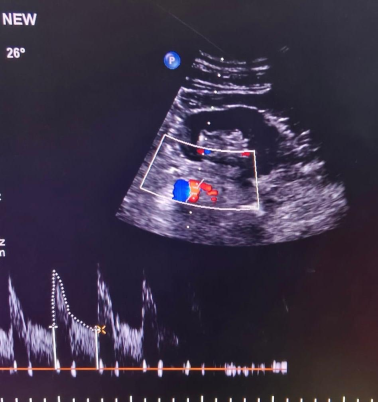
Pancreas appeared atrophic with calcifications. MPD measures 5.5mm. A well-defined heterogeneous hypoechoic collection with a well-defined wall ~4mm in perinephric region of the left kidney ~16×9×11 cm with no internal vascularity noted along with minimal ascites. Renal artery doppler was normal. (Figure 1)
Figure 1. USG doppler showing a normal (low resistance, monophasic and continuous forward flow) waveform of the renal artery.
2.2. CECT (Contrast-Enhanced Computed Tomography)
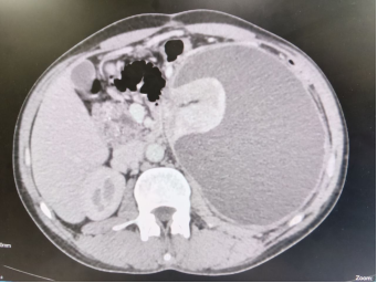
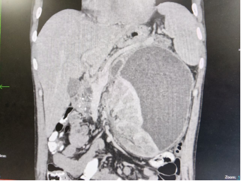
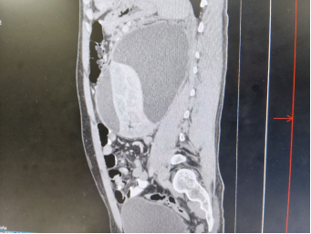
CECT of the abdomen (Figures 2-4) demonstrated an atrophic pancreas with scattered parenchymal calcifications, dilated and irregular main pancreatic duct (MPD) measuring 6.5 mm, consistent with chronic calcific pancreatitis.
A large, unilocular, peripherally enhancing cystic collection measuring approximately 13.4×12.8×16.5 cm (estimated volume ~1500 mL) was identified arising from the pancreatic tail, extending posteriorly through the posterior renal fascia into the left perirenal space. The collection showed no internal solid components or hyperdense content. Cyst wall thickness measured 4-6 mm.
Figure 2. CECT abdomen (Axial section): Atrophic pancreas with calcifications and a large homogeneous hypodense pseudocyst occupying the left hemiabdomen, near-completely encasing the left kidney with demonstrable mass effect on the renal parenchyma.
Figure 3. CECT abdomen (Coronal section): The massive pseudocyst (~1500 mL) arising from the pancreatic tail and extending inferiorly into the left perirenal space, displacing and compressing the left kidney. The kidney is sandwiched between the cyst and the posterior abdominal wall.
Figure 4. CECT abdomen (Sagittal section): The pseudocyst spans the entire left flank in the sagittal plane, demonstrating the extent of perirenal extension and the compressed, displaced left kidney (visible as a flattened parenchymal rim). The persistent nephrogram is appreciable on the delayed phase as reduced cortical opacification relative to the contralateral side.
The lesion exerted significant mass effect, near-completely encasing the left kidney and compressing the renal hilum and intrarenal vasculature. On the delayed nephrographic phase, a persistent nephrogram was observed on the affected side characterized by prolonged cortical enhancement with delayed excretion of contrast into the collecting system- consistent with a Page kidney. The right kidney appeared normal. Rest of the structures were normal.
2.3. Surgical Findings
Given the large size of the pseudocyst (~1500 mL), its mature wall, chronic symptomatic nature, extensive perirenal extension with near-complete encasement of the kidney, and the need for definitive long-term drainage in a young patient with chronic pancreatitis, primary internal surgical drainage was favoured over percutaneous drainage. Percutaneous drainage was considered less suitable because of the possibility of incomplete drainage, external pancreatic fistula formation, and recurrence. Therefore, Roux-en-Y cystojejunostomy was planned as definitive treatment. On laparotomy via a midline vertical incision, a massive pseudocyst was identified in the pancreatic tail extending posteroinferiorly into the left perirenal space, in direct contact with the left renal capsule and compressing the renal cortex and hilar vessels. The operative findings were consistent and correlating with the radiological findings. Needle aspiration was performed for confirmation and approximately 1500 mL of turbid, dark-brown pancreatic fluid was evacuated. Cyst fluid and pseudocyst wall were sent for cytological analysis, microbiology and histopathology. Cyst fluid amylase sent intraoperatively was ~22800 IU/L.
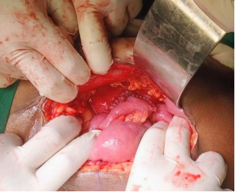
Figure 5. Intraoperative photograph showing the completed Roux-en-Y cysto-jejunostomy. Note the anastomosis fashioned at the most inferior point of the collection to ensure dependent gravitational drainage of the perirenal pseudocyst.
The pseudocyst was meticulously dissected from surrounding structures both anteriorly and posteriorly. The adjacent small bowel and its mesenteric vascular arcades were carefully preserved. A 5 cm wide side-to-side cysto-jejunostomy (Figure 5), was fashioned at the most inferior and dependent portion of the pseudocyst to ensure complete gravitational drainage and decompression of the compressed renal bed. An isoperistaltic Roux-en-Y jejunal limb was constructed by dividing the proximal jejunum 25 cm distal to the ligament of Treitz and bringing the distal limb antecolic through a mesenteric window. A two-layer hand-sewn anastomosis was done using 3-0 polydioxanone suture. Entero-enterostomy was fashioned 40 cm distal to the cysto-jejunal anastomosis. Mesenteric defects were closed and abdomen was closed in layers. Post-operatively, patient was monitored with 4-hourly blood pressure recordings. From postoperative day (POD) 2 onwards, blood pressure demonstrated a consistent downward trend: POD 2: 138/88 mmHg; POD 4: 128/82 mmHg. Patient was discharged on postoperative day-4.
2.4. Histopathology
Gross examination of the excised cyst wall specimen revealed a thick, fibrous, rubbery wall without a mucosal lining, hemorrhage, or necrosis.
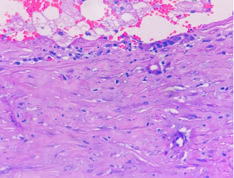
Histopathological examination (Figure 6), demonstrated a thick fibrous capsule composed of densely packed collagen fibers with interspersed chronic inflammatory cells, predominantly lymphocytes and plasma cells. Granulation tissue with proliferating capillaries and fibroblasts was identified at the inner surface. Microscopy showed chronic inflammatory infiltration with fibroblasts and myofibroblasts predominant in the wall, consistent with a mature pancreatic pseudocyst.
Figure 6. Microscopic image of wall of a pseudocyst of pancreas showing chronic inflammatory infiltration with fibroblasts and myofibroblasts.
2.5. Follow-up
At 2-week follow-up, surgical site was healthy and blood pressure was: 122/78 mmHg. No antihypertensive medications were administered at any point. At 6-month outpatient follow-up, he remained entirely asymptomatic. There was no abdominal pain, diarrhoea, steatorrhea, early satiety, abdominal distension, or unintentional weight loss. Blood pressure remained normal at 118/76 mmHg without antihypertensive medication; all blood parameters including renal function tests within normal limits i.e., eGFR of 120ml/min/1.73m2 and serum creatinine of 0.4mg/dl. USG abdomen showed a normal left kidney with preserved cortical echogenicity. He was advised follow up on an as-need basis.
3. Discussion
Pancreatic pseudocysts occur as a result of sequelae of acute or chronic inflammation of the pancreas leading to accumulation of enzyme rich pancreatic fluid enclosed by a layer of non-epithelial granulation tissue and is therefore termed a ‘false cyst’ or a ‘pseudocyst’. Such fluid collections can occur anywhere in the abdomen after an insult to the pancreas. In certain rare situations, pancreatic secretions may extend into the perirenal space causing autolysis of the perirenal fat and fascia leading to formation of a pseudocyst around the kidney. Despite the high global prevalence of pancreatic pseudocysts, their perirenal extension causing Page kidney with secondary hypertension remains extraordinarily rare and only five cases (Table 1) have been reported in the indexed literature. Aswani et al., was the first to report this phenomenon to occur in a case secondary to a pancreatic pseudocyst in their case report in 2015.
Page kidney phenomenon was first experimentally introduced by Dr. Irvine H. Page in 1939 through application of cellophane wrappings around canine kidneys, the condition results in microvascular ischemia at the level of the intrarenal arterioles and juxtaglomerular apparatus, with subsequent renin release and systemic hypertension. Page demonstrated that circumferential perinephric constriction, independent of primary renal vascular disease, was sufficient to produce persistent severe hypertension and, critically, that relief of compression reversed it, establishing the curative principle that governs management to this day.
In contemporary practice, most frequent etiologies of Page kidney are traumatic subcapsular hematoma and iatrogenic perinephric hematoma following renal biopsy, nephrostomy, and percutaneous lithotripsy for stone disease. However, the compressive perinephric agent need not necessarily be hemorrhagic: urinomas, lymphoceles, abscesses, and critically pancreatic pseudocysts extending into the retroperitoneal perirenal space have each been documented as rare causative agents. The common denominator is a space-occupying lesion generating a compartment syndrome-like effect within the perirenal fascia, compromising intrarenal microvascular autoregulation without occluding the renal artery or vein.
Patients with such phenomenon have a history of pancreatitis either acute or chronic along with symptoms and signs of a pancreatic pseudocyst i.e., early satiety, fullness, abdominal distension, dull aching pain, palpable lump etc. along with new onset hypertension after the attack of pancreatitis as seen in our case. Normal serum amylase and lipase do not exclude chronic pancreatitis; indeed, enzyme levels are often normal in established chronic disease because progressive fibrosis and acinar cell destruction reduce pancreatic enzyme production, unlike acute pancreatitis. Radiological imaging modalities play a major role in their diagnosis. Ultrasound studies may help diagnose the presence of a cyst around the kidney. Doppler studies of the renal vessels are usually normal and provide indirect evidence of secondary HTN due to mechanical activation of RAAS. CECT and an MRI-abdomen help in confirming the diagnosis, delineating the cyst, providing the nature of the pancreas and ruling out any other pathologies. There is no clear evidence of which is superior, however MRI is usually preferred to help delineating the anatomy of the pancreatic duct and its communication with the pseudocyst. Presence of a persistent nephrogram on delayed-phase of a CECT of abdomen with a pseudocyst compressing the kidney on the affected side is the pathognomonic radiological sign of the Page kidney similar to our case.
Management in cases with a pseudocyst causing Page kidney is usually conservative and non-surgical as reported in the available literature. Symptomatic management with antihypertensive agents (ACE inhibitors or angiotensin receptor blockers) and radiologically guided drainage of the pseudocyst was the mainstay of management in previously documented cases, as summarized in Table 1.
Definitive surgical internal drainage as primary therapy for Page kidney secondary to pancreatic pseudocyst has not been previously reported. To the best of our knowledge, this is the first reported case of successful management of a page kidney by surgical drainage procedure. We suggest considering surgical management in selected patients with progressive symptoms, persistent hypertension despite medical therapy, good performance status, and suitable operative risk profiles. We hypothesize that surgical intervention achieves definitive internal drainage of the pseudocyst while relieving extrinsic renal parenchymal compression, resulting in sustained suppression of the RAAS. The progressive normalization of blood pressure without the need for antihypertensive therapy following surgical decompression provides compelling evidence for a mechanically mediated, RAAS-driven pathophysiology. This provides symptomatic relief and may reduce recurrence and long-term anti-hypertensive requirements. Surgery involves drainage of the pseudocyst into the bowel lumen depending upon the location. Large cysts in proximity to the stomach or duodenum may be drained via cystogastrostomy or a cysto-duodenostomy. Cysts that are not amenable to cystogastrostomy or cystoduodenostomy may require cystojejunostomy as performed in our case (Figure 5). The type of anastomosis (single vs two layered) depends on the surgeon’s preference where most of them prefer doing a two layered anastomosis as done in our case. The approach also depends upon the surgeon’s expertise or the institutional preference where both open approach or minimally invasive techniques (laparoscopic or robotic) can be used. Our case was done by an open approach based on the surgeon’s preference. Minimally invasive techniques have advantages of shorter hospital stay, reduced wound related complications, early mobilization and tolerance to oral feeds, when compared to open techniques.
Although the aetiology of chronic pancreatitis remained idiopathic in our patient, the possibility of recurrence of pancreatitis and pseudocyst formation cannot be completely excluded. Long-term clinical and radiological surveillance is therefore warranted. At 6 months follow-up, there was no evidence of recurrent pseudocyst formation or recurrent symptoms. Patients who are not potential candidates for surgery i.e., with multiple comorbidities, organ dysfunction, on ventilator support, etc., medical management with USG guided drainage is the only option. However, the potential complications of any procedure in such a complicated physiology may exist and should be managed accordingly. Follow-up of such patients should involve serial recording of blood pressure 4th hourly in the early postoperative period till its normalization and routine surgical care, followed by 3-6 monthly follow up initially for two to three visits and then yearly if symptomatic. Asymptomatic patients can be observed on an as-need basis.
Table 1. Reported cases of pancreatic pseudocyst causing Page kidney.

Author / Year

Aetiology

Presentation

Imaging

Management

Outcome

Aswani et al., 2015

Alcoholic CP

HTN + Abdominal pain

CECT

Percutaneous drainage

Normotensive

Hiremath et al., 2022

Acute pancreatitis + PRP fistula

HTN + Weakness

CECT

USG aspiration + Pigtail

Normotensive

Sharma et al., 2022

Alcoholic CP

Hypertensive crisis

CECT

ACEi + Observation

Improved

Thakur et al., 2024

Acute pancreatitis

HTN

CECT+MRCP

ACEi+ Percutaneous drainage

Normotensive

Gandhi et al., 2024

CP

HTN + Abdominal pain

CECT

Percutaneous drainage

Normotensive

Present case, 2026

Idiopathic CP

HTN + Lump

CECT

Roux-en-Y Cysto-jejunostomy

Normotensive (no medications)

CP = Chronic Pancreatitis; HTN = Hypertension; PRP = Pancreatico-perirenal; ACEi = Angiotensin-converting enzyme inhibitor; CECT = Contrast-enhanced CT.
4. Conclusion
Page kidney secondary to a pancreatic pseudocyst is an exceptionally uncommon surgically treatable cause of secondary hypertension. To the best of our knowledge, ours is the first to report management of page kidney secondary to a pancreatic pseudocyst by surgery. We recommend clinicians to maintain a high index of suspicion for this phenomenon in young patients with pancreatitis and unexplained new-onset hypertension, and consideration of surgical internal drainage as non-surgical modalities may not provide long term relief. Continued case reporting of such cases with standardized documentation is essential to build the evidence base for management guidelines for this rare entity.
Abbreviations

ACEi

Angiotensin-Converting Enzyme Inhibitor

aPTT

Activated Partial Thromboplastin Time

CECT

Contrast-Enhanced Computed Tomography

eGFR

Estimated Glomerular Filtration Rate

IU/L

International Units Per Liter

MPD

Main Pancreatic Duct

MRCP

Magnetic Resonance Cholangiopancreatography

PT-INR

Prothrombin Time-International Normalized Ratio

RAAS

Renin-Angiotensin-Aldosterone System

USG

Ultrasonography

Author Contributions
Venugopal H. G.: Conceptualization, Project administration
Mukund Mangarai: Writing – original draft, Writing – review & editing
Akshay Patil: Resources, Software, Data curation
Conflicts of Interest
The authors declare no conflicts of interest or competing interests with respect to this study.
References
[1] Koo JG, Liau MYQ, Kryvoruchko IA, et al. Pancreatic pseudocyst: The past, the present, and the future. World J Gastrointest Surg. 2024; 16(7): 1986-2002.
[2] Banks PA, Bollen TL, Dervenis C, et al. Classification of acute pancreatitis - 2012: revision of the Atlanta classification and definitions by international consensus. Gut. 2013; 62(1): 102-111.
[3] Vaidya PN, Rathi BM, Finnigan NA. Page Kidney. In: StatPearls [Internet]. Treasure Island (FL): StatPearls Publishing; Updated January 2023. Available from:
[4] Aswani Y, Anandpara KM, Hira P. Page kidney due to a renal pseudocyst in a setting of pancreatitis. BMJ Case Rep. 2015; 2015: bcr2014207436.
[5] Page IH. The production of persistent arterial hypertension by cellophane perinephritis. JAMA. 1939; 113(23): 2046-2048.
[6] Smyth A, Collins CS, Thorsteinsdottir B, et al. Page kidney: etiology, renal function outcomes and risk for future hypertension. J Clin Hypertens (Greenwich). 2012; 14(4): 216-221.
[7] Thakur S, Kapila S, Makhaik S, Jhobta A. Unusual presentation of subcapsular renal pseudocyst as page kidney - A case report. Case Rep Clin Radiol. 2024; 2: 95-8.
[8] Ray MS, Raval A, Yadav P, et al. Large pseudocyst of pancreas: open surgical drainage by Roux-en-Y cystojejunostomy -- experience of 28 cases over 18 years. Int Surg J. 2025; 12(7): 1112-1118.
[9] Badgurjar MK, Mandovra P, Mathur SK, Patankar R. Laparoscopic loop cystojejunostomy: An alternative to Roux-en-Y cystojejunostomy for pancreatic pseudocyst. Journal of Minimal Access Surgery. 2021 Apr-Jun; 17(2): 221-225.
[10] Hiremath R, Kuttancheri T, Gurumurthy B, et al. A new page in the literature of pancreatic pseudocyst: case report on perirenal pseudocyst presenting as Page kidney. Egypt J Radiol Nucl Med. 2022; 53: 240.
[11] Sharma R, Attarha B, Rechcigl K, Aung WM. Spontaneous Page kidney as a complication of a pancreatic pseudocyst. ACG Case Rep J. 2022; 9(7): e00813.
[12] Gandhi M, Patel S, Shah N, et al. Pancreatic pseudocysts: a complication leads to Page kidney due to external compression of the kidney. J Krishna Inst Med Sci Univ. 2024; 13(4): 175-179.
Cite This Article
  • APA Style

    G., V. H., Mangarai, M., Patil, A. (2026). Page Kidney Secondary to Pancreatic Pseudocyst: Definitive Surgical Management with Sustained Normotension: A Case Report with Literature Review. International Journal of Gastroenterology, 10(2), 67-73. https://doi.org/10.11648/j.ijg.20261002.11

    Copy | Download

    ACS Style

    G., V. H.; Mangarai, M.; Patil, A. Page Kidney Secondary to Pancreatic Pseudocyst: Definitive Surgical Management with Sustained Normotension: A Case Report with Literature Review. Int. J. Gastroenterol. 2026, 10(2), 67-73. doi: 10.11648/j.ijg.20261002.11

    Copy | Download

    AMA Style

    G. VH, Mangarai M, Patil A. Page Kidney Secondary to Pancreatic Pseudocyst: Definitive Surgical Management with Sustained Normotension: A Case Report with Literature Review. Int J Gastroenterol. 2026;10(2):67-73. doi: 10.11648/j.ijg.20261002.11

    Copy | Download

  • @article{10.11648/j.ijg.20261002.11,
      author = {Venugopal H. G. and Mukund Mangarai and Akshay Patil},
      title = {Page Kidney Secondary to Pancreatic Pseudocyst: Definitive Surgical Management with Sustained Normotension: A Case Report with Literature Review},
      journal = {International Journal of Gastroenterology},
      volume = {10},
      number = {2},
      pages = {67-73},
      doi = {10.11648/j.ijg.20261002.11},
      url = {https://doi.org/10.11648/j.ijg.20261002.11},
      eprint = {https://article.sciencepublishinggroup.com/pdf/10.11648.j.ijg.20261002.11},
      abstract = {Background: Page kidney is an uncommon but reversible cause of secondary hypertension due to extrinsic compression of the renal parenchyma, triggering activation of the renin-angiotensin-aldosterone system (RAAS). Pancreatic pseudocysts are a well-recognized complication of acute or chronic pancreatitis and are a rare cause of this phenomenon. Case Presentation: A 21-year-old previously normotensive male presented to the Surgical Gastroenterology (SGE) Out-patient Department (OPD) with dull aching upper abdominal pain for 8 days and progressive abdominal distension for 6 months. At presentation patient had new-onset hypertension (150/100 mmHg) incidentally detected on admission. Contrast-enhanced computed tomography (CECT) of the abdomen revealed chronic calcific pancreatitis with a massive pancreatic tail pseudocyst (~1500 mL) extending into the left perirenal space, near-complete encasement of the left kidney, with a persistent nephrogram on delayed phase imaging, a pathognomonic radiological feature of ‘Page kidney’. The patient underwent laparotomy and Roux-en-Y cysto-jejunostomy in view of persistent abdominal symptoms and showed complete normalization of blood pressure. Patient remained normotensive and asymptomatic at 6 months on follow-up. Discussion: Page kidney secondary to a pancreatic pseudocyst is an exceptionally rare cause of secondary hypertension with very few cases reported in the world literature. All the patients reported in literature were managed conservatively by non-operative management, involving anti-hypertensives and USG (ultrasound) guided pigtail insertion. However, our case describes the management of a page kidney secondary to a pseudocyst by Roux-en-Y cysto-jejunostomy as a definitive therapy, achieving sustained medication-free normotension. Conclusion: Page kidney is a rare, curable cause of secondary hypertension. Early management helps prevent deterioration and salvage of renal function. Surgical drainage procedures are a feasible and durable alternative to conservative management in selected patients, particularly those with recurrent or complex pancreatic pseudocysts.},
     year = {2026}
    }
    

    Copy | Download

  • TY  - JOUR
    T1  - Page Kidney Secondary to Pancreatic Pseudocyst: Definitive Surgical Management with Sustained Normotension: A Case Report with Literature Review
    AU  - Venugopal H. G.
    AU  - Mukund Mangarai
    AU  - Akshay Patil
    Y1  - 2026/08/10
    PY  - 2026
    N1  - https://doi.org/10.11648/j.ijg.20261002.11
    DO  - 10.11648/j.ijg.20261002.11
    T2  - International Journal of Gastroenterology
    JF  - International Journal of Gastroenterology
    JO  - International Journal of Gastroenterology
    SP  - 67
    EP  - 73
    PB  - Science Publishing Group
    SN  - 2640-169X
    UR  - https://doi.org/10.11648/j.ijg.20261002.11
    AB  - Background: Page kidney is an uncommon but reversible cause of secondary hypertension due to extrinsic compression of the renal parenchyma, triggering activation of the renin-angiotensin-aldosterone system (RAAS). Pancreatic pseudocysts are a well-recognized complication of acute or chronic pancreatitis and are a rare cause of this phenomenon. Case Presentation: A 21-year-old previously normotensive male presented to the Surgical Gastroenterology (SGE) Out-patient Department (OPD) with dull aching upper abdominal pain for 8 days and progressive abdominal distension for 6 months. At presentation patient had new-onset hypertension (150/100 mmHg) incidentally detected on admission. Contrast-enhanced computed tomography (CECT) of the abdomen revealed chronic calcific pancreatitis with a massive pancreatic tail pseudocyst (~1500 mL) extending into the left perirenal space, near-complete encasement of the left kidney, with a persistent nephrogram on delayed phase imaging, a pathognomonic radiological feature of ‘Page kidney’. The patient underwent laparotomy and Roux-en-Y cysto-jejunostomy in view of persistent abdominal symptoms and showed complete normalization of blood pressure. Patient remained normotensive and asymptomatic at 6 months on follow-up. Discussion: Page kidney secondary to a pancreatic pseudocyst is an exceptionally rare cause of secondary hypertension with very few cases reported in the world literature. All the patients reported in literature were managed conservatively by non-operative management, involving anti-hypertensives and USG (ultrasound) guided pigtail insertion. However, our case describes the management of a page kidney secondary to a pseudocyst by Roux-en-Y cysto-jejunostomy as a definitive therapy, achieving sustained medication-free normotension. Conclusion: Page kidney is a rare, curable cause of secondary hypertension. Early management helps prevent deterioration and salvage of renal function. Surgical drainage procedures are a feasible and durable alternative to conservative management in selected patients, particularly those with recurrent or complex pancreatic pseudocysts.
    VL  - 10
    IS  - 2
    ER  - 

    Copy | Download

Author Information
  • Department of Surgical Gastroenterology, Bangalore Medical College and Research Institute, Bangalore, India
  • Department of Surgical Gastroenterology, Bangalore Medical College and Research Institute, Bangalore, India

  • Department of Surgical Gastroenterology, Bangalore Medical College and Research Institute, Bangalore, India