In dermatology clinics we routinely come across cases of lesions on the hands. Patients may present with papules, vesicles, nodules on the hands. Such lumps and bumps on hands can be a manifestation of various diseases, ranging from benign lesions to systemic conditions. Routinely the rash on hands indicates primary skin disease. But lesions on hands can be seen in inflammatory conditions like psoriasis, eczema, rheumatoid arthritis, gout or infective diseases like bacterial (folliculitis, anthrax), viral (pox virus, herpetic whitlow, molluscum, wart), mycobacterial (tuberculosis verrucose cutis, lupus vulgaris), fungal infections (sporotrichosis). Few benign lesions like keratoacanthoma, ganglion or malignant conditions like SCC, Melanoma may present. A thorough examination and diagnosis of these lesions can provide valuable insights into underlying conditions. Here I am sharing interesting clinical cases highlighting the significance of hand lesions in diagnosing diseases like infective (secondary syphilis) and inflammatory conditions (gout, psoriatic arthritis) as well as clues to variety of systemic diseases (leprosy, tuberous xanthoma). Lumps and bumps on hands can serve as a valuable diagnostic clue to various diseases. The diagnosis of hand lesions is mostly clinical and depends on the clinical history and exploratory objective findings. A proper history taking, thorough clinical and systemic examination, combined with laboratory tests can aid in accurate diagnosis and timely management. Hand lesions may be a window of systemic disease.
| Published in | International Journal of Clinical Dermatology (Volume 8, Issue 1) |
| DOI | 10.11648/j.ijcd.20250801.12 |
| Page(s) | 6-10 |
| Creative Commons |
This is an Open Access article, distributed under the terms of the Creative Commons Attribution 4.0 International License (http://creativecommons.org/licenses/by/4.0/), which permits unrestricted use, distribution and reproduction in any medium or format, provided the original work is properly cited. |
| Copyright |
Copyright © The Author(s), 2025. Published by Science Publishing Group |
Lumps, Bumps, Hands
Case Number | Title | Introduction | Case presentation | Investigations | Management |
|---|---|---|---|---|---|
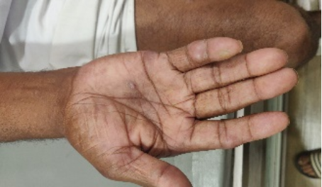
1 | Gouty nodule on hands a case report | A 52-years-old man presented with painful, swollen nodules on his hands [2, 3] (Figure 1). Laboratory investigations revealed elevated serum uric acid levels. the patient was diagnosed with gout and treated with allopurinol. | Laboratory investigations revealed elevated serum uric acid levels (10.73), elevated CRP | Gout is a common form of inflammatory arthritis characterized sudden, severe attacks of pain, swelling, redness and tenderness in one or more joints. gouty typhi are deposits of monosodium urate crystals that can form in various tissues, including the skin, joints, and bones. Patient was treated with allopurinol. | |
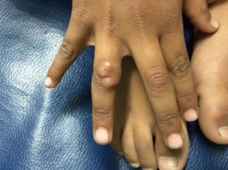
2 | Tuberous xanthoma on the hands [4] a case report | Tuberous xanthoma is a form of xanthoma characterized by the deposition of lipid-laden macrophages in the skin. It is often associated with hyperlipidemia. | A 3-years-boy brought by parents to our clinic with a history of few, painless yellowish nodules on his knuckles (Figure 2). The nodules were firm, measured 0.5-2 cm in diameter, and were located on the dorsal aspect of hands mainly knuckles. | On investigations there were elevated LDL cholesterols and elevated triglycerides. | The patient was diagnosed with tuberous xanthoma [5] and treated under guidance of cardiologist. |
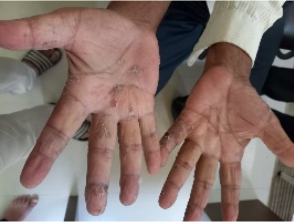
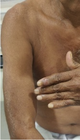
3 | Secondary Syphilis Presenting as Palmoplantar Rash | Secondary syphilis is a systemic infection caused by the bacterium Treponema pallidum [7]. It can present with a wide range of symptoms, including skin rashes, fever, and, lymphadenopathy. | A 38-year-old man presented to our clinic with a 2 weeks history of multiple, asymptomatic papulosquamous rash over palms [7] . The lesions were measured approximately 0.5-1 cm in diameter, and were arranged in a symmetrical pattern (Figure 3). Biett’s sign is characterized by erythematous macules with peripheral scaling [6] . | Laboratory tests revealed Positive TPHA TEST (1: 320) million units (IM). | The patient was diagnosed with secondary syphilis and treated with benzathine penicillin (2.4) [8] |
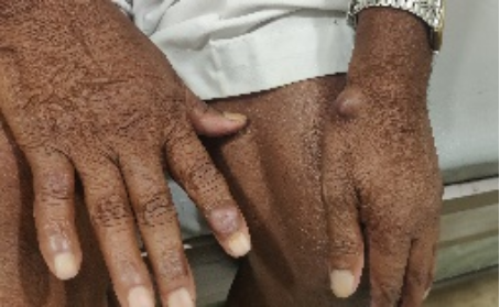
4 | Hansen’s disease presented as hand nodule | A 56 years old male presented with lesion hand for 2-3 months. On examination there was a nodule on left wrist and second on rt index finger on distal interphalangeal joint [9] (Figure 4). The nodules were painless, hard to firm in consistency [10, 12]. Non-oozy, no regional lymphadenopathy. On detail examination there was hypopigmented, hyposthetic patch over left arm (figure 5). | On histopatholgy, it was confirmed to have leprosy [11] . | The patient was started on multi-drug therapy. | |
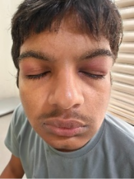
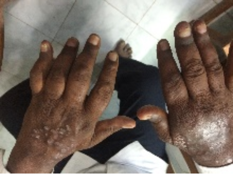
5 | Dermatomyositis [13] | A 15-years-old student visited with complaints of recurrent periorbital swelling and rash over hands. On examination there was left sided periorbital swelling (figure 6) and papulosquamous rash over knuckles (Figure 7). There violaceous appearance over the rash, which gave the clue. No other major complaints were present like arthritis, no proximal weakness. | On investigations there were raised liver enzymes and lactate dehydrogenase (1104 IU/I). | The patient was referred to rheumatologist for further management. | |
6 | Psoriasis | A 64-years male presented with complaints of inability to close fingers [14] . On examination there was swelling, stiffness at interphalangeal joints(figure 8). Also, a deformity was noted at distal little finger [15] . | On x-ray there was pencil-in-cup like deformity. | He was started with methotrexate pulse therapy. |
IPJ | Interphalangeal Joint |
SCC | Squamous Cell Carcinoma |
| [1] | TriHealth Orthopedic & Sports Institute: Lumps and Bumps of the Fingers and Hand. |
| [2] | Hiroyuki Yano, MD and Mitsuyo Kinjo, MD, MPH. Cutaneous digital papules. Cleveland Clinic Journal of Medicine February 2021, 88(2) 73-74. |
| [3] | Brian F. Mandell. Finger nodules: Tip of the gouty iceberg. Cleveland Clinic Journal of Medicine, 2023. |
| [4] | Timur Selcuk Akpinar, Murat Kose, Samim Emet. A Rare Manifestation of Tuberous Xanthomas: J Gen Intern Med. 2014 Dec 18; 30(5): 691. |
| [5] | Dan Li a, Longfei You b, Songqing Fan c, Lihua Tan. Xanthomatosis in bilateral hands mimicking rheumatoid arthritis: A case report. Medicine (Baltimore). 2017 Dec 22; 96(51). |
| [6] | Zheng, y. Xu, m. Biett’s sign in secondary syphilis. QJM 2024; 117 55-56. |
| [7] | Konstantopoulou M, Andrady U, Lord MG, Macfarlane AW. Papulosquamous dermatitis. Syphilis: a forgotten disease? Dermatol Online J. 2006 Dec 10; 12(7): 27. PMid: 17459313. |
| [8] | Mattei PL, Beachkofsky TM, Gilson RT, Wisco OJ. Syphilis: A Reemerging Infection. Am Fam Physician. 2012 Sep 1; 86(5): 433–40. PMid: 22963062. |
| [9] | Diana A Yens 1, Dimitrios J Asters, Ariel Teitel. Subcutaneous nodules and joint deformity in leprosy: case report and review: J Clin Rheumatol 2003 june; 9(3): 181-6. |
| [10] | Gao, Le-Nyu MD; Zhong, Bing MD; Wang, Yong MD*. Rheumatoid arthritis-like features in Hansen disease: A case report: Medicine 97(29): July 2018. |
| [11] | Pankaj Das et al. An unusual case of tenosynovitis in Hansen’s disease: Eur J Rheum.2022. |
| [12] | Puniya Suwarna et al. Hansen’s Disease presenting as tenosynovitis: A Case Report. Lepr rev (2020) 91; 204-208. |
| [13] | Qudsiya Z, Waseem M. Dermatomyositis. In: StatPearls. StatPearls Publishing; 2023. |
| [14] | F. Cantini, C. Salvarani, I. Olivieri, L. Macchioni, et al. Distal extremity swelling with pitting edema in psoriatic arthritis: A case-control study. Clinical and Experimental Rheumatology 2001; 19: 291-296. |
| [15] | Dactylitis: A pictorial review of key symptoms. Diagnostic and Interventional imaging vol 101, issue 4, April 2020. 193-207. |
| [16] | A Offidani 1, A Cellini, G Valeri, A Giovagnoni. Subclinical joint involvement in psoriasis: magnetic resonance imaging and X-ray findings. Acta Derm Venerol. 1998 Nov; 78(6): 463-5. |
APA Style
Bhokare, A. B. (2025). Lumps and Bumps on Hands: A Diagnostic Clue. International Journal of Clinical Dermatology, 8(1), 6-10. https://doi.org/10.11648/j.ijcd.20250801.12
ACS Style
Bhokare, A. B. Lumps and Bumps on Hands: A Diagnostic Clue. Int. J. Clin. Dermatol. 2025, 8(1), 6-10. doi: 10.11648/j.ijcd.20250801.12
@article{10.11648/j.ijcd.20250801.12,
  author = {Anil Balkrishna Bhokare},
  title = {Lumps and Bumps on Hands: A Diagnostic Clue},
  journal = {International Journal of Clinical Dermatology},
  volume = {8},
  number = {1},
  pages = {6-10},
  doi = {10.11648/j.ijcd.20250801.12},
  url = {https://doi.org/10.11648/j.ijcd.20250801.12},
  eprint = {https://article.sciencepublishinggroup.com/pdf/10.11648.j.ijcd.20250801.12},
  abstract = {In dermatology clinics we routinely come across cases of lesions on the hands. Patients may present with papules, vesicles, nodules on the hands. Such lumps and bumps on hands can be a manifestation of various diseases, ranging from benign lesions to systemic conditions. Routinely the rash on hands indicates primary skin disease. But lesions on hands can be seen in inflammatory conditions like psoriasis, eczema, rheumatoid arthritis, gout or infective diseases like bacterial (folliculitis, anthrax), viral (pox virus, herpetic whitlow, molluscum, wart), mycobacterial (tuberculosis verrucose cutis, lupus vulgaris), fungal infections (sporotrichosis). Few benign lesions like keratoacanthoma, ganglion or malignant conditions like SCC, Melanoma may present. A thorough examination and diagnosis of these lesions can provide valuable insights into underlying conditions. Here I am sharing interesting clinical cases highlighting the significance of hand lesions in diagnosing diseases like infective (secondary syphilis) and inflammatory conditions (gout, psoriatic arthritis) as well as clues to variety of systemic diseases (leprosy, tuberous xanthoma). Lumps and bumps on hands can serve as a valuable diagnostic clue to various diseases. The diagnosis of hand lesions is mostly clinical and depends on the clinical history and exploratory objective findings. A proper history taking, thorough clinical and systemic examination, combined with laboratory tests can aid in accurate diagnosis and timely management. Hand lesions may be a window of systemic disease.},
 year = {2025}
}
TY  - JOUR
T1  - Lumps and Bumps on Hands: A Diagnostic Clue
AU  - Anil Balkrishna Bhokare
Y1  - 2025/02/26
PY  - 2025
N1  - https://doi.org/10.11648/j.ijcd.20250801.12
DO  - 10.11648/j.ijcd.20250801.12
T2  - International Journal of Clinical Dermatology
JF  - International Journal of Clinical Dermatology
JO  - International Journal of Clinical Dermatology
SP  - 6
EP  - 10
PB  - Science Publishing Group
SN  - 2995-1305
UR  - https://doi.org/10.11648/j.ijcd.20250801.12
AB  - In dermatology clinics we routinely come across cases of lesions on the hands. Patients may present with papules, vesicles, nodules on the hands. Such lumps and bumps on hands can be a manifestation of various diseases, ranging from benign lesions to systemic conditions. Routinely the rash on hands indicates primary skin disease. But lesions on hands can be seen in inflammatory conditions like psoriasis, eczema, rheumatoid arthritis, gout or infective diseases like bacterial (folliculitis, anthrax), viral (pox virus, herpetic whitlow, molluscum, wart), mycobacterial (tuberculosis verrucose cutis, lupus vulgaris), fungal infections (sporotrichosis). Few benign lesions like keratoacanthoma, ganglion or malignant conditions like SCC, Melanoma may present. A thorough examination and diagnosis of these lesions can provide valuable insights into underlying conditions. Here I am sharing interesting clinical cases highlighting the significance of hand lesions in diagnosing diseases like infective (secondary syphilis) and inflammatory conditions (gout, psoriatic arthritis) as well as clues to variety of systemic diseases (leprosy, tuberous xanthoma). Lumps and bumps on hands can serve as a valuable diagnostic clue to various diseases. The diagnosis of hand lesions is mostly clinical and depends on the clinical history and exploratory objective findings. A proper history taking, thorough clinical and systemic examination, combined with laboratory tests can aid in accurate diagnosis and timely management. Hand lesions may be a window of systemic disease.
VL  - 8
IS  - 1
ER  -
Apex Skin & Laser Clinic, Malegaon, India
Information