Few studies have reported the isolation of microorganisms from mining sites in Nicaragua. The objective of this study is to isolate autochthonous fungi from mining sediments of Santo Domingo, Chontales in the central region of Nicaragua and assess them for the tolerance to chromium (Cr) and lead (Pb). For the isolation of fungi, serial dilution and plate seeding on solid cultivation of Potato Dextrose Agar (PDA) was used. The microorganisms were identified by macroscopic observation and microscopy based on the colony colour, shape, hyphae, conidia and spore arrangement. Molecular identification was performed by polymerase chain reaction (PCR) analysis, extracting DNA for amplification of internal transcribed spacer (ITS) regions for ITS1-STS4 for fungi. The PCR product was sequenced and compared with other sequences int the GenBank (NCBI). The fungal genomes Fusarium oxysporum, Pichia kudriavzevii, Trichoderma harzianum and Aspergillus awamori were identified. The tolerance index (TI) was determined from different concentrations of Cr and Pb, demonstrating that Fusarium oxysporum, Trichoderma harzianum and Aspergillus awamori are tolerant in the range of 1 to 5 mg L-1 for Cr and 52 to 207 mg. L-1 for Pb, according to the analysis of variance with the Duncan test. Since the tested species are autochthonous to the contaminated environment in Santo Domingo, they are interesting as a point of departure for soil remediation endeavours in the area.
| Published in | Frontiers in Environmental Microbiology (Volume 11, Issue 1) |
| DOI | 10.11648/j.fem.20251101.12 |
| Page(s) | 10-18 |
| Creative Commons |
This is an Open Access article, distributed under the terms of the Creative Commons Attribution 4.0 International License (http://creativecommons.org/licenses/by/4.0/), which permits unrestricted use, distribution and reproduction in any medium or format, provided the original work is properly cited. |
| Copyright |
Copyright © The Author(s), 2025. Published by Science Publishing Group |
Mycoremediation, Polluted Soil, Tolerance Index, Mining Sites, Nicaragua
Sequence code | Taxonomy species | Closet | Identity | Max Score | Percentage Similarity (%) |
|---|---|---|---|---|---|
E10-H1 | Fusarium oxysporum | Fusarium oxysporum281 (JN232163.1) | 510/515 | 920 | 99 |
G10-H2 | Pichia kudriavzevii | Pichia kudriavzevii L5983 (MT731410.0) | 486/491 | 881 | 99 |
A8-3 | Trichoderma harzianum | Trichoderma harzianum Th43-14 (MF078649.1) | 581/585 | 1055 | 99 |
B8-4 | Aspergillus awamori | Aspergillus awamori CBS 115.52 (MH856950.1) | 550/550 | 1016 | 100 |
CIB/UNAN | Microbiology Laboratory of the Biotechnology Research Center of the National Autonomous University of Nicaragua |
BLAST | Basic Local Alignment Search Tool |
CFU | Colony-Forming Unit |
DNA | Deoxyribonucleic Acid |
EDTA | Ethylenediaminetetraacetic Acid |
FASTA | Software Package for DNA and Protein Sequence Alignment |
ITS | Internal Transcribed Spacer |
MEM | Ministry of Energy and Mines |
NCBI | National Center for Biotechnology Information |
NJ | Neighbour-Joining |
PCR | Polymerase Chain Reaction |
PDA | Potato Dextrose Agar |
TI | Tolerance Index |
| [1] | Ali, M., et al., Influence of the artisanal gold mining on soil contamination with heavy metals: A case study from Dar-Mali locality, North of Atbara, River Nile State, Sudan. Eurasian Journal of Soil Science, 2017. 6(1): p. 28-36. |
| [2] | Nouri, J., et al., Phytoremediation potential of native plants grown in the vicinity of Ahangaran lead–zinc mine (Hamedan, Iran). Environmental Earth Sciences, 2011. 62: p. 639-644. |
| [3] | Ali, H., E. Khan, and I. Ilahi, Environmental chemistry and ecotoxicology of hazardous heavy metals: environmental persistence, toxicity, and bioaccumulation. Journal of chemistry, 2019. 2019(1): p. 6730305. |
| [4] | De Lacerda, L. D. and W. Salomons, Mercury from gold and silver mining: a chemical time bomb? 2012: Springer Science & Business Media. |
| [5] | Järup, L., Hazards of heavy metal contamination. British medical bulletin, 2003. 68(1): p. 167-182. |
| [6] | Gremion, F., et al., Impacts of heavy metal contamination and phytoremediation on a microbial community during a twelve-month microcosm experiment. FEMS Microbiology Ecology, 2004. 48(2): p. 273-283. |
| [7] | Sobolev, D. and M. F. Begonia, Effects of heavy metal contamination upon soil microbes: lead-induced changes in general and denitrifying microbial communities as evidenced by molecular markers. Int J Environ Res Public Health, 2008. 5(5): p. 450-6. |
| [8] | Ezzouhri, L., et al., Heavy metal tolerance of filamentous fungi isolated from polluted sites in Tangier, Morocco. Afr J Microbiol Res, 2009. 3(2): p. 35-48. |
| [9] | Muñoz, A., et al., Heavy metal tolerance of microorganisms isolated from wastewaters: Identification and evaluation of its potential for biosorption. Chemical Engineering Journal, 2012. 210: p. 325-332. |
| [10] | Ding, Z., et al., Isolation of heavy metal-resistant fungi from contaminated soil and co-culturing with rice seedlings. African Journal of Microbiology Research, 2016. 10(28): p. 1080-1085. |
| [11] | Alvarado-Campo, K. L., et al., Heavy metal tolerance of microorganisms isolated from coastal marine sediments and their lead removal potential. Microorganisms, 2023. 11(11): p. 2708. |
| [12] | De Padua, J. C. and T. E. E. dela Cruz, Isolation and characterization of nickel-tolerant Trichoderma strains from marine and terrestrial environments. Journal of Fungi, 2021. 7(8): p. 591. |
| [13] | Jiménez-Fernández, D., et al., Identification and quantification of Fusarium oxysporum in planta and soil by means of an improved specific and quantitative PCR assay. Applied Soil Ecology, 2010. 46(3): p. 372-382. |
| [14] | Mishra, N., S. Khan, and S. K. Sundari, Native isolate of Trichoderma: a biocontrol agent with unique stress tolerance properties. World Journal of Microbiology and Biotechnology, 2016. 32: p. 1-23. |
| [15] | Gowthami, S., M. Thirumarimurugan, and V. Sivakumar, Heavy metal tolerance potential of fungus isolated from copper smelting industry. Int Res J Pharm, 2017. 8(6): p. 120-125. |
| [16] | Priyadarshini, E., et al., Metal-Fungus interaction: Review on cellular processes underlying heavy metal detoxification and synthesis of metal nanoparticles. Chemosphere, 2021. 274: p. 129976. |
| [17] | Nariyampet, S. A., S. Raman, and A. W. M. Pakir, Isolation of an ascomycota fungus from soil and its identification using DNA barcode. Journal of Advanced Scientific Research, 2022. 13(04): p. 19-22. |
| [18] | Olisedeme, C., et al., Molecular characterization of fungi associated with dump site soil. Journal of Advances in Biology & Biotechnology, 2021. 24(9): p. 19-30. |
| [19] | Fernández Linares, L. C., et al., Manual de técnicas de análisis de suelos aplicadas a la remediación de sitios contaminados. 2006. |
| [20] | Oladipo, O. G., et al., Heavy metal tolerance traits of filamentous fungi isolated from gold and gemstone mining sites. Brazilian journal of microbiology, 2018. 49(1): p. 29-37. |
| [21] | Akhtar, S., et al., Metal tolerance potential of filamentous fungi isolated from soils irrigated with untreated municipal effluent. Soil Environ, 2013. 32(1): p. 55-62. |
| [22] | Barnett, H. and B. Hunter, Illustrated genera of imperfect fungi. 1972. |
| [23] | Almaraz-Sanchez, A., et al., Identificacion de hongos antagonistas a phytophthora cinnamomi rands en bosques de encino de el Arrayanal, Colima y Tecoanapa y Guerrero, Revista Chapingo. Serie Ciencias Forestales y del Ambiente, 2012. 18(3): p. 341-355. |
| [24] | Toju, H., et al., High-coverage ITS primers for the DNA-based identification of ascomycetes and basidiomycetes in environmental samples. PloS one, 2012. 7(7): p. e40863. |
| [25] | Khan, A. M. and S. Bhadauria, Molecular characterization of keratin degrading fungi isolated from semi-arid soil by PCR using ITS4 and ITS5 primers. Journal of King Saud University-Science, 2019. 31(4): p. 1418-1423. |
| [26] | Muñoz-Silva, L., et al., Microorganismos tolerantes a metales pesados del pasivo minero Santa Rosa, Jangas (Perú). Revista peruana de biología, 2019. 26(1): p. 109-118. |
| [27] | Abdullahi, M. and A. Ibrahim, Bioaccumulation of lead (Pb), chromium (Cr) and cadmium (Cd) by Aspergillus flavus and Fusarium oxysporum isolated from tannery wastewater. J. Environ. Toxicol. Public Heal, 2018. 3: p. 18-24. |
| [28] | Iram, S., et al., Heavy metal tolerance of filamentous fungal strains isolated from soil irrigated with industrial wastewater. Biologija, 2012. 58(3). |
| [29] | Iram, S., et al., Heavy metal tolerance of fungus isolated from soil contaminated with sewage and industrial wastewater. Polish Journal of Environmental Studies, 2013. 22(3). |
| [30] | Miranda, M. D. S., L. F. M. Mayorga, and L. A. P. Aguilera, Identificación morfológica y molecular de especies autóctonas Trichoderma spp., aisladas de suelos de importancia agrícola. El Higo Revista Científica, 2021. 11(1): p. 26-42. |
| [31] | Tansengco, M., et al., Heavy Metal Tolerance and Removal Capacity of Trichoderma species Isolated from Mine Tailings in Itogon, Benguet. Environment & Natural Resources Journal, 2018. 16(1). |
| [32] | Nongmaithem, N., A. Roy, and P. M. Bhattacharya, Screening of Trichoderma isolates for their potential of biosorption of nickel and cadmium. brazilian journal of microbiology, 2016. 47: p. 305-313. |
| [33] | Menolli Jr, N. and M. Sanchez-Garcia, Brazilian fungal diversity represented by DNA markers generated over 20 years. Brazilian Journal of Microbiology, 2020. 51(2): p. 729-749. |
| [34] | Qayyum, S., et al., Isolation and Characterization of Heavy Metal Resistant Fungal Isolates from Industrial Soil in China. Pakistan journal of zoology, 2016. 48(5). |
| [35] | Iskandar, N. L., N. A. I. M. Zainudin, and S. G. Tan, Tolerance and biosorption of copper (Cu) and lead (Pb) by filamentous fungi isolated from a freshwater ecosystem. Journal of Environmental Sciences, 2011. 23(5): p. 824-830. |
| [36] | Rose, P. K. and R. Devi, Heavy metal tolerance and adaptability assessment of indigenous filamentous fungi isolated from industrial wastewater and sludge samples. Beni-Suef University Journal of Basic and Applied Sciences, 2018. 7(4): p. 688-694. |
| [37] | Sule, A., et al., Isolation, characterization and heavy metals tolerance indices of indigenous fungal flora from a tannery located at Challawa Industrial Estate of Kano State, Nigeria. Journal of Applied Sciences and Environmental Management, 2022. 26(7): p. 1289-1298. |
| [38] | Wang, J. and C. Chen, Biosorbents for heavy metals removal and their future. Biotechnology advances, 2009. 27(2): p. 195-226. |
| [39] | Wu, B., et al., Mycoextraction by Clitocybe maxima combined with metal immobilization by biochar and activated carbon in an aged soil. Science of the Total Environment, 2016. 562: p. 732-739. |
APA Style
Pascua, M. J., Oviedo, M. T. P., Romero, M. L., Haller, H. (2025). Chromium and Lead Tolerance of Fungi Isolated from Mining Sites in Santo Domingo, Chontales Nicaragua. Frontiers in Environmental Microbiology, 11(1), 10-18. https://doi.org/10.11648/j.fem.20251101.12
ACS Style
Pascua, M. J.; Oviedo, M. T. P.; Romero, M. L.; Haller, H. Chromium and Lead Tolerance of Fungi Isolated from Mining Sites in Santo Domingo, Chontales Nicaragua. Front. Environ. Microbiol. 2025, 11(1), 10-18. doi: 10.11648/j.fem.20251101.12
AMA Style
Pascua MJ, Oviedo MTP, Romero ML, Haller H. Chromium and Lead Tolerance of Fungi Isolated from Mining Sites in Santo Domingo, Chontales Nicaragua. Front Environ Microbiol. 2025;11(1):10-18. doi: 10.11648/j.fem.20251101.12
@article{10.11648/j.fem.20251101.12,
  author = {Martha Jarquín Pascua and María Teresa Plata Oviedo and Martha Lacayo Romero and Henrik Haller},
  title = {Chromium and Lead Tolerance of Fungi Isolated from Mining Sites in Santo Domingo, Chontales Nicaragua},
  journal = {Frontiers in Environmental Microbiology},
  volume = {11},
  number = {1},
  pages = {10-18},
  doi = {10.11648/j.fem.20251101.12},
  url = {https://doi.org/10.11648/j.fem.20251101.12},
  eprint = {https://article.sciencepublishinggroup.com/pdf/10.11648.j.fem.20251101.12},
  abstract = {Few studies have reported the isolation of microorganisms from mining sites in Nicaragua. The objective of this study is to isolate autochthonous fungi from mining sediments of Santo Domingo, Chontales in the central region of Nicaragua and assess them for the tolerance to chromium (Cr) and lead (Pb). For the isolation of fungi, serial dilution and plate seeding on solid cultivation of Potato Dextrose Agar (PDA) was used. The microorganisms were identified by macroscopic observation and microscopy based on the colony colour, shape, hyphae, conidia and spore arrangement. Molecular identification was performed by polymerase chain reaction (PCR) analysis, extracting DNA for amplification of internal transcribed spacer (ITS) regions for ITS1-STS4 for fungi. The PCR product was sequenced and compared with other sequences int the GenBank (NCBI). The fungal genomes Fusarium oxysporum, Pichia kudriavzevii, Trichoderma harzianum and Aspergillus awamori were identified. The tolerance index (TI) was determined from different concentrations of Cr and Pb, demonstrating that Fusarium oxysporum, Trichoderma harzianum and Aspergillus awamori are tolerant in the range of 1 to 5 mg L-1 for Cr and 52 to 207 mg. L-1 for Pb, according to the analysis of variance with the Duncan test. Since the tested species are autochthonous to the contaminated environment in Santo Domingo, they are interesting as a point of departure for soil remediation endeavours in the area.},
 year = {2025}
}
TY  - JOUR
T1  - Chromium and Lead Tolerance of Fungi Isolated from Mining Sites in Santo Domingo, Chontales Nicaragua
AU  - Martha Jarquín Pascua
AU  - María Teresa Plata Oviedo
AU  - Martha Lacayo Romero
AU  - Henrik Haller
Y1  - 2025/02/11
PY  - 2025
N1  - https://doi.org/10.11648/j.fem.20251101.12
DO  - 10.11648/j.fem.20251101.12
T2  - Frontiers in Environmental Microbiology
JF  - Frontiers in Environmental Microbiology
JO  - Frontiers in Environmental Microbiology
SP  - 10
EP  - 18
PB  - Science Publishing Group
SN  - 2469-8067
UR  - https://doi.org/10.11648/j.fem.20251101.12
AB  - Few studies have reported the isolation of microorganisms from mining sites in Nicaragua. The objective of this study is to isolate autochthonous fungi from mining sediments of Santo Domingo, Chontales in the central region of Nicaragua and assess them for the tolerance to chromium (Cr) and lead (Pb). For the isolation of fungi, serial dilution and plate seeding on solid cultivation of Potato Dextrose Agar (PDA) was used. The microorganisms were identified by macroscopic observation and microscopy based on the colony colour, shape, hyphae, conidia and spore arrangement. Molecular identification was performed by polymerase chain reaction (PCR) analysis, extracting DNA for amplification of internal transcribed spacer (ITS) regions for ITS1-STS4 for fungi. The PCR product was sequenced and compared with other sequences int the GenBank (NCBI). The fungal genomes Fusarium oxysporum, Pichia kudriavzevii, Trichoderma harzianum and Aspergillus awamori were identified. The tolerance index (TI) was determined from different concentrations of Cr and Pb, demonstrating that Fusarium oxysporum, Trichoderma harzianum and Aspergillus awamori are tolerant in the range of 1 to 5 mg L-1 for Cr and 52 to 207 mg. L-1 for Pb, according to the analysis of variance with the Duncan test. Since the tested species are autochthonous to the contaminated environment in Santo Domingo, they are interesting as a point of departure for soil remediation endeavours in the area.
VL  - 11
IS  - 1
ER  -
Biothecnology Research Center, Nacional Autonomous University of Nicaragua, Managua, Nicaragua
Biothecnology Research Center, Nacional Autonomous University of Nicaragua, Managua, Nicaragua
Biothecnology Research Center, Nacional Autonomous University of Nicaragua, Managua, Nicaragua
Department of Natural Science, Sustainable Development & Design, Mid Sweden University, Östersund, Sweden
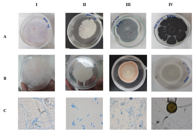
Figure 1. Macroscopic and microscopic characteristic of fungi isolated from sediments mining (I, II, III y IV). Colonies show front (A) and reverse (B) side of different isolated fungi on PDA medium after five day of incubation and show in the optical microscopic (C) 40 X and 100 X.
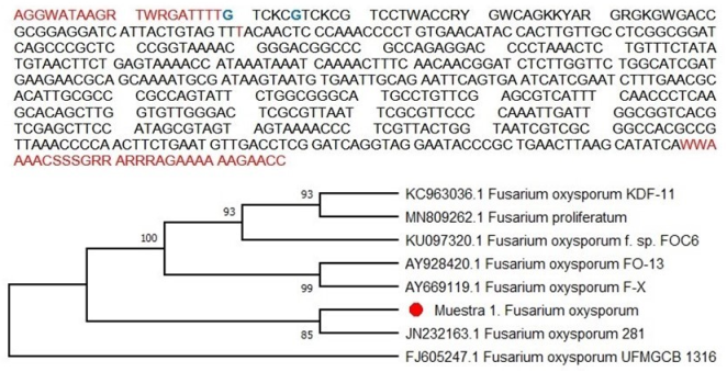
Figure 2. Sequence and phylogenetic trees of Fusarium oxysporum.

Figure 3. Sequence and phylogenetic trees of Pichia kudriavzevii.
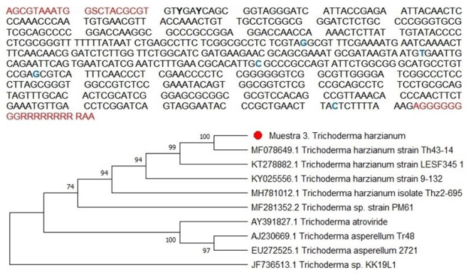
Figure 4. Sequence and phylogenetic trees of Trichoderma harzianum.
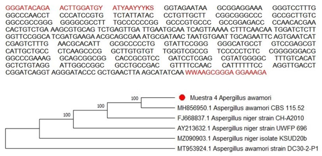
Figure 5. Sequence and phylogenetic trees of Aspergillus awamori.
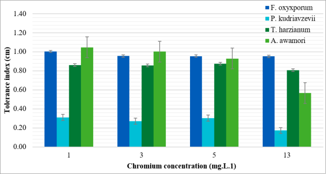
Figure 6. Chromium tolerance index of the isolated fungi.
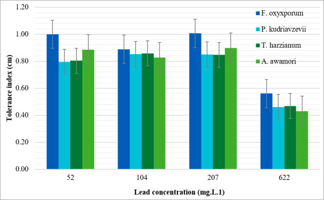
Figure 7. Lead tolerance index of the isolated fungi.
Information

