Abstract
This review summarized the mechanism of a novel fusion protein (PHPV fusion protein) containing membrane penetrating peptide and oligopeptide-1 (EGF). The new PHPV fusion protein contains two kinds of polypeptides: membrane penetrating peptide and oligopeptide-1, so it has a good function of preventing and treating HPV virus. PHPV fusion protein not only has the multi effect function of membrane penetrating peptide and oligopeptide-1, but also the negative charge (anion) C-terminal of the cross-linked and fused new protein surface interacts with the positive charge (cation) N-terminal on HPV particles. Through the combination of the N-terminal of the positive charge on its surface with the negative charge of the hydrophobic region in the middle and the C-terminal of the recognition region site containing peptidase, the membrane translocation signal is coupled with the NLS of the nuclear localization signal, thus blocking the invasion of human papillomavirus into host cells. Through the interaction of cell membrane, it can penetrate the natural barrier of cell membrane and play a role in destroying the viral membrane shell to inactivate the virus, so as to prevent cervical precancerous lesions and treat HPV infection. PHPV fusion protein can repair damaged cells, enhance the elasticity of loose vaginal mucosa, tighten the inner wall of the vagina, improve the vaginal wetness, and reduce the vaginitis reaction.
Keywords
PHPV Fusion Protein, Transmembrane Peptide, Oligopeptide-1 (EGF), Human Papilloma Virus (HPV), Cervical Cancer
1. Introduction
(1) Background of the Research:
Cervical cancer is the third largest malignant tumor in women in the world, accounting for more than half of the malignant tumors of female reproductive organs, and is also the main malignant tumor threatening women's health in China. Cervical cancer is the most common gynecological malignant tumor, and it is also one of the few malignant tumors with a relatively clear etiology. According to the statistics of the World Health Organization in 2018, there are nearly 570,000 new cases of cervical cancer and about 311,000 deaths every year worldwide. Among them, there are 106,000 new cases of cervical cancer and about 48,000 deaths in China every year.
Human papilloma virus (HPV)
| [1] | Kranjee C, Banks L A systematic canalysis of human papilloma virus (HPV) E6 PDZ substrates identifies MAGI-1 as a Major target of HPV type 16 (HPV-16) and HPV-18. Whose loss accompanies disruption of tight junctions. J Virol, 2011, 85: 1757-1764. |
| [2] | You J. Papilloma virus interaction with cellular chromatin. Biochim Biophys Acta. 2010, 1799: 192-199. |
[1, 2]
persistent infection is the main cause of cervical cancer, but HPV infection does not mean cervical cancer, most HPV infection will be cleared by the human immune system. Only persistent infection with high-risk HPV can cause cervical cancer.
Epidemiological studies show that the high-risk HPV infection rate in the population is about 15%~20%, more than 50% of women will develop HPV infection after experiencing the first sexual life, and 80% of women have been infected with HPV virus in their lifetime.
However, more than 90% of women can be cleared by the immune system within 3 years after HPV infection, only 10% of patients may have persistent infection, and <1% of those with persistent infection eventually develop cervical cancer.
(2) The Purpose of the Research
HPV infection has been identified as a necessary condition for the occurrence of cervical precancerous lesions and invasive cervical cancer
| [3] | Yonemura Y, Sugivama K, Fujimura T, et al. Epidermal growth factor receptor status and S- phasefractions in gastric carcicoma. Oncology, 1989, 16: 158-164. |
| [4] | Lee CM, Lee RJ, Hammond E, et al. Expression of HER-neu (cerbB-2) and epidermal growth factor receptor in cervical cancer: Prognostic correlation with clinical characteristics, and comparison of manual and automated imaging analysis [J]. Gynecol Oncol, 2004, 93(1): 209-214. |
[3, 4]
. A large number of research data have shown that HPV is closely related to cervical cancer and precancerous lesions, and high-risk HPV (HRHPV) infection is most closely related to cervical cancer
| [5] | El-Andaloussi S, Johansson HJ, Holm T, et al. A novel cell penetrating peptide, M918, for efficient delivery of proteins and peptide nucleic acids [J]. Mol Ther, 2007, 15: 1820−1826. |
[5]
.
HPV infection with the cervix usually does not show any discomfort, and the virus can lie dormant for years before causing changes in cervical cells, known as precancerous lesions. Precancerous lesions can develop to cervical cancer for more than 10 years, and there can be no symptoms during this period. Therefore, early screening is very important, and both primary and secondary prevention can greatly reduce the incidence and mortality of cervical cancer
.
(3) The Significance of the Research
Although the number of female cervical cancer patients in China in five years ranks the fourth among all tumors in China, due to the large population base, late vaccination and small population coverage, the number of deaths and 5-year morbidity of cervical cancer in China will remain the highest in the world in 2020. Therefore, it is very important to develop innovative design strategy of anti HPV infection agents.
2. HPV Infection and Cervical Cancer
Human papilloma virus (HPV)
HPV infection is the main cause of cervical lesions
| [1] | Kranjee C, Banks L A systematic canalysis of human papilloma virus (HPV) E6 PDZ substrates identifies MAGI-1 as a Major target of HPV type 16 (HPV-16) and HPV-18. Whose loss accompanies disruption of tight junctions. J Virol, 2011, 85: 1757-1764. |
[1]
. HPV is a kind of papilloma virus A belonging to the Pleuroviridae family. It is a common small DNA double stranded virus. It is a sexually transmitted disease caused by spherical DNA virus infection. The main types are HPV1, 2, 6, 11, 16, 18, 31, 33 and 35. The long-term infection of HPV16 and HPV18 may be related to female cervical cancer. The genome DNA length of each type of HPV is between 722 and 8000 bp. Research shows that there are more than 100 HPV subtypes, and about 40 HPV subtypes are related to reproductive tract diseases
| [2] | You J. Papilloma virus interaction with cellular chromatin. Biochim Biophys Acta. 2010, 1799: 192-199. |
[2]
. High risk HPV DNA can be detected in 99.7% of cervical cancer patients. Persistent high risk HPV infection is an essential factor in the occurrence of cervical cancer. Persistent viral infection is directly related to the interaction of viral gene proteins.
The main infection area of the virus is human epidermis and mucosal squamous epithelium. Up to now, more than 130 kinds of HPV sub-types have been isolated. HPV only invades human beings and has no pathogenicity to other animals. After regular systematic treatment, HPV will be cleared by the human body. For a long time, it has been known that HPV can cause human benign tumors and warts, such as human verruca vulgaris, condyloma acuminatum and papilloma on the skin and mucosa near the reproductive organs. Like hepatitis B virus, HPV is also a DNA virus. It used to be a member of the family polyoncovacuoviridae. In 1999, International Committee on Taxonomy of Viruses (ICTV) abolished the family polyoncovacuoviridae and replaced it with the family papillomavirus. Therefore, HPV belongs to this family
| [8] | Lehtinen M, Iacopo Baussano I, Paavonen J, Simopekka Vänskä S, Dillner J. Eradication of human papillomavirus and elimination of HPV-related diseases - scientific basis for global public health policies. Expert Rev Vaccines. 2019; 18(2): 153-160. |
[8]
.
HPV is a kind of species-specific epitheliotropic virus, which belongs to small DNA virus with double stranded closed loop and contains about 8000 base pairs. It includes 8 early open reading frames (E1-E8), 2 late reading frames and 1 non coding long control area. In the early open reading frame, E6 and E7 genes are the most important to stimulate cell growth. E6 and E7 proteins encoded cause immortalization of cervical epithelial cells
| [3] | Yonemura Y, Sugivama K, Fujimura T, et al. Epidermal growth factor receptor status and S- phasefractions in gastric carcicoma. Oncology, 1989, 16: 158-164. |
[3]
. The late reading frame L1 and L2 genes encode the major and minor capsid proteins of HPV, respectively, and assemble the capsid of HPV.
When we "dissect" HPV, we will find that it is composed of the protein coat and the wrapped core. The "raw materials" of capsid are mainly capsid protein (L1) and secondary capsid protein (L2); The core of shedding the coat is the real body that can cause human diseases - small double stranded circular DNA without envelope and a small amount of HIV, HSV, HPV. In 1949, Strauss first observed HPV particles under an electron microscope. It was spherical, symmetrical with 20 facets, and about 45-55 nm in diameter. According to the difference of HPV gene sequence structure, people divide HPV into nearly 130 genotypes, of which nearly 100 genes such as HIV and HSV have been identified.
Human being is the only host of HPV, and it likes to place its home in the skin and mucous membrane, so it has a high host specific affinity
| [4] | Lee CM, Lee RJ, Hammond E, et al. Expression of HER-neu (cerbB-2) and epidermal growth factor receptor in cervical cancer: Prognostic correlation with clinical characteristics, and comparison of manual and automated imaging analysis [J]. Gynecol Oncol, 2004, 93(1): 209-214. |
[4]
. HPV is a kind of papillomavirus A belonging to the Pleuroviridae family. It is a spherical DNA virus, which can cause the proliferation of squamous epithelium of human skin and mucous membrane. By 2013, more than 130 kinds of HPV have been isolated. The virus only invades human beings and has no pathogenicity to other animals. Once it is infected, human beings will carry it for life.
Because of different HPV viruses, the harm to patients is also different. Some viruses will bring malignant tumors to patients, eventually leading to canceration, which seriously threatens the lives of patients. Some diseases will lead to the death of pregnant women's fetuses or become deformed children, and some diseases will lead to male infertility
| [5] | El-Andaloussi S, Johansson HJ, Holm T, et al. A novel cell penetrating peptide, M918, for efficient delivery of proteins and peptide nucleic acids [J]. Mol Ther, 2007, 15: 1820−1826. |
[5]
.
3. Clinical Manifestations and Types of HPV Virus and Diseases Caused by Infection
Different types cause different clinical manifestations, which can be divided into:
1) Low risk type of skin: including HPV-1, 2, 3, 4, 7, 10, 12, 15, which are related to common warts, flat warts, plantar warts, etc;
2) skin high-risk type: including hpv-5, 8, 14, 17, 20, 36 and 38, which are related to verrucous epidermal dysplasia, and other malignant tumors that may be related to HPV infection include vulvar cancer, penile cancer, anal cancer, prostate cancer and bladder cancer;
3) Low risk type of mucosa: such as HPV-6, 11, 13, 32, 34, 40, 42, 43, 44, 53, 54, and infected genital, anal, oropharynx, and esophageal mucosa;
4) Mucosa high-risk type: HPV-16, 18, 30, 31, 33, 35, 39 and cervical cancer, rectal cancer, oral cancer, tonsil cancer, etc
| [9] | Hortlund M, van Mol T, Van de Pol F, Bogers J, Joakim Dillner J. Human papillomavirus load and genotype analysis improves the prediction of invasive cervical cancer. Int J Cancer. 2021; 149(3): 684-691. |
[9]
.
The biological activities of HPV virus include: HPV has strong resistance, can withstand drying and long-term storage, can be inactivated by heating or formalin treatment, so high temperature disinfection and 2% glutaraldehyde disinfection can inactivate
| [9] | Hortlund M, van Mol T, Van de Pol F, Bogers J, Joakim Dillner J. Human papillomavirus load and genotype analysis improves the prediction of invasive cervical cancer. Int J Cancer. 2021; 149(3): 684-691. |
[9]
.
4. HPV Infection Induces Epithelial Transformation Pathway
HPV infects basal cells through small wounds in epithelial tissue, and gradually replicates and releases as cells proliferate and differentiate into keratinocytes/mucosal cells. Its replication in host cells can be divided into five stages: 1) Adsorption; 2) Integration; 3) Proliferation; 4) Assembly; 5) Release.
HPV recognizes and binds cell surface receptors
1) In normal epithelial cells: E6/E7 is not expressed or low expressed, which does not lead to high grade pathological changes of cells, HPV can be cleared, and transient infection.
2) In normal epithelial cells: E6/E7 oncogene expression: 10% of infected people transform into persistent infection, E6 inhibits P53, inhibits apoptosis signal, E7 inhibits PRb, and activates growth signal.
3) Expression of other oncogenes: viral genes are gradually integrated into the cell genome, and oncogenes are continuously expressed to inhibit the cancer suppression mechanism, and cervical intraepithelial becomes neoplasia (CIN1/2/3).
4) Synergistic effects of other cancer promoting factors: HIV infection, multiple sexual partners, smoking and drinking, chemicals, immunosuppression, bacterial infection, environmental infection.
5) Cervical epithelial tumor can be transformed into cervical cancer continuously. Removing HPV can inhibit the canceration process.
Although it is clear that the cause of cervical cancer is HPV infection, the tumorigenic mechanism of HPV is still being studied and explored. The key processes include HPV life cycle and HPV immune escape.
HPV virus first enters the tissues through minor damage to the cervical epithelium. The immature metaplasia in the cervical transformation area is the most vulnerable period for HPV to invade. At this time, the basal cells are the first infected by HPV, and HPV enters the cells mainly through multi-step endocytosis.
The life cycle of HPV can be divided into three stages: establishment, maintenance and expansion. The establishment stage is to transfect the basement membrane cells. The HPV genome in the maintenance stage can be maintained in the basement cells for years to decades. Some of them have precancerous lesions or reverse themselves, and a few of them progress to tumors.
Amplification stage: the infected basal cells differentiate to the epithelial surface, and the number of viral copies is expanded to thousands. The expression of rich E6 and E7 genes activates the replication of host cell DNA, so that the viral DNA continues to synthesize. The expression of HPV L1 and L2 promotes the assembly of HPV genome, the release of mature virus particles from outer epithelial cells, and the leaving of cells in the form of infectious virus particles.
CIN1 (mild atypical hyperplasia) cells are slightly heterotypic, irregularly arranged, but still maintain polarity, and the abnormal proliferation cells are limited to the lower 1/3 of the epithelial layer.
CIN2 (moderate atypical hyperplasia) cells are obviously heterotypic and arranged in disorder. Abnormal proliferated cells occupy 2/3 of the lower epithelium.
CIN3 (severe atypical hyperplasia and carcinoma in situ) The epithelial cells of severe atypical hyperplasia are significantly atypical and lose their polarity. The abnormally proliferated cells expand to 2/3 of the epithelium or almost the whole layer, which is difficult to distinguish from carcinoma in situ. Epithelial heterotypic cells of carcinoma in situ were involved in the whole layer, polarity disappeared, nuclear heterotypic was significant, and mitotic phase was common. The epithelial basement membrane is intact without interstitial infiltration
.
5. Design Ideas and Research Progress of Anti HPV Infection Agents
(1). Structural modification molecules activate immune cell response
The three aspects of treatment target, delivery carrier and dosage form selection are the core issues of scientific design of new drugs.
The premise of HPV infection of vaginal and cervical epithelium is micro trauma, so promoting wound repair is a point ignored by other drugs.
In addition, although the absorption of local mucosa is faster than that of intact skin, it is still less efficient than other methods. Therefore, how to improve the absorption and deliver effective substances to the virus location is also a difficulty in research and development.
Finally, immune escape is the fundamental reason why HPV still exists after evolution, so how to improve the local immune response is also a very important aspect.
On the basis of previous work, scientists at home and abroad cooperating with Fleming have found a natural macromolecular defense substance, which has a variety antiviral and antibacterial effects, and can also activate the immune response, but it also has shortcomings. Therefore, the structural transformation of defensive substances has been carried out, and new preparations have been innovated.
The in vitro culture of a variety of tumor cell lines and the in vivo transplantation tumor model experiment show that the peptide segment with structural modification can promote cell death, promote the role of reactive oxygen species, activate apoptosis signals, and activate the signal transduction of immune response pathway, which also shows that the peptide segment with structural modification can inhibit the growth of tumor cells.
It shows that the modified peptide does not promote the malignant transformation of cervical precancerous lesions, but has the potential role of inhibiting cell transformation, which reduces the possibility of formation of side effects of possible growth factor signals promoting transformation to a certain extent
.
(2) Structural peptide promotes epithelial damage repair -- EGF effect
In 1962, American scientist Dr. Stanley Cohen and Professor Rita Levi Montalcini, an Italian scientist, extracted a polypeptide from the mandible of mice. This polypeptide is widely present in humans or other animals. A very small amount can strongly stimulate cell growth and inhibit aging, so it is named epidermal growth factor (EGF). In 1986, this discovery was awarded the Nobel Prize in Physiology and Medicine for opening up a new field of life science.
Human epidermal growth factor (hEGF), purified from human urine in 1974, is widely used in the medical field to treat corneal injury, burns, scalds, surgery and other wounds due to its highly effective repair function
| [3] | Yonemura Y, Sugivama K, Fujimura T, et al. Epidermal growth factor receptor status and S- phasefractions in gastric carcicoma. Oncology, 1989, 16: 158-164. |
[3]
.
The huge market needs to stimulate the industrial development of hEGF. Before the 1980s, EGF was mainly extracted from tissues and body fluids. Because the content of hEGF is extremely small, the extraction cost is high and the product purity is low
| [4] | Lee CM, Lee RJ, Hammond E, et al. Expression of HER-neu (cerbB-2) and epidermal growth factor receptor in cervical cancer: Prognostic correlation with clinical characteristics, and comparison of manual and automated imaging analysis [J]. Gynecol Oncol, 2004, 93(1): 209-214. |
[4]
. After the 1980s, with the development of genetic engineering technology, the scientific research field succeeded in producing recombinant human epidermal growth factor (hEGF) using Escherichia coli through gene recombination technology, laying a scientific and technological foundation for industrial production of EGF.
Recombinant human epidermal growth factor has also become oligopeptide-1, which can directly promote the repair of skin mucosa and the growth of epidermal cells, reverse differentiate aging cells into young cells, promote the synthesis of collagen and elastin, promote the regeneration of granulation tissue, delay cell aging, nourish female reproductive organs, and fade wrinkles and stains. It is one of the greatest discoveries in the history of human medicine and aesthetics
| [4] | Lee CM, Lee RJ, Hammond E, et al. Expression of HER-neu (cerbB-2) and epidermal growth factor receptor in cervical cancer: Prognostic correlation with clinical characteristics, and comparison of manual and automated imaging analysis [J]. Gynecol Oncol, 2004, 93(1): 209-214. |
[4]
.
EGF is composed of 53 amino acids and includes three disulfide bonds. The molecular weight of EGF precursor is 130KD, and its volume is 300 times smaller than that of bacteria. The human EGF gene is located in the long arm of chromosome 4.
EGF can promote cell proliferation and differentiation, so as to replace aging cells and dead cells with new cells. Traditional cosmetics play an external modifying role and cannot fundamentally solve skin problems. EGF is used in cosmetics to repair and adjust cells at the molecular level based on the physiological structure of human skin.
EGF is the only cosmetic ingredient that has won the Nobel Prize for Science. In 1999, the research achievements of EGF were rated as the top ten scientific achievements of the year by the world authoritative journal Science. It is praised as a "beauty factor" by the beauty industry.
Oligopeptide-1 (EGF) is an active substance in human body. By stimulating tyrosine phosphorylation of oligopeptide-1 receptor, it can repair the hyperplastic skin surface cells, and has excellent effects on damaged skin such as wounds and ulcers. Its greatest feature is to promote the proliferation and reverse differentiation of cells, so as to replace aging and dead cells with new cells.
Clinical role of oligopeptide-1 (EGF): Epidermal growth factor is of great significance in the field of medicine. Research shows that it can promote the growth of injured skin tissue, inhibit the secretion of gastric acid, promote the growth of burned skin and the healing of skin ulcers. It will play an important role in the treatment of skin diseases, gastric diseases, and corneal transplantation. (1) Quickly repair damaged skin, and improve skin repair and care. (2) It can smooth wrinkles around the eyes, tighten the skin, reduce the average age of skin tissue, moisturize the skin, and enhance skin elasticity. (3) Whitening and freckle removing, reduce the content of melanin and colored cells in skin cells, and accelerate the metabolism and shedding of melanocytes. (4) Sun protection and post sun repair can reduce the damage of ultraviolet radiation to the skin and reduce the growth of dark spots on the skin after sun exposure. (5) It can prevent acne, remove scars, shorten the time of wound healing and reduce the formation of scars, which has a good effect on the prevention and care of acne.
Through EGF structural reconstruction, only the functional fragments of wound repair were retained, and the area where EGF promotes cell growth was removed. Through EGF structural transformation, macromolecules are cut into small peptides that only retain the function of wound repair, and the signal structure area for promoting cell growth is removed, without causing excessive growth signals.
EGF can block the recognition, integration, replication, assembly and release of viral receptors, activate host immune response, and promote epithelial wound repair. EGF is a very mature epithelial damage repair factor. Through the structural transformation of EGF, it can improve the absorption rate and reduce the cell growth promoting signal.
Compared with the control peptide, the modified EGF peptide has better wound repair function. Compared with EGF before structural modification, the modified EGF peptide has the same effect as EGF. It also shows that EGF external use only promotes damage repair, but does not promote cell proliferation. It shows that EGF is applied externally to the epidermis for a short period of time. Without other cancer promoting mechanisms, a single growth promoting signal is not enough to cause malignant transformation of epithelial cells.
Membrane penetrating peptide is a kind of high efficient protein carrier macromolecule, which has anti-virus and antibacterial effects.
The special peptide segment of membrane penetrating peptide can effectively transport macromolecules and also can destroy viruses.
6. The Peptides Transport Function by the Cell Membrane Penetrating Peptide (CPP)
Cell Membrane Penetrating Peptide is a kind of short peptide that can carry macromolecules into cells, and its transmembrane ability does not depend on classical endocytosis. Cell penetrating peptide is a small peptide with cell penetrating function composed of less than 30 amino acids. It can cross the natural barrier of cell membrane by cross linking with cell membrane
| [12] | Zhang P, Moreno R, Lambert PL, DiMaio D. Cell-penetrating peptide inhibits retromer-mediated human papillomavirus trafficking during virus entry. Proc Natl Acad Sci U S A. 2020 Mar 17; 117(11): 6121-6128. |
| [13] | Morris MC, Depollier J, Mery J, Heitz F, Divita G. A peptide carrier for the delivery of biologically active proteins into mammalian cells. Nat Biotechnol. 2001; 12: 1173-6. |
[12, 13]
.
Through the research on the biochemical properties of naturally occurring cell penetrating peptides, we have gradually mastered some common characteristics of cell penetrating peptides. These substances are polypeptide fragments with positive charges of different lengths, which are rich in arginine, lysine and other basic amino acid residues, and have secondary structures α- The spatial conformation of helix.
With these characteristics, membrane penetrating peptides PEp-1 and MPG with stronger penetrability and higher efficiency have been artificially synthesized, and have successfully carried macromolecular substances into cells to play biological activities. Some scholars also call such short peptides as protein transduction domains or Trojan peptides. At present, scientists have applied it to gene therapy.
The recently designed ELP (Val Pro Gly Xaa Gly) transmembrane peptide can guide the loaded oligopeptide-1 repair protein to penetrate the cell membrane and penetrate through the epidermis. It is rich in basic amino acids and contains more positively charged peptide fragments, which can be targeted into the skin basal layer receptor
| [14] | Esbjorner EK, Lincoln P, Norden B. Counterion-mediated membrane penetration: cationic cell-penetrating peptides overcome born energy barrier by ion-pairing with phospholipids [J]. Biochim Biophys Acta, 2007, 1768: 1550−1558. |
| [15] | Torchilin VP. Tat peptide-mediated intracellular delivery of pharmaceutical nanocarriers [J]. Adv Drug Deliv Rev, 2008, 60: 548−558. |
| [16] | Nakase I, Takeuchi T, Tanaka G, et al. Methodological and cellular aspects that govern the internalization mechanisms of arginine-rich cell-penetrating peptides [J]. Adv Drug Deliv Rev, 2008, 60: 598−607. |
[14-16]
.
The membrane penetrating peptide has the following characteristics: 1) It has net positive charge and amphiphilic; 2) High transport efficiency through membrane; 3) It can import nearly all cells; 4) It can carry a variety of active substances into cells; 5) It can target and penetrate the cell membrane to inactivate multiple viruses without causing cell damage.
A large number of studies have shown that the transmembrane peptide has a strong transport potential. Compared with other transport modes of biological macromolecules, the transmembrane peptide has many unique features
| [17] | Hansen M, Kilk K, Langel U. Predicting cell-penetrating peptides [J]. Adv Drug Deliv Rev, 2008, 60: 572−579. |
| [18] | Maiolo JR, Ferrer M, Ottinger EA. Effects of cargo molecules on the cellular uptake of arginine-rich cell-penetrating peptides [J]. Biochim Biophys Acta, 2005, 1712: 161−172. |
| [19] | Wang P, Liu M, Guo YM. Development of cell-penetrating peptide applying as a vector in molecular imaging [J]. J Med Imaging, 2008, 18: 565−568. |
[17-19]
. The substances carried by membrane penetrating peptides can be proteins (green fluorescent protein, RNA enzyme, galactosidase, etc.), polypeptides (less than 100 amino acid residues
| [20] | Zorko M, Langel B. Cell-penetrating peptides: mechanism and kinetics of cargo delivery [J]. Adv Drug Deliv Rev, 2005, 57: 529−545. |
[20]
), DNA, chemical small molecule drugs, oligonucleosides (less than 100 bp
| [20] | Zorko M, Langel B. Cell-penetrating peptides: mechanism and kinetics of cargo delivery [J]. Adv Drug Deliv Rev, 2005, 57: 529−545. |
[20]
), antisense nucleic acids, peptide nucleic acids, nanoparticles, fluorescein, organic molecules, adenovirus carriers, imaging materials, liposomes, iron particles, etc.
The carrying capacity
| [21] | Ziegler A. Thermodynamic studies and binding mechanisms of cell-penetrating peptides with lipids and glycosaminoglycans [J]. Adv Drug Deliv Rev, 2008, 60: 580−597. |
[21]
can reach 120kDa (such as galactosidase) and 40nm (such as iron particles), and there is no obvious evidence to indicate the carrying limit. Cell penetrating peptides can carry various biological macromolecules through chemical binding or gene fusion
| [21] | Ziegler A. Thermodynamic studies and binding mechanisms of cell-penetrating peptides with lipids and glycosaminoglycans [J]. Adv Drug Deliv Rev, 2008, 60: 580−597. |
[21]
. There are two ways to connect transmembrane peptides with drugs: non covalent connection and covalent connection. The use of CPPs can also increase the activity of peptide drugs and carry out specific transportation of cells and organs
| [22] | Herbig ME, Fromm U, Leuenberger J, et al. Bilayer interaction and localization of cell penetrating peptides with model membranes: a comparative study of a human calcitonin (hCT) derived peptide with pVEC and pAntp (43–58) [J]. Biochim Biophys Acta, 2005, 1712: 197−211. |
[22]
.
The use of foreign protein or peptide drugs for many diseases is a very valuable treatment. Wang et al.
| [19] | Wang P, Liu M, Guo YM. Development of cell-penetrating peptide applying as a vector in molecular imaging [J]. J Med Imaging, 2008, 18: 565−568. |
[19]
used the protein transduction function of transmembrane peptide to mediate the signal peptide green fluorescent protein transmembrane peptide NT4-GFP Ant fusion protein to reach target cells through cell membrane and blood brain barrier.
Tat (Tat48 − 60) can transport the cytotoxic peptide analog P21EAF1/CIP1 of cyclin dependent kinase inhibitor into the nucleus, and studies
| [23] | Shishido T, Yonezewa D, Iwata K, et al. Construction of arginine-rich peptide displaying bionanocapsules [J]. Bioorg Med Chem Lett, 2009, 19: 1473−1476. |
[23]
found that Tat48 − 60 – P10 can induce apoptosis. Zhou et al.
| [24] | Zhou LH, Thakur CS, Molinaro RJ, et al. Delivery of 2-5A cargo into living cells using the Tat cell penetrating peptide: 2-5A-tat [J]. Bioorg Med Chem, 2006, 14: 7862−7874. |
[24]
according to one α structure of spiral peptide CAI has designed a cell penetrating peptide NYAD-1, which is more stable than CAI α helical structure. It was found that NYAD-1 could pass through the cell membrane and co locate on the plasma membrane with Gag poly protein, destroying the self-assembly of virus particles.
Researchers
| [25] | Marshall NB, Oda SK, London, CA, et al. Arginine-rich cellpenetrating peptides facilitate delivery of antisense oligomers into murine leukocytes and alter pre-mRNA splicing [J]. J Immunol Methods, 2007, 325: 114−126. |
| [26] | Lindgren M, Rosenthal-Aizman K, Saar K, et al. Overcoming methotrexate resistance in breast cancer tumour cells by the use of a new cell-penetrating peptide [J]. Biochem Pharmacol, 2006, 71: 416−425. |
| [27] | Kamei N, Morishita M, Eda Y, et al. Usefulness of cell penetrating peptides to improve intestinal insulin absorption [J]. J Control Release, 2008, 132: 21−25. |
| [28] | Khafagy ES, Morishita M, Isowa K, et al. Effect of cell penetrating peptides on the nasal absorption of insulin [J]. J Control Release, 2009, 133: 103−108. |
| [29] | Tunnemann G, Karezewski P, Haase H, etal. Modulation of muscle contractionby a cell-permeable peptide [J]. J Mol Med, 2007, 85: 1405−1412. |
| [30] | Kloss A, Henklein P, Siele D, et al. The cell-penetrating peptide octa-arginine is a potent inhibitor of proteasome activities [J]. Eur J Pharm Biopharm, 2009, 72: 219−225. |
[25-30]
designed a binding polypeptide containing membrane penetrating peptide, elastin like polypeptide ELP (Val Pro Gly Xaa Gly) and a cyclin dependent kinase inhibitor P21. This polypeptide can slow down the growth rate of SKOV-3 ovarian cancer cells and HeLa uterine cancer cells and inhibit their proliferation. HIV Tat combines with Tyler's disease antigen Tp2 to form a recombinant Tat Tp2 fusion protein, which was found to enhance the excitability of the dull CD8+T cell response
| [26] | Lindgren M, Rosenthal-Aizman K, Saar K, et al. Overcoming methotrexate resistance in breast cancer tumour cells by the use of a new cell-penetrating peptide [J]. Biochem Pharmacol, 2006, 71: 416−425. |
[26]
.
The carrier peptide segment effectively transports anti HPV substances into the cell (competes with the virus to reverse the binding site of the transporter, thus inhibiting the virus from entering the cell), and plays an anti viral role. The dose effect curve well shows that this anti infective effect is specific. If the carrier peptide segment carrying anti infective substances is removed, the cell resumes HPV infection. It also shows that the anti infection effect depends on the carrier peptide.
7. PHPV Fusion Factor
Scientists from Ruixin Pharmaceutical Co., Ltd., Guangdong Medical Insurance Pharmaceutical Co., Ltd. and Hong Kong Pharmaceutical Co., Ltd., together with the Chinese Academy of Biomedical Sciences, the American Nanomedicine Institute, the International Anti viral Innovation Alliance and South China University of Technology, spent ten years of dedicated research and found a new method that can effectively block the HPV infection cell pathway.
This method is based on the principle of synthetic biology, using genetic engineering technology, through the fusion expression of special plasmids, the polypeptides with different lengths of positive and negative charges are fused into a new biological defense factor with stronger cell penetration and higher protein transport efficiency. It is named "PHPV fusion factor".
PHPV fusion factor vaginal bacteria inhibiting gel has the following characteristics: one major contribution, two major innovations and three major functions. The first contribution is that the fusion factor provides the first line of defense for the prevention and treatment of HPV infection, or can fill the blank of HPV treatment. Two major innovations are: (1) one is sequence innovation, a fusion molecule of new design; (2) second, expression innovation, fusion expression of special plasmid fermentation system; (3) the three major functions include blocking HPV infection, reducing HPV load, preventing cervical precancerous lesions, and can also be used for skin diseases caused by HPV, reducing the recurrence rate of condyloma acuminatum.
At present, many hospitals have joined in the clinical observation and cooperative research of fusion factors, among which the representative hospitals are the Xiangya Second Affiliated Hospital of Central South University, the First Affiliated Hospital of Zhengzhou University, the First Affiliated Hospital of Zhongshan University, etc.
8. Action Principle of PHPV Fusion Factor Vaginal Bacteria Inhibiting Gel
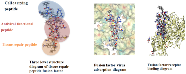
PHPV fusion protein contains oligopeptide-1 and transmembrane peptide, so it has a good function of preventing and treating HPV virus. PHPV fusion protein is a membrane penetrating peptide that is creatively introduced on the basis of oligopeptide-1. PHPV fusion protein not only has the multiple functions of membrane penetrating peptide and oligopeptide, but also complexes the negative charge (anion) C-terminal of the cross-linked fusion new protein surface with the positive charge (cation) N-terminal of HPV particles, By combining the N-terminal of the positive charge on its surface with the negative charge on the hydrophobic region in the middle and the C-terminal of the recognition region site containing peptidase, the membrane translocation signal is coupled with the NLS of the nuclear localization signal, so as to block the invasion of human papillomavirus into the host cell, and then penetrate the natural barrier of the cell membrane through the interaction of the cell membrane, playing a role in destroying the viral membrane shell to inactivate the virus, To prevent cervical precancerous lesions and treat HPV infection.
The unique structural design of the fusion factor enables it to physically adsorb the virus and inhibit viral transcription and replication; It can also compete with viruses for cell surface receptors, so that virus particles cannot bind to receptor proteins, inhibit virus adsorption, inactivate virus infection, and play a role in preventing cervical lesions.
Figure 1. The structure diagram of PHPV peptide.
9. Clinical Application Effect of PHPV Fusion Factor
(1) Antiviral principle of PHPV fusion factor
HPV virus only inhabits the epithelial cells of skin and mucous membrane, and does not enter the blood. Local topical products are easier to reach the focus.
Its main antiviral mechanism is: by intertwining the positive charge part of the surface conformation of the fusion factor with the negative charge part of the virus particles, the negative charge on the HPV virus surface is combined with the positive charge of the peptidase recognition site in the middle hydrophobic region of the fusion factor, and the membrane transposition signal is coupled with the nuclear localization signal. Based on the principle of physical adsorption of positive and negative charges, it blocks viral transcription and replication. It can inhibit the assembly and release of virus particles and reduce the viral load of HPV.
The fusion factor can also prevent the virus from entering the host cell by competing for the viral binding receptor site on the cell membrane; Activate host immune response signal pathway, regulate and improve immune cells' defense against viruses. PHPV fusion factor vaginal bacteria blocking gel can form a protective film on the inner wall of the genital tract, block the contact between the virus and the inner wall cell surface, inhibit the adsorption of the virus, and passivate the virus infection ability.
(2) Fusion factor blocks HPV infection pathway
It can block the recognition, invasion, integration, replication, assembly and release of the virus to the host by physical action, and activate the immune response signal pathway.
1) Adsorption: the gel layer fills the cell gap, changes the cell surface charge, and physically hinders HPV from contacting cells and their surface receptors.
2) Infection: The fusion factor competes for the binding site of HPV receptor on the cell membrane, and physical isolation prevents the virus entering the host cell.
3) Integrated replication: The positive charge part of the surface conformation of the fusion factor interacts with the negative charge part of HPV nucleic acid based on the principle of positive and negative charge affinity and physical adsorption to prevent viral transcription and replication.
4) Proliferation assembly: inhibit virus assembly into particles.
5) Release fragmentation: block the release process of virus particles.
6) Systemic immune response: the fusion factor activates the host cell immune response signal pathway and improves the immune cells' defense against viruses.
10. Conclusion
This review summarized the mechanism of a novel fusion protein (PHPV fusion protein) containing membrane penetrating peptide and oligopeptide-1 (EGF). The new PHPV fusion protein contains two kinds of polypeptides: membrane penetrating peptide and oligopeptide-1 (EGF), so it has a good function of preventing and treating HPV virus. PHPV fusion protein can repair damaged cells, enhance the elasticity of loose vaginal mucosa, tighten the inner wall of the vagina, improve the vaginal wetness, and reduce the vaginitis reaction.
Abbreviations
HPV | Human Papilloma Virus |
HRHPV | High-Risk Human Papilloma Virus |
HIV | Human Immunodeficiency Virus |
HSV | Herpes Simplex Virus |
ICTV | International Committee on Taxonomy of Viruses |
CIN1 | Mild Atypical Hyperplasia |
CIN2 | Moderate Atypical Hyperplasia |
CIN3 | Severe Atypical Hyperplasia And Carcinoma in Situ |
EGF | Epidermal Growth Factor |
hEGF | Human Epidermal Growth Factor |
CPP | Cell Membrane Penetrating Peptide |
Institutional Review Board Statement
Not applicable.
Informed Consent Statement
Not applicable.
Author Contributions
Zixuan Lyu: Conceptualization, Data curation, Writing—original draft
Yulin Chen: Conceptualization, Data curation, Writing—original draft
Chiming Wei: Conceptualization, Writing—review and editing
Funding
This research received no external funding.
Data Availability Statement
Not applicable.
Conflicts of Interest
The authors declare no conflict of interest.
References
| [1] |
Kranjee C, Banks L A systematic canalysis of human papilloma virus (HPV) E6 PDZ substrates identifies MAGI-1 as a Major target of HPV type 16 (HPV-16) and HPV-18. Whose loss accompanies disruption of tight junctions. J Virol, 2011, 85: 1757-1764.
|
| [2] |
You J. Papilloma virus interaction with cellular chromatin. Biochim Biophys Acta. 2010, 1799: 192-199.
|
| [3] |
Yonemura Y, Sugivama K, Fujimura T, et al. Epidermal growth factor receptor status and S- phasefractions in gastric carcicoma. Oncology, 1989, 16: 158-164.
|
| [4] |
Lee CM, Lee RJ, Hammond E, et al. Expression of HER-neu (cerbB-2) and epidermal growth factor receptor in cervical cancer: Prognostic correlation with clinical characteristics, and comparison of manual and automated imaging analysis [J]. Gynecol Oncol, 2004, 93(1): 209-214.
|
| [5] |
El-Andaloussi S, Johansson HJ, Holm T, et al. A novel cell penetrating peptide, M918, for efficient delivery of proteins and peptide nucleic acids [J]. Mol Ther, 2007, 15: 1820−1826.
|
| [6] |
Harald zur Hausen – Facts. NobelPrize.org. Nobel Prize Outreach AB 2022. Tue. 15 Feb 2022.
https://www.nobelprize.org/prizes/medicine/2008/hausen/facts/
|
| [7] |
Global cancer statistics 2020: GLOBOCAN estimates of incidence and mortality worldwide for 36 cancers in 185 countries. CA Cancer J Clin. 2021: 71: 209–49.
https://doi.org/10.3322/caac.21660
|
| [8] |
Lehtinen M, Iacopo Baussano I, Paavonen J, Simopekka Vänskä S, Dillner J. Eradication of human papillomavirus and elimination of HPV-related diseases - scientific basis for global public health policies. Expert Rev Vaccines. 2019; 18(2): 153-160.
|
| [9] |
Hortlund M, van Mol T, Van de Pol F, Bogers J, Joakim Dillner J. Human papillomavirus load and genotype analysis improves the prediction of invasive cervical cancer. Int J Cancer. 2021; 149(3): 684-691.
|
| [10] |
Medda A. Human Papillomavirus and Cellular Pathways: Hits and Targets. Pathogens. 2021; 10(3): 262.
https://doi.org/10.3390/p athogens10030262
|
| [11] |
Human Papillomavirus (HPV) Infection. Sexually Transmitted Infections Treatment Guidelines, 2021.
https://www.cdc.gov/std/treatment-guidelines/hpv.htm
|
| [12] |
Zhang P, Moreno R, Lambert PL, DiMaio D. Cell-penetrating peptide inhibits retromer-mediated human papillomavirus trafficking during virus entry. Proc Natl Acad Sci U S A. 2020 Mar 17; 117(11): 6121-6128.
|
| [13] |
Morris MC, Depollier J, Mery J, Heitz F, Divita G. A peptide carrier for the delivery of biologically active proteins into mammalian cells. Nat Biotechnol. 2001; 12: 1173-6.
|
| [14] |
Esbjorner EK, Lincoln P, Norden B. Counterion-mediated membrane penetration: cationic cell-penetrating peptides overcome born energy barrier by ion-pairing with phospholipids [J]. Biochim Biophys Acta, 2007, 1768: 1550−1558.
|
| [15] |
Torchilin VP. Tat peptide-mediated intracellular delivery of pharmaceutical nanocarriers [J]. Adv Drug Deliv Rev, 2008, 60: 548−558.
|
| [16] |
Nakase I, Takeuchi T, Tanaka G, et al. Methodological and cellular aspects that govern the internalization mechanisms of arginine-rich cell-penetrating peptides [J]. Adv Drug Deliv Rev, 2008, 60: 598−607.
|
| [17] |
Hansen M, Kilk K, Langel U. Predicting cell-penetrating peptides [J]. Adv Drug Deliv Rev, 2008, 60: 572−579.
|
| [18] |
Maiolo JR, Ferrer M, Ottinger EA. Effects of cargo molecules on the cellular uptake of arginine-rich cell-penetrating peptides [J]. Biochim Biophys Acta, 2005, 1712: 161−172.
|
| [19] |
Wang P, Liu M, Guo YM. Development of cell-penetrating peptide applying as a vector in molecular imaging [J]. J Med Imaging, 2008, 18: 565−568.
|
| [20] |
Zorko M, Langel B. Cell-penetrating peptides: mechanism and kinetics of cargo delivery [J]. Adv Drug Deliv Rev, 2005, 57: 529−545.
|
| [21] |
Ziegler A. Thermodynamic studies and binding mechanisms of cell-penetrating peptides with lipids and glycosaminoglycans [J]. Adv Drug Deliv Rev, 2008, 60: 580−597.
|
| [22] |
Herbig ME, Fromm U, Leuenberger J, et al. Bilayer interaction and localization of cell penetrating peptides with model membranes: a comparative study of a human calcitonin (hCT) derived peptide with pVEC and pAntp (43–58) [J]. Biochim Biophys Acta, 2005, 1712: 197−211.
|
| [23] |
Shishido T, Yonezewa D, Iwata K, et al. Construction of arginine-rich peptide displaying bionanocapsules [J]. Bioorg Med Chem Lett, 2009, 19: 1473−1476.
|
| [24] |
Zhou LH, Thakur CS, Molinaro RJ, et al. Delivery of 2-5A cargo into living cells using the Tat cell penetrating peptide: 2-5A-tat [J]. Bioorg Med Chem, 2006, 14: 7862−7874.
|
| [25] |
Marshall NB, Oda SK, London, CA, et al. Arginine-rich cellpenetrating peptides facilitate delivery of antisense oligomers into murine leukocytes and alter pre-mRNA splicing [J]. J Immunol Methods, 2007, 325: 114−126.
|
| [26] |
Lindgren M, Rosenthal-Aizman K, Saar K, et al. Overcoming methotrexate resistance in breast cancer tumour cells by the use of a new cell-penetrating peptide [J]. Biochem Pharmacol, 2006, 71: 416−425.
|
| [27] |
Kamei N, Morishita M, Eda Y, et al. Usefulness of cell penetrating peptides to improve intestinal insulin absorption [J]. J Control Release, 2008, 132: 21−25.
|
| [28] |
Khafagy ES, Morishita M, Isowa K, et al. Effect of cell penetrating peptides on the nasal absorption of insulin [J]. J Control Release, 2009, 133: 103−108.
|
| [29] |
Tunnemann G, Karezewski P, Haase H, etal. Modulation of muscle contractionby a cell-permeable peptide [J]. J Mol Med, 2007, 85: 1405−1412.
|
| [30] |
Kloss A, Henklein P, Siele D, et al. The cell-penetrating peptide octa-arginine is a potent inhibitor of proteasome activities [J]. Eur J Pharm Biopharm, 2009, 72: 219−225.
|
Cite This Article
-
ACS Style
Lyu, Z.; Chen, Y.; Wei, C. Innovative Design Strategy and Research Progress of Anti Human Papilloma Virus Infection Agents. Eur. J. Clin. Biomed. Sci. 2024, 10(1), 15-22. doi: 10.11648/j.ejcbs.20241001.12
 Copy
|
Copy
|
 Download
-
@article{10.11648/j.ejcbs.20241001.12,
  author = {Zixuan Lyu and Yulin Chen and Chiming Wei},
  title = {Innovative Design Strategy and Research Progress of Anti Human Papilloma Virus Infection Agents
},
  journal = {European Journal of Clinical and Biomedical Sciences},
  volume = {10},
  number = {1},
  pages = {15-22},
  doi = {10.11648/j.ejcbs.20241001.12},
  url = {https://doi.org/10.11648/j.ejcbs.20241001.12},
  eprint = {https://article.sciencepublishinggroup.com/pdf/10.11648.j.ejcbs.20241001.12},
  abstract = {This review summarized the mechanism of a novel fusion protein (PHPV fusion protein) containing membrane penetrating peptide and oligopeptide-1 (EGF). The new PHPV fusion protein contains two kinds of polypeptides: membrane penetrating peptide and oligopeptide-1, so it has a good function of preventing and treating HPV virus. PHPV fusion protein not only has the multi effect function of membrane penetrating peptide and oligopeptide-1, but also the negative charge (anion) C-terminal of the cross-linked and fused new protein surface interacts with the positive charge (cation) N-terminal on HPV particles. Through the combination of the N-terminal of the positive charge on its surface with the negative charge of the hydrophobic region in the middle and the C-terminal of the recognition region site containing peptidase, the membrane translocation signal is coupled with the NLS of the nuclear localization signal, thus blocking the invasion of human papillomavirus into host cells. Through the interaction of cell membrane, it can penetrate the natural barrier of cell membrane and play a role in destroying the viral membrane shell to inactivate the virus, so as to prevent cervical precancerous lesions and treat HPV infection. PHPV fusion protein can repair damaged cells, enhance the elasticity of loose vaginal mucosa, tighten the inner wall of the vagina, improve the vaginal wetness, and reduce the vaginitis reaction.
},
 year = {2024}
}
Copy
|
Download
-
TY  - JOUR
T1  - Innovative Design Strategy and Research Progress of Anti Human Papilloma Virus Infection Agents

AU  - Zixuan Lyu
AU  - Yulin Chen
AU  - Chiming Wei
Y1  - 2024/06/21
PY  - 2024
N1  - https://doi.org/10.11648/j.ejcbs.20241001.12
DO  - 10.11648/j.ejcbs.20241001.12
T2  - European Journal of Clinical and Biomedical Sciences
JF  - European Journal of Clinical and Biomedical Sciences
JO  - European Journal of Clinical and Biomedical Sciences
SP  - 15
EP  - 22
PB  - Science Publishing Group
SN  - 2575-5005
UR  - https://doi.org/10.11648/j.ejcbs.20241001.12
AB  - This review summarized the mechanism of a novel fusion protein (PHPV fusion protein) containing membrane penetrating peptide and oligopeptide-1 (EGF). The new PHPV fusion protein contains two kinds of polypeptides: membrane penetrating peptide and oligopeptide-1, so it has a good function of preventing and treating HPV virus. PHPV fusion protein not only has the multi effect function of membrane penetrating peptide and oligopeptide-1, but also the negative charge (anion) C-terminal of the cross-linked and fused new protein surface interacts with the positive charge (cation) N-terminal on HPV particles. Through the combination of the N-terminal of the positive charge on its surface with the negative charge of the hydrophobic region in the middle and the C-terminal of the recognition region site containing peptidase, the membrane translocation signal is coupled with the NLS of the nuclear localization signal, thus blocking the invasion of human papillomavirus into host cells. Through the interaction of cell membrane, it can penetrate the natural barrier of cell membrane and play a role in destroying the viral membrane shell to inactivate the virus, so as to prevent cervical precancerous lesions and treat HPV infection. PHPV fusion protein can repair damaged cells, enhance the elasticity of loose vaginal mucosa, tighten the inner wall of the vagina, improve the vaginal wetness, and reduce the vaginitis reaction.

VL  - 10
IS  - 1
ER  -
Copy
|
Download