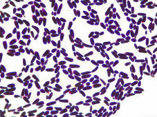
One of the infection that affects women of reproductive age which is difficult to hide based on the discomfort presented is vaginal candidiasis. This study was conducted to investigate the level of vaginal discharge causing fungal infection among undergraduate female students of the university. High vaginal swab (HVS) were collected from 150 female students of the Federal university of Technology Akure. Ondo State, Nigeria. between September 2024 to May 2025, aged 16 – 30 years that visited the University Health Centre with complaints and symptoms viz a viz; discomfort, painful urination, discharge and intense itching in the vagina, etc. Laboratory analysis was conducted, HVS samples were processed, identified microscopically, culturally and biochemically using standard procedure. The prevalence of vaginal candidiasis among female undergraduate students diagnosed was 32 (21.3%) of the 150 participants. Out of the 32 positive samples 28 (87.5%) were Candida albicans while 4 (12.5%) were Candida tropicalis. In the age distribution of the study, 16-18 years was 8 (25%), 19 -24 years was 18 (56.25%) and 25 -30 years was 6 (18.75%). The most highest distribution of the study were within 19 – 24 years which accounted for 56.25% (18) while age 25 -30 years accounted for 6 (18.75%) which indicated the least distribution. The findings indicate low prevalence of vulvovaginal candidiasis among female students of the Federal university of Technology, Akure. Nigeria. However, though the prevalence was low but significant, therefore efforts to enhance and improve hostel sanitation, personal hygiene and minimize use of tight pants are better approach to reduce it occurrence thereby curtail the risk of the fungal infection.
| Published in | Biomedical Sciences (Volume 12, Issue 2) |
| DOI | 10.11648/j.bs.20261202.13 |
| Page(s) | 38-43 |
| Creative Commons |
This is an Open Access article, distributed under the terms of the Creative Commons Attribution 4.0 International License (http://creativecommons.org/licenses/by/4.0/), which permits unrestricted use, distribution and reproduction in any medium or format, provided the original work is properly cited. |
| Copyright |
Copyright © The Author(s), 2026. Published by Science Publishing Group |
Undergraduate Female Students, Prevalence, Vulvovaginal Candidiasis, Discharge, Infection
Age (years) | No Analyzed | No Positive | Candida albicans | Candida tropicalis |
|---|---|---|---|---|
16 -18 | 48 | 8 (25%) | 7 | 1 |
19 -24 | 66 | 18 (56.25%) | 15 | 3 |
25 -30 | 36 | 6 (18.75) | 6 | -- |
Total | 150 | 32 (21.3%) | 28 (87.5%) | 4 (12.5%) |
Morpholeogy | Sugar Fermentation | Sugar Assimilation | Other reactions | ||||||||
|---|---|---|---|---|---|---|---|---|---|---|---|
G | M | L | S | G | M | L | S | U | GT | ||
Candida albicans | 4-6um white, smooth, creamy hyphae spherical budding | AG | AG | _ | A* | + | + | _ | + | - | + |
Candida tropicalis | 4-8um, white creamy, slightly wrinkled edge, pseudohyphae and spherical budding | AG | AG | _ | AG | + | + | _ | + | + | + |
Ages | Vaginal discharge | Discomfort | Total |
|---|---|---|---|
16 -18 | 30 | 15 | 45 |
19 -25 | 44 | 26 | 70 |
26 -30 | 23 | 12 | 35 |
Total | 97 (65%) | 53 (35%) | 150 |
VVC | Vulvovaginal Candidiasis |
GTT | Germ Tube Test |
G | Glucose |
L | Lactose |
M | Maltose |
S | Sucrose |
A & G | Acid and Gas |
U | Urease |
HVS | High Vaginal Swab |
HIV | Human Immunodeficiency Virus |
| [1] | Abdul-Aziz, M. A. K., Abdul-Ghani, N. A., Alhilali, Al-Mjahedl. K. A. and Alabsi S. A. (2019). Bacterial vaginosis, vulvovaginal candidiasis. A Trichomonal vaginitis among reproductive aged women seeking primary Healthcare in Sana’a city Yemen. BMC Infec. Disease 19(1): 879 |
| [2] | Akingbade, O. A., Akinjimi, A. A. Okerentugba, P. O. and Okonko, I. O. (2013). Prevalence of Candida albicans amongst women attending health centre in Abeokuta. Ogun state. Nigeria. New York Science Journal 6: 53-59. |
| [3] | Ali, J., Okonko, l. O., Odu, N. N., Kolade, A. F. and Nwanze, J. O. (2011). Detection and prevalence of Candida albicans among patients in lbadan. South-Western, Nigeria. Journal of Microbiology and Biotechnology Research 1 (13): 176-184. |
| [4] | Bays, D. T., Jenkin, E. N. Lupman, M. Chiller, T. Strong, N. Ostrosky-Zeichner, L., Hoenighn, M. Papas, P. G. and Thompson Iii G. R. (2024). Epidemiology of lnvasive Candidiasis. Clin. Epidemiol. 16: 547 -566. |
| [5] | Benedict, K., Jackson B. R., Chiller, T., and Beerk, D. (2019). Estimation of direct healthcare costs of fungal diseases in the United State. Clic. lnfect. Dis. 68: (11): 1791-1797. |
| [6] | Bignoumba, M., Onanga, R., Bivigou, Mbomba, B., Gafou, A., Mouanga, Ndizime, Y. Lendamba R. W. et al. (2019) Vulvovaginal candidiasis among symptomatic women of childbearing age attended at a medical analysis Laboratory in Franceville, Gabon. J. de Mycologie Medicale 29(4): 317-319 |
| [7] |
Vulvovaginal Candidiasis – STI Treatment Guildelines. Available on line.
https://www.cdc.gov/stdtreatment–guildelines/candidiasis.htm (accessed on 30 January 2025). |
| [8] | Grando, D., and Watson, J. (2025) Perspective on vaginal ecology and management of recurrent vulvovaginal candidiasis: A narrative Review. Journal of Fungi (Basel) Nov. 13: 11. |
| [9] | Grigoriou, O., Baka, S. Makrakis, E. Hassiakos, D., Kapparos, G. and Kouskouni, E. (2006) Prevalence of clinical vaginal candidiasis in a university hospital and possible risk factors. Eur. J. Obstet. Gynecol. Reprod. Biol. 26(1): 121-125. |
| [10] | Guzel, A. B. IIkit, M., and Akar, T. (2011). Evaluation of risk factors in patients with vulvovaginal candidiasis and the value of Chromlb candia agar versus CHRO magar. Candida for recovery and presumptive identification of vaginal yeast species. Medical Mycology 49: 16-25. |
| [11] | Hamad, M., Kazandi, N., Awadallah, S. and Allam, H. (2014). Prevalence and epidemiological characteristics of vaginal candidiasis in the United Arab Emirate (UAE) Mycose: 57(3): 184 -190. |
| [12] | Jumbo, G. T. Opajobi, S. O. Egar, D. Z., Banwat, E. B. and Denet, A. P. (2010) Symptomatic vulvovaginal candidiasis and genital colonization by Candida species in Nigeria. Journal of Public Health Epidemiology 2 (6): 141-151. |
| [13] | Konadu, D. G., Owuzu-Ofori A., Yidana Z., Boadu, F., Iddrisu, L. F., and Adu-Gyasi, D., et al. (2019). Prevalence of vulvovaginal candidiasis, bacterial vaginosis and trichomoniasis in pregnant women attending antenatal clinic in the middle belt of Ghana. BMC Pregnancy Childbirth 19(1): 341. |
| [14] | Linhare, L. M., Witkin, S. S., Miranda, S. D., Fonsera, A. M., Pirotti, J. A. and Ledger, W. (2001). Diffenciation between women with vulvovaginal symptoms who are positive or negative for Candida species by culture. lnfect. Dis. Obstet Gynecol. 9(4): 221-225. |
| [15] |
Makanjuola, O., Bongomin, F., and Fayemiwo S. A. (2018). An update on the roles ofnon- candida albicans species in vulvovaginalis Journal of Fungi 4, 121.
https://doi.org/10.3390/jof 4040121 |
| [16] | Nwobike, B. C. Ibekwe N. E., Nwosu, A. C. Ezeh, B. A., and Ngwogu A. C. (2025) Prevalence of vulvovaginal candidiasis among female students in Abia State University Teaching Hospital Aba. Nigeria. South Asian Journal of Research in Microbiology. Volume 19, lssue 8, page 1-10. |
| [17] | Rathod, S. D., Kiousner, J. D., Krupp, K., Reingold, A. L. and Machivanan (2012) Epidemic features of vulvovaginal candidiasis among reproductive age women in lndia. lnfect. Dis. Obstret Gynecol. 2012: 85907. |
| [18] | Srb, N., Talapko, J. Mestrovic, J., Fures, r., Stupnisek, M. Srb, A. M., and Skrlec, l. A. (2025). A comprehensive overview of Candida albicans as the leading pathogen in vulvovaginal candidiasis. J. Fungi (Basel). Aug 28: 11(9). |
| [19] | Sustr, V. Foessleitner, P. Kiss, H., and Farr, A. (2020). Vulvovaginal candidosis: Current concepts, challenges and perspective J. Fungi 6(4): 267. |
| [20] | Varnabiri, M., Salmanzadeh, S, Mahmoudabadi, A. Z. Halvaeezadeh, M. Taghipour, S., Molavi, S., Alavi, S. M. Nezhad, K. H and Choghakabodi, P. M. 9 (2020). The occurrence of vulvovaginal Candida species and their Antifungal susceptibility pattern in HIV Seropositive women in Ahvaz, Southwest lran. Clin. Epidemiol. Glob. Health 8, 903-907. |
| [21] | Willem, H. M. E., Ahmed, S. S., Liu, J., XuZ and Peter, B. M. (2020). Vulvovaginal candidiasis A current understanding and burning questions. J. Fungi 6(1): 27. |
| [22] | Yan L Wang, X. D., Segedmousavi, S., Yuan, H. N. Abulize, P., and Pan, W. H. (2019). Antifungal susceptibility profile of Candida albicans isolated from vulvovaginal candidiasis in Xinjung province of China. Mycopathologia 184(3): 413-433. |
| [23] | Zeng, X., Zhang, Y., Zhang, T., Xue, Y., Xu, H. and An, R. (2018). Risk factors of vulvovaginal candidiasis among women of reproductive age in Xian, A cross sectional study. Biomed. Res. Int. 2018: 9703754. |
APA Style
Akindele, A. A., Erinle, B. A., Oyedeji, B. R., Adebolu, T. T. (2026). Prevalence of Vulvovaginal Candidiasis Amongst Undergraduate Female Students of a Southwest University in Nigeria. Biomedical Sciences, 12(2), 38-43. https://doi.org/10.11648/j.bs.20261202.13
ACS Style
Akindele, A. A.; Erinle, B. A.; Oyedeji, B. R.; Adebolu, T. T. Prevalence of Vulvovaginal Candidiasis Amongst Undergraduate Female Students of a Southwest University in Nigeria. Biomed. Sci. 2026, 12(2), 38-43. doi: 10.11648/j.bs.20261202.13
@article{10.11648/j.bs.20261202.13,
  author = {Abiodun Akeem Akindele and Babatunde Adedapo Erinle and Bosede Rachel Oyedeji and Tokunbo Tinuade Adebolu},
  title = {Prevalence of Vulvovaginal Candidiasis Amongst Undergraduate Female Students of a Southwest University in Nigeria},
  journal = {Biomedical Sciences},
  volume = {12},
  number = {2},
  pages = {38-43},
  doi = {10.11648/j.bs.20261202.13},
  url = {https://doi.org/10.11648/j.bs.20261202.13},
  eprint = {https://article.sciencepublishinggroup.com/pdf/10.11648.j.bs.20261202.13},
  abstract = {One of the infection that affects women of reproductive age which is difficult to hide based on the discomfort presented is vaginal candidiasis. This study was conducted to investigate the level of vaginal discharge causing fungal infection among undergraduate female students of the university. High vaginal swab (HVS) were collected from 150 female students of the Federal university of Technology Akure. Ondo State, Nigeria. between September 2024 to May 2025, aged 16 – 30 years that visited the University Health Centre with complaints and symptoms viz a viz; discomfort, painful urination, discharge and intense itching in the vagina, etc. Laboratory analysis was conducted, HVS samples were processed, identified microscopically, culturally and biochemically using standard procedure. The prevalence of vaginal candidiasis among female undergraduate students diagnosed was 32 (21.3%) of the 150 participants. Out of the 32 positive samples 28 (87.5%) were Candida albicans while 4 (12.5%) were Candida tropicalis. In the age distribution of the study, 16-18 years was 8 (25%), 19 -24 years was 18 (56.25%) and 25 -30 years was 6 (18.75%). The most highest distribution of the study were within 19 – 24 years which accounted for 56.25% (18) while age 25 -30 years accounted for 6 (18.75%) which indicated the least distribution. The findings indicate low prevalence of vulvovaginal candidiasis among female students of the Federal university of Technology, Akure. Nigeria. However, though the prevalence was low but significant, therefore efforts to enhance and improve hostel sanitation, personal hygiene and minimize use of tight pants are better approach to reduce it occurrence thereby curtail the risk of the fungal infection.},
 year = {2026}
}
TY  - JOUR
T1  - Prevalence of Vulvovaginal Candidiasis Amongst Undergraduate Female Students of a Southwest University in Nigeria
AU  - Abiodun Akeem Akindele
AU  - Babatunde Adedapo Erinle
AU  - Bosede Rachel Oyedeji
AU  - Tokunbo Tinuade Adebolu
Y1  - 2026/06/29
PY  - 2026
N1  - https://doi.org/10.11648/j.bs.20261202.13
DO  - 10.11648/j.bs.20261202.13
T2  - Biomedical Sciences
JF  - Biomedical Sciences
JO  - Biomedical Sciences
SP  - 38
EP  - 43
PB  - Science Publishing Group
SN  - 2575-3932
UR  - https://doi.org/10.11648/j.bs.20261202.13
AB  - One of the infection that affects women of reproductive age which is difficult to hide based on the discomfort presented is vaginal candidiasis. This study was conducted to investigate the level of vaginal discharge causing fungal infection among undergraduate female students of the university. High vaginal swab (HVS) were collected from 150 female students of the Federal university of Technology Akure. Ondo State, Nigeria. between September 2024 to May 2025, aged 16 – 30 years that visited the University Health Centre with complaints and symptoms viz a viz; discomfort, painful urination, discharge and intense itching in the vagina, etc. Laboratory analysis was conducted, HVS samples were processed, identified microscopically, culturally and biochemically using standard procedure. The prevalence of vaginal candidiasis among female undergraduate students diagnosed was 32 (21.3%) of the 150 participants. Out of the 32 positive samples 28 (87.5%) were Candida albicans while 4 (12.5%) were Candida tropicalis. In the age distribution of the study, 16-18 years was 8 (25%), 19 -24 years was 18 (56.25%) and 25 -30 years was 6 (18.75%). The most highest distribution of the study were within 19 – 24 years which accounted for 56.25% (18) while age 25 -30 years accounted for 6 (18.75%) which indicated the least distribution. The findings indicate low prevalence of vulvovaginal candidiasis among female students of the Federal university of Technology, Akure. Nigeria. However, though the prevalence was low but significant, therefore efforts to enhance and improve hostel sanitation, personal hygiene and minimize use of tight pants are better approach to reduce it occurrence thereby curtail the risk of the fungal infection.
VL  - 12
IS  - 2
ER  -
Department of Medical Laboratory Science, Ladoke Akintola University, Ogbomoso, Nigeria
Department of Medical Laboratory, Federal University of Technology, Akure, Nigeria
Department of Microbiology and Parasitology, Afe Babalola University, Ado-Ekiti, Nigeria
Department of Microbiology, Federal University of Technology, Akure, Nigeria
Information