Research Article | | Peer-Reviewed

Identification of Essential Components of RNA Binding Domain of TLS/FUS

Received: 5 April 2024     Accepted: 18 April 2024     Published: 10 May 2024
Views:       Downloads:
Abstract

TLS/FUS is RNA-binding protein having multiple functions of regulations of genes, homeostasis, and cellular growth. Recent studies show that TLS is involved in phase separation and occasionally forms precipitation related to neurodegenerative diseases like amyotrophic lateral sclerosis (ALS). RNA has been reported to suppress phase separation, droplet formation, and concomitant precipitation of TLS, suggesting that RNA is a possible candidate for ALS drug discovery. Our experiments demonstrated that a long noncoding RNA, promoter-associated noncoding RNA (pncRNA-D), specifically binds TLS and represses its phase separation and precipitation. To obtain competent drug seeds, it is essential to reveal mechanism of action of lncRNAs with specificity to TLS and inhibitory activity on phase separation and related precipitation. For this purpose, several lncRNAs (lncRNAs 1 to 6) were selected upon assays with GST-TLS binding and inhibition on the precipitation. With criteria of binding specificity for TLS, lncRNA3 has been selected for further analysis for RNA-binding ability. Initially, RNA-binding region at TLS amino acid sequence was identified from four fragments of TLS. RNA binding assay with biotinylated lncRNA3 precipitated with avidin magnetic beads indicated clearly that TLS binds the fragment 4 (373-526 aa), C-terminus end of TLS. Then, dissecting fragment 4 presents four regions, RGG2, zinc finger, RGG3, and the nuclear localization signal (NLS) region in this order. Experiments with extensive deletion mutants indicated that just one deletion out of the four regions irs not enough to delete the TLS binding, although combinatorial deletion of zinc finger with other three regions almost wiped off the lncRNA3 binding. Remarkably, each of four regions alone has no binding to TLS, either. Collectively, RGG2, zinc finger, RGG3, and NLS all are essential for binding to lncRNA3, but are required to work synergistically for full binding. These data indicate that dynamic assembly of RNA-binding domain works for action of lncRNAs and possibly has allosteric effect on intrinsically disordered region (IDR) of N-terminus of TLS, implying relation of RNA-binding with phase separation and the resultant precipitation.

Published in Biomedical Sciences (Volume 10, Issue 2)
DOI 10.11648/j.bs.20241002.13
Page(s) 30-43
Creative Commons

This is an Open Access article, distributed under the terms of the Creative Commons Attribution 4.0 International License (http://creativecommons.org/licenses/by/4.0/), which permits unrestricted use, distribution and reproduction in any medium or format, provided the original work is properly cited.

Copyright

Copyright © The Author(s), 2024. Published by Science Publishing Group

Keywords

TLS/FUS, pncRNA-D, GST-TLS, Long Noncoding RNA, Phase Separation, Intrinsically Disordered Region, IDP

1. Introduction
At the beginning of our TLS studies, it has been performed to search for interacting molecules with CREB-binding protein (CBP). Using baculovirus-expressed FLAG-tagged CBP, we detected RNA-binding protein TLS in the CBP-bound fraction. Then, following experiments showed that TLS exerts inhibitory activity against histone acetyltransferase (HAT) of CBP. Furthermore, TLS represses transcription from a promoter of a target gene, cyclin D1 upon binding to a long noncoding RNA (lncRNA), promoter-associated ncRNA-D (pncRNA-D) . In this promoter, pncRNA-D specifically binds TLS and induces allosteric conformational alteration of TLS resulting in the inhibition of HAT activity of cognate coactivators, CBP and p300. TLS plays a role in regulation of transcription through modulation of HAT activity in a particular promoter. These data demonstrate that TLS functions as a transcription regulator, modulating production of RNA.
It has been shown that TLS also functions as a causative gene for amyotrophic lateral sclerosis (ALS) because the TLS is leaked from nucleus to cytoplasm and forms toxic precipitates . This mislocalization of TLS induces its precipitation in cytosolic compartment, leading to the ALS and other neurodegenerative diseases like frontotemporal lobar degeneration (FTLD) . Mislocalization of the mutated TLSs is supposed to prompt formation of toxic aggregation of TLS, and also loss of function of TLS, leading to the diseases. It largely remains uncovered how dysregulation of function of TLS should cause the disease. For clear understanding mechanisms of the onset of ALS, more analysis of TLS roles in neuronal functions should be demanded.
Deletion of TLS using gene editing technology with CRISPR/Cas9 in organoids derived from human induced pluripotent stem cells (iPS cells) induces proliferation and differentiation of neuronal cells in cortical brain-organoids, but impairs these phenotypes in spinal cord organoids . These reciprocal responses in cortical and spinal cord- organoids, mediated through cellular signaling with neurotrophic factors regulated with TLS. These experiments present direct evidence of the role of TLS in the neuronal development in the human central nervous system (CNS). Mutated TLS-P525L knock in experiment generated mice with TLS-P525L . The mutated mice gained toxic aggregates of TLS, and lost the motor neuron dose-dependently with the mutated TLS. Furthermore, antisense oligonucleotide against the TLS, ION363, efficiently silenced the TLS mutant and reduced degeneration of the motor neurons in the brain and also spinal cord. These data indicate that the TLS mutant cause neurodegeneration and ALS, with positive data of ION363 as a potent drug candidate.
Pilot test of a patient with the TLS-P525L showed that intrathecal injections of ION363 reduced expression of wild type and TLS mutant and also the load of the pathological precipitation in the CNS. The set of experiments using mouse systems and a human subject demonstrate that reduction of the affected TLS with treatment of ION363 is efficient strategy of therapeutics against ALS caused by TLS mutations .
Initiating from early 21 century, radical alterations have occurred in wide ranges of RNA investigations in biomedical sciences, transforming in a view points of Central Dogma which sets RNA as just a stage of gene expression . Massive analyses of transcriptomes in the human genome have showed that numerous RNA molecules transcribed from the noncoding regions of DNA sequences . Mostly, the unidentified transcripts are found to be long noncoding RNAs (lncRNAs) of which length is more than 200 nucleotides and their biological activity remains largely undocumented . Then, lncRNAs require RBPs for exerting their functions . RBP makes complexes with other RBP to be involved in gene expression at transcription, translation, and maturation of RNAs . RBP needs to bind cognate RNA for its function through RNA interacting regions including RNA recognition motif (RRM) , RGG, and DEAD box helicase domain . Recent investigation on structures of high-molecular weight RBP complexes including ribosome and spliceosome revealed previously unidentified interactions between proteins and RNAs with unconventional RNA binding regions . These data indicate more non-canonical RNA binding motif might work in cellular environments.
Most of RBPs have been found to bear intrinsically disordered regions (IDRs) . IDRs in RBP play crucial roles in cellular structures including RNA granules and paraspeckles . Actual function of RBP IDRs remains unrevealed. The gel electrophoresis mobility shift assay with 32P-RNA probes showed that the RGG domains of TLS, FMRP, and hnRNPU recognize RNA in a moderate specificity . It has been shown that the RGG domain increases binding affinity of TLS to RNA and destabilizes RNA conformation, generating novel binding surfaces in a sequence-independent manner . The series of data present a possibility that IDR works for another binding surface to RNA, meaning that IDR should be a novel motif of RBPs for RNA binding .
Our recent experiments indicated that lncRNA pncRNA-D represses phase separation and resultant aggregation. This means that lncRNA could be a drug seed for ALS. Searching ALS drug seed requires a good library of TLS-specific lncRNAs. For this purpose, we have developed and established a systematic method of identifying TLS-bound lncRNAs . Our affinity purification experiments with bacterially expressed GST-TLS presented 1743 long noncoding RNAs using human lncRNA microarray with cutting length as 60 mers. Further analysis revealed that six lncRNAs (lncRNA1, lncRNA2, lncRNA3, lncRNA4, lncRNA5 and lncRNA 6) have specific binding to TLS and also exert inhibitory activity against phase separation-dependent precipitation of TLS. From these lncRNAs, we selected lncRNA3 for its highest specificity for TLS, suggesting that lncRNA3 should be a possible candidate for the ALS drug. In this manuscript, we set a series of experiments to analyze RNA-binding domains of TLS and extensive dissection of the RNA-binding abilities of TLS using lncRNA3. The sets of data present a molecular modeling of TLS RNA binding domains. These data should be beneficial for future design of unprecedented molecular therapeutics against ALS based upon lncRNA functions.
2. Materials and Methods

2.1. Antibodies and Reagents

Mouse anti-TLS/FUS antibody (611385, Lot no. 2209827) was purchased from BD Biosciences (New Jersey, USA). Rabbit anti-TLS/FUS antibody (11570-1-AP) was purchased from Protein Tech (Illinois, USA). Rabbit anti-mouse HRP conjugated IgG (P0161, 20017456) was purchased from Dako (Glostrup, Denmark). Goat anti-rabbit HRP-conjugated IgG (7074S, 25) was purchased from Cell Signaling Technology (Massachusetts, USA). HeLa cell nuclear extract (NE) was prepared as previously described protocol . RNeasy plus Mini Kit was purchased from Qiagen (Düsseldorf, Germany). Polyguanylic acid (poly (G)) potassium salt, Poly (U) Sepharose, 1, 6-hexanediol (1,6-HD), and Glutathione-agarose beads were purchased from Sigma-Aldrich Co. (Missouri, USA). Poly (G) Sepharose was prepared as described previously .

2.2. Affinity Purification of Bacterially-Expressed GST-TLS

GST-TLS and related constructs were expressed in E. coli and prepared into lysates described previously . The lysates bearing GST-TLS or GST-TLS fragments were incubated with glutathione agarose beads for 60 mins at 4°C upon rotation. In most of experiments, GST-TLS was utilized on the glutathione agarose beads. Occasionally, bound GST-TLS was purified with elution by 20 mM glutathione in 120 mM Tris-HCl buffer (pH 8.0) with 150 mM NaCl. Both of the preparations of GST-TLS were adjusted as 1µg.

2.3. RNA Binding Assay

The RNA binding assays were performed previously described . Briefly, Dynabeads-M280 (Thermo Fisher) was washed with PBS containing 0.02% Tween 20. One µmol of biotinylated RNA oligos or RNAs were added to the beads and incubated for 15 min at room temperature with rotation. Subsequently, the beads were incubated with bacterial lysates of GST-TLS or related mutations of GST-TLS, or HeLa cell NE for 1 h at 4°C with rotation. Beads were then washed three times in 1 ml of WCE buffer (25 mM HEPES, pH 7.9, 50 mM NaCl, 2.5 mM MgCl, 0.1 mM EDTA, and 0.05% Triton X100) and resuspended in SDS sample buffer and boiled at 100°C for 2 min. Finally, the Dynabeads were removed and supernatants were analyzed by an SDS-polyacrylamide gel electrophoresis (SDS-PAGE) following for stain with the coomassie brilliant blue (CBB) of a SimplyBlue™ SafeStain (Thermo Fisher) or Western blotting analyses. The pncRNA-D (32 to 62; 31mer) is shown as (1-1) and the pncRNA-D (32 to 44, 13mer) is shown as 5(1-1). Then, (1-1) and 5(1-1) are used as an equivalent positive control for RNA binding assay.

2.4. Phase Separation and Precipitation Assay of GST-TLS

Purified GST-TLS with glutathione-agarose beads was employed for development of assays to observe the phase separation mediated precipitation. Firstly, biotinylated isoxazole (BISOX) is added to GST-TLS solution at 50 µM and incubated at 4°C for 60 mins to give a precipitation. Furthermore, procedure without any chemical has been developed. WCE buffer with 1 µg of GST-TLS is incubated at 4°C for 60 mins, and centrifuged at 1000 rpm or 3000 rpm for 5 mins to generate precipitation. The precipitation was washed with15% 1,6-HD or H2O once. The precipitation was recovered by centrifugation at 10000 rpm for 1 mins. The precipitation was analyzed by SDS-PAGE following the coomassie brilliant blue (CBB) staining procedure to visualize.

2.5. Protein Analysis

SDS-PAGE was performed with 10% polyacrylamide gels following CBB staining . Western blotting was done with anti-TLS monoclonal antibody with the dilution ratio 1:2000 using standard protocol shown previously.

2.6. RNA Sequences Used in the Experiments

lncRNA1 60 mers: UUCUCCUCCAAGAACCUUGGCAUCCAGGCGGCCCCCUAACCUGGCAGCUGCAGGAUGGAU
lncRNA2 60 mers: UAUAACCACUGUAACUCUGCUGUCCGUAGGGCUGACUGCUCUGCUGGGAAUAGCCCUGCC
lncRNA3 60 mers: UAAACUUUCCUAACCUGGGCUCAACCUUGGUUUCGUCUCUCAGUCUUAAUUUUGCUUCAG
lncRNA4 60 mers: GUUGCGUUUUCGUACGGCUGACUAAAGCGGAUACCGGUGGCGACUCAUUUCUCGUUUUAU
lncRNA5 60 mers: UGUCCUCCAGCAGCUCUAGCCUGGAUGCGGUCCCAGAGAUAAAUCAUAUCUCUUUAAAAA
lncRNA6 60 mers: GGGGUCAAAUCCAUCCCUAGUCAUGGCCCCCUGGAGAAGUGGCAAGCCUUGUACUCAUGA
(1-1): pncRNA-D-32-62 (31 mers): GUUAAGAGGGUACGGUGGUUUGAUGACACUG
5(1-1): pncRNA-D-32-44 (13 mers): GUUAAGAGGGUAC
lncRNA3-1(1-15) (15mers): UAAACUUUCCUAACC
lncRNA3-2(16-33) (18mers): UGGGCUCAACCUUGGUUU
lncRNA3-3(34-47) (14mers): CGUCUCUCAGUCUU
lncRNA3-4(48-60) (13mers): AAUUUUGCUUCAG
3. Results

3.1. LncRNAs with Preferential Interaction with TLS and Other Proteins

Our series of experiments demonstrated that pncRNA-D represses phase separation, droplet formation, and precipitation. These data imply that lncRNAs is a possible drug seeds for ALS. Exploring dug candidates needs a good library of TLS bound RNAs. For this purpose, a systematic protocol of searching TLS-specific lncRNAs has been desighed with inhibitory activity against phase separation-based precipitation. Actually, using bacterially expressed GST-TLS was incubated with HeLa cell total RNA and separated GST-TLS bound fractions. Screening the lncRNAs bound to TLS with human lncRNA microarray assay presents six lncRNA with binding to TLS and inhibitory activity against phase separation-induced precipitation.
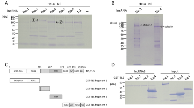
Figure 1. LncRNAs with preferential interaction with specific proteins including TLS.
(A) Detection of proteins bound to the lncRNA1 through lncRNA6, and 1-1 of pncRNA-D using SDS-PAGE gel and CBB staining. The experimental procedures are shown in text.
(*) indicates non-specific binding
Most of RNA-bindings are promiscuous. Therefore, we carefully select lncRNAs which bind less numbers of proteins. Our RNA binding assays of lncRNA 1 through lncRNA 6 showed that lncRNA3 alone has a specific protein band1, but other lncRNAs share binding to protein 2 (Figure 1A). Then, we selected lncRNA3 for further analysis of RNA binding mechanisms. Mass spectrometric analysis of these protein bands recognized by lncRNA 3 and lncRNA 4 indicated nuclear matrix protein matrin3 and nucleolus protein nucleolin, respectively (Figure 1B). We performed mapping experiments using four fragments across full-length TLS (Figure 1C). The RNA binding assay of lncRNA3 to the fragments of TLS showed remarkable binding at the fragment 4 and slight binding on fragment 1 (Figure 1D). Therefore, we performed extensive mapping experiments using the fragment 4 of TLS.

3.2. Extensive Mapping of RNA-Binding Ability on the Fragment 4 of TLS

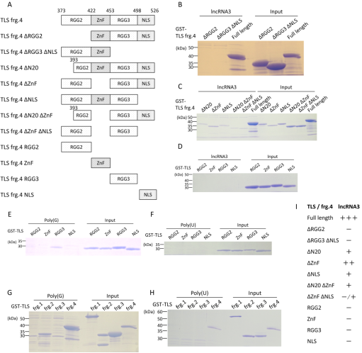
Figure 2. Extensive mapping of RNA-binding ability on the fragment 4 of TLS.
(A) The domain structures of the fragment 4 of TLS and the constructs of fragment 4 which are employed for mapping studies.
(I) Summary of RNA binding assays on every component of fragment 4
Initially, we employed the sets of constructs (Figure 2A) which split the TLS fragment 4 into two sub-fragments, the fragment 4 ΔRGG2 and the fragment 4 ΔRGG3 and ΔNLS. The RNA binding assay indicated no binding on these two deletion mutants upon basis of visibility of CBB-stained protein bands, suggesting that RGG2 and RGG3-NLS are essential for binding of lncRNA3 (Figure 2B). Using next sets of constructs containing deletion at N-terminus 20 amino acids (ΔN20), at the zinc finger (ΔZnF), at NLS and combination of these deletion mutants, we tested which of N20, zinc finger, and NLD mostly contribute to RNA binding. The experiments showed that deletion of zinc finger exhibits relatively weak impact on the RNA binding of TLS, while deletion of N20 and NLS has stronger effect on the RNA binding (Figure 2CI).
Double deletions at N20-zinc finger and zinc finger-NLS almost completely wipe off the binding of lncRNA3 to these mutants (Figure 2C), suggesting N20, zinc finger, and NLS work cooperatively for lncRNA3 binding. Extensive experiments using just constructs from RGG2, zinc finger, RGG3, and NLS showed no remarkable binding to lncRNA3 (Figure 2D). Collectively, these sets of data suggest that RGG2, zinc finger, RGG3, and NLS are essential for TLS binding to lncRNA 3, but these regions alone have no binding (Figure 2I). The reason why no binding was detected is that bindings of each component might not be firm enough for accommodating binding to lncRNA3.
Therefore, we designed the binding assay with a potent binder for TLS, poly (G) RNA which forms G-quadruplex structure. Previous our data showed that poly (G) RNA binds TLS strongly . Using poly (G) RNA, moderate binding on RGG3 was detected (Figure 2E), while poly (U) RNA has no significant binding, working as a negative control (Figure 2F). Four regions of fragment 4 need to work cooperatively to exert effective RNA binding. Additional experiments of poly (G) RNA with four fragments over full-length TLS indicated that additional binding on fragment 2 is detected with the fragment 4 binding (Figure 2G). Contrast to the data with poly (G), poly (U) had binding to the fragment 4 alone as same as that of lncRNA3 (Figure 2H). The TLS binding to poly (G) RNA might be slightly different mode compared to lncRNA3.

3.3. Dissection of lncRNA 3 into Four Fragments and Identification of TLS Binding Site

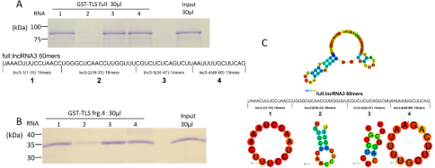
Figure 3. Dissection of lncRNA 3 into four fragments and distinctive bindings to TLS.
(A) Full-length TLS binds to distinctive fragments of lncRNA3
(C) Predicted secondary structure of four fragments of lncRNA3. CentroidFold generates images.
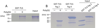
Figure 4. Poly (G) RNA promotes phase-separation induced precipitation of TLS.
(A) The effect of poly (G) RNA on phase separation and resultant precipitation of GST-TLS is examined with previously established condition.
(B) The effect of poly (G) RNA on precipitation of the two fragments of GST-TLS is examined.
The mapping experiments confirm four domains on fragment 4 is major target site of TLS. Next, we explored functional units of lncRNA which interact with TLS. Then, we tentatively divided lncRNA 3 into four fragments (13mer through 18mer), and performed RNA binding assay with full-length TLS (Figure 3A). The experiments showed that specific interacting fragments, lncRNA 3-1, lncRNA3-3 and lncRNA3-4. Previous our data indicated that G-rich RNA prefers binding to TLS. Unexpectedly, one of specific binding was observed at the lncRNA 3-1 which does not contain any G residue and only has UAC residues on its sequence (Figure 3A). In contrast to lncRNA3-1, another binding target of TLS, lncRNA 3-3 has GUC residues which are typical target consensus by TLS (Figure 3A). Further analysis over the lncRNA fragments with the sets of four TLS fragments showed that the TLS fragment 4 exhibits similar binding patter to that of full-length TLS (Figure 3BC), while the fragments 1, 2, 3 have no significant binding to any of lncRNA fragments (data not shown). These data confirm the fragment 4 of TLS plays a central role on the lncRNA 3 binding, and might exert pivotal effect on the inhibition of TLS precipitation. Predicted confirmation of these four fragments of lncRNA3 is depicted, although any functional or physiological clue has not been found (Figure 3C).

3.4. Poly (G) RNA Promotes Precipitation of TLS

Our RNA binding experiments of TLS with Poly (G) indicated solid binding of TLS to poly (G) RNA (figure 2 GH). Recently, it has been reported that G4-RNA promotes phase separation, droplets, and resultant aggregation or precipitation . Upon based on the data, we examined effect of poly (G) RNA on the precipitation of GST-TLS with protocol previously developed. Impressively, poly (G) RNA strongly induced the precipitation of GST-TLS (Figure 4A). This fits previous report , but poly (G) RNA should be a novel agent stimulant for the precipitation of TLS. RNA binding assay of poly (G) RNA was indicated to bind the fragments 2 and 4 of GST-TLS (Figure 2G). On this data, we tested effect of poly (G) RNA on precipitation of the four fragments of GST-TLS. It demonstrated that poly (G) RNA promotes precipitation of GST-TLS fragments 2 and 4 (Figure 4B), but did not induce any precipitation of the fragments 1 and 3 (data not shown), indicating that only the fragments bound to poly (G) RNA were precipitated.
4. Discussion
Initial experiment of the manuscript is to select a lncRNA which binds less numbers of proteins beside of TLS. In lncRNA 1 through 6, lncRNA 3 alone has a unique protein band 1, while all other lncRNAs do not have this band 1 although these lncRNAs reciprocally bind to the band 2 that lacks binding of lncRNA3. Then, further analysis has been performed on lncRNA3. This binding assay of HeLa cell NE and lncRNAs was performed with low sensitivity of detection technique, just CBB-staining. This is just criteria for selection to lncRNAs with strong interaction. The band 1 is found to be matrin-3, which forms a complex with TLS and is related to ALS , while the band 2 is nucleolin related to diseases including ALS and cancers , with which is occasionally associate TLS (unpublished data).
Mapping experiments of lncRNA3 with GST-TLS four fragments showed specific binding to GST-TLS fragment 4 (Figure1CD). The data fits our previous binding profile of four fragments with pncRNA-D . Based upon the binding of lncRNA3 to the TLS fragment 4, we split it to two subfragments, the fragment 4 ΔRGG2 and the fragment 4 ΔRGG3 and ΔNLS (Figure 2AB). Split of the fragment 4 into the two fragments completely abolished binding activity to lncRNA 3, indicating that RGG2 and RGG3-NLS are essential regions for binding to lncRNA3. Therefore, we generated more deletion mutants on the fragment 4. In contrast to the split mutants of the fragments, deletions of zinc finger, N-terminus of RGG2, and NLS did not completely remove the binding activity. Furthermore, combinatorial deletions of zinc finger with other components of the fragment 4 significantly reduced the binding, but did not completely abolish it like the split mutants (Figure 2I). Taken together, RGG2 and possibly RGG3 have crucial function on binding of TLS to lncRNA3. Remarkable data from binding experiment of all four components of fragment 4 demonstrated no significant binding to lncRNA3 (Figure 2D), suggesting that RGG2, zinc finger, RGG3, and NLS all are required for their binding, but each alone does not function at all. Mixing all four fragments did not work either. These four components, RGG2, zinc finger, RGG3 and NLS are supposed to be aligned in this order for binding to lncRNA3. A noteworthy point is that NLS functions as RNA binding region. This is unexpected one, because more than half of NLS (498-526) bears nuclear localization ability, and only three RGG repeats. The amino acid sequence of NLS implies that NLS might have RNA binding activity, which is another noncanonical RNA-binding domain.
Functional regions of lncRNA3 were divided into four parts to identify its functional region. Then, only one fragment (lncRNA3-2) does not have significant binding to TLS and the fragment 4 (Figure 3AB). Unexpectedly, lncRNA3-1 containing AAACUUU has solid binding to TLS. Our previous experiments indicated TLS prefers to bind to RNA sequence with GUC instead of AU. Published data showed the AUUAUU as components of the stem-loop RNA structure instead of sequence specificity . However, this lncRNA 3-1 should form a possible circular configuration of RNA (Figure 3C), but not has stem-loop, suggesting novel RNA sequence preference to TLS.
RNA-binding proteins (RBPs) have diverse functions compared to DNA-binding proteins (DBPs) . DNA-binding transcription factors recognize specific binding sites or responsive elements in a promoter or enhancer, like AGAACA for glucocorticoid receptor (GR) belonging to nuclear receptor family, usually bearing several base pairs in bases of DNA with consensus motifs, and activate transcription of the target genes . Transcription factors precisely recognize specific binding sites, and stringently regulate transcription upon binding to the sites . For example, mutation of AGAACA responsive element into GGACCA resulted in abolish of specific binding of GR to its responsive elements . Therefore, transcription factors is need to have high affinity binding to responsive DNA elements for its appropriate missions to regulate multiple gene networks. Contrarily, RBPs play multiple roles in RNA metabolism, splicing, also construction of membrane less organelles. . Occasionally, RBPs form complexes with RNA to form cellular structures . Relatively low RNA affinity of RBPs fits diverse missions in cellular environments .
TLS has been shown to interact with 5500-6845 RNA species . It is likely that TLS should have multiple interaction domains to each specific surface of RNAs. Divergent interactions of RBPs with RNA are partly forced by flexible conformations of RNA. Such kinds of protein-RNA interactions might be accommodated by IDR because of also its elasticity.
Our mapping experiment indicated that the fragment 1 of TLS exhibited marginal bindings to RNAs with low affinity . This wobbly binding of the fragment 1 should be based upon its IDR. It has been reported that IDRs function as binding interfaces to RNAs with specific properties and exert biological activities through their RNA binding abilities .
Polyguanylic acid (poly (G) RNA) was shown to bind TLS, known to form G-quadruplex (G4) with divergent activities . Additional experiments with poly (G) RNA which has distinctive binding to TLS were performed with four components of the fragment 4 (Figure 2E). Then, RGG3 is just only one binding with poly (G) RNA while poly (U) RNA has no binding as negative control (Figure 2EF), suggesting that RGG3 might be core for synergistic assembly for all four components to the bona fide RNA-binding domain. Remarkable binding data using poly (G) RNA, which forms G-quadruplex (G4) structure with the four fragments of TLS displayed that poly (G) RNA binds the fragment 2 in addition to the fragment 4 (Figure 2GH). The fragment 2 contains C-terminal half of the RGG1 region with IDR. poly (G) RNA could connect RNA-binding domain of the fragment 4 to IDR from RGG1, inspiring glue of RNA-binding domain to IDR.
Frontotemporal degeneration (FTLD) causes frontotemporal dementia (FTD), the most common form of dementia after Alzheimer’s disease, and is also associated with motor disorders . The pathological characters of FTLD are neuronal inclusions of specific, abnormally assembled proteins. Most of cases of the inclusions contain amyloid filaments assemblies of TAR DNA-binding proteins 43 (TDP-43) or tau, with distinct structures characterizing distinct FTLD subtypes . The presence of amyloid filaments and their identities in the remaining 10% of FTLD cases are unknown but are widely believed to be composed of TLS. Therefore, these cases are commonly referred to as FTLD-FUS (or TLS) . Then, cryogenic electron microscope observation was performed to determine the structures of amyloid filaments extracted from the prefrontal and temporal cortices of four distinct individuals bearing FTLD-FUS. Unexpectedly, abundant amyloid filaments of the homologue TATA-binding protein-associated factor 15 (TAF15) was identified instead of TLS itself , suggesting now FTLD-TAF15. The formation of TAF15 amyloid filaments with a characteristic fold in FTLD settles TAF15 retinopathy in neurodegenerative disease. These data regarding TAF15 amyloid filaments stimulate understanding of pathology of neurodegenerative disease, and also plans for diagnostic and therapeutic technologies against TAF15 retinopathy.
Moreover, these data present a crucial event that TAF15 alone forms amyloid filaments in FTLD-TAF15 of the environment with FUS and EWS . It also indicates two essential items regarding onset of neurodegenerative diseases. First, in this context TAF15 has no genetic mutation on its amino acid sequence, meaning that wild type TAF15 precipitates in this lesion. This implicates that something there stimulates precipitation of TAF15. This is a fundamental issue of the onset of the diseases. Second, there is specificity for RBPs. In FTLD-TAF15, TAF15 lives in neuronal cells with TLS and EWS, but only TAF15 forms precipitation there. There is some preference for each RBP regarding induction of forming amyloid filaments. Some kinds of the preference of TLS, EWS, and TAF15 regarding precipitation should be dependent on each character and cellular environment of the lesion of the disease. This is also fundamental factors for onset of ALS and also related neurodegenerative diseases. The data from FTLD-TAF15 present possibility regarding onset of ALS, implying that somehow wild type TLS forms precipitation and also TLS is selected to generate inclusions even with other species of RBPs with uncovered mechanisms. It should be emerging issue for developing therapeutics against TLS-induced ALS elucidation of molecular mechanisms of phase separation, droplet formation and precipitation of TLS in particular cellular environments associated with ALS.
Onset of familial ALS is induced by mutation in TLS. Mutation in TLS results in its translocation from nuclei to cytosolic compartment, inducing aberrant precipitation of TLS and damaging motor neuron, causing ALS . However, 90% of patients are sporadic ALS (sALS) without description of familial history, or genetic alteration on TLS . It has been shown that sALS patients has cytoplasmic accumulation of inclusions containing aggregates of wild type TLS . Wild type TLS precipitates in physiological environment of living cells and mechanism of the precipitation remains elusive.
It has been shown that some events or stimulants induce precipitation of wild type TLS, which are potential pathogen for causing sALS. First, biotinylated isoxazole (BISOX) has been shown to induce phase separation and precipitation of TLS . Kato et al. found that BISOX is a stimulant for phase separation and precipitation of TLS, establishing the phase separation research field. Second, Removal of RNA bound to TLS causes precipitate of TLS. Actually, RNA interacts with TLS and prevents it from its phase separation and precipitation. In nucleus, TLS is in solution, because nucleus is at high concentration of RNA (>200 ng/µl as total RNA) that binds and prevents TLS from phase separation and resultant precipitation . Accidentally, wild type TLS is transferred to cytosolic compartment in which are at lower concentration of RNA, and precipitated with removal of the TLS-bound RNA . These series of events should induce formation of inclusion bodies in cytosol and initial event of ALS onset. Third, recent reports indicated that G4-cotaining mRNAs promote phase separation and precipitation of TLS . Then, we tested effect of poly (G) RNA on precipitation of TLS, and demonstrated that poly (G) RNA effectively induces precipitation of TLS (Figure 4AB). It has been reported that dendric mRNAs like postsynaptic density protein 95 (PSD-95) and Ca/calmodulin-dependent protein kinase type II subunit alpha (CamKIIa) have G4s at 3-untranslated region involved in neuronal functions, implying possible roles of G4 RNA on onset of ALS . These data suggests that G4-RNA is a possible candidate for pathogen for sporadic ALS.
Elucidation of wild type TLS precipitation regular cellular environment is central question that will present a clue to make an efficient therapeutic against sporadic ALS. Actually, we have developed a compound against BISOX, Biotin-Lys-His, BLH, which blocks the BISOX-induced precipitation of TLS . BLH is a potent candidate for the ALS drug. We have identified that pncRNA-D has inhibitory effect on the precipitation of TLS in physiological conditions and also more species of lncRNAs has similar inhibitory effect on the TLS precipitation . These RNAs might be a seed for RNA medicine against ALS. Related to action of G-4 RNA on the TLS precipitation, blocking of G4-RNA might be also a seed for ALS therapeutics. Some sequences with poly (C) might be annealed to poly (G) sequence and suppress pathogenic activity of poly (G) sequence.
5. Conclusions
The pool of lncRNAs has been generated with TLS binding and inhibition of phase separation, and precipitation of TLS, a major cause for ALS. After identification of TLS-specific lncRNA, further analysis has been executed on lncRNA3 regarding mechanism of RNA binding. Mapping experiments showed that the C-terminus of TLS, the fragment4, works as RNA-binding domain for lncRNA 3. The data confirm previous our result with pncRNA-D, although the GGUG consensus RNA oligos bind the fragment 2 comprising of RGG in addition to the fragment 4. Furthermore, TLS has been reported to have capacity of binding to more than 5000 species of RNA, indicating that multiple surfaces of TLS could bind RNA. It is likely that a TLS molecule accommodates several RNA molecules simultaneously. Then an open question is how TLS could match such numerous RNAs. Fluctuation, flexibility and sustainability of RNA-binding domain of TLS should present a clue to a solution. The RNA binding experiments of lncRNA3 with the RGG2, zinc finger, RGG3, and NLS in the fragment 4 presented a working model for action of the TLS RNA-binding domain. These four components need to be assembled as in this order, although each one does not have enough potency on the RNA binding. A key region for RNA-binding should be IDR that generates multiple surfaces for RNAs and endows flexible confirmation of TLS on RNA-binding. We might take advantage of IDR action for making efficient inhibitor for TLS precipitation, a potent drug seed for ALS.
Abbreviations
ALS: Amyotrophic Lateral Sclerosis
BISOX: Biotinylated Isoxazole
CamKIIa: Ca/calmodulin-Dependent Protein Kinase Type II subunit Alpha
CBB: Coomassie Brilliant Blue
CBP: CREB-binding Protein
CNS: Central Nervous System
DBPs: DNA-Binding Proteins
FTD: Frontotemporal Dementia
FTLD: Frontotemporal Lobar Degeneration
G4: G-quadruplex
GR: Glucocorticoid Receptor
HAT: Histone Acetyltransferase
IDRs: Intrinsically Disordered Regions
iPS cells: Induced Pluripotent Stem Cells
lncRNA: Long Noncoding RNA
NLS: Nuclear Localization Signal
pncRNA-D: Promoter-Associated ncRNA-D
poly (G): Polyguanylic Acid
RBPs: RNA-binding Proteins
RRM: RNA Recognition Motif
SDS-PAGE: SDS-polyacrylamide Gel Electrophoresis
Acknowledgments
The authors would thank for fruitful and constructive discussion by all members of the Kurokawa laboratory. The authors also thank Christopher K. Glass and Michael Geoffrey Rosenfeld for profound insight into development of projects. This study was supported by Grant-in-Aid for Scientific Research (C: 18K06939; C 22K06904). This work was supported by the Joint Usage/Research Program on Zero-Emission Energy Research, Institute of Advanced Energy, Kyoto University (2023A-12).
Author Contributions
Naomi Ueda: Data curation, Formal Analysis, Investigation, Methodology, Project administration, Resources, Validation, Visualization, Writing – review & editing
Ryoma Yoneda: Formal Analysis, Investigation, Resources, Validation, Writing – review & editing
Riki Kurokawa: Conceptualization, Data curation, Formal Analysis, Funding acquisition, Investigation, Methodology, Project administration, Resources, Supervision, Validation, Visualization, Writing – original draft, Writing – review & editing
Conflicts of Interest
The authors declare no conflicts of interest.
References
[1] Wang, X., et al., Induced ncRNAs allosterically modify RNA-binding proteins in cis to inhibit transcription. Nature, 2008. 454(7200): p. 126-30
[2] Vance, C., et al., Mutations in FUS, an RNA processing protein, cause familial amyotrophic lateral sclerosis type 6. Science, 2009. 323(5918): p. 1208-11
[3] Kwiatkowski, T. J., Jr., et al., Mutations in the FUS/TLS gene on chromosome 16 cause familial amyotrophic lateral sclerosis. Science, 2009. 323(5918): p. 1205-8
[4] Lagier-Tourenne, C. and D. W. Cleveland, Rethinking ALS: the FUS about TDP-43. Cell, 2009. 136(6): p. 1001-4
[5] Da Cruz, S. and D. W. Cleveland, Understanding the role of TDP-43 and FUS/TLS in ALS and beyond. Current Opinion in Neurobiology, 2011. 21(6): p. 904-919
[6] Taylor, J. P., R. H. Brown Jr, and D. W. Cleveland, Decoding ALS: from genes to mechanism. Nature, 2016. 539(7628): p. 197-206
[7] Van Langenhove, T., et al., Genetic contribution of FUS to frontotemporal lobar degeneration. Neurology, 2010. 74(5): p. 366-71
[8] Broustal, O., et al., FUS mutations in frontotemporal lobar degeneration with amyotrophic lateral sclerosis. Journal of Alzheimer's disease: JAD, 2010. 22(3): p. 765-9.
[9] Martinelli, I., et al., G507D mutation in FUS gene causes familial amyotrophic lateral sclerosis with a specific genotype-phenotype correlation. Neurobiology of aging, 2022. 118: p. 124-128
[10] Zou, H., et al., The function of FUS in neurodevelopment revealed by the brain and spinal cord organoids. Molecular and Cellular Neuroscience, 2022. 123: p. 103771
[11] Korobeynikov, V. A., et al., Antisense oligonucleotide silencing of FUS expression as a therapeutic approach in amyotrophic lateral sclerosis. Nature Medicine, 2022. 28(1): p. 104-116
[12] Kurokawa, R., ed. Long Noncoding RNAs. 2015, Springer. 257.
[13] Lipovich, L., et al., Developmental changes in the transcriptome of human cerebral cortex tissue: long noncoding RNA transcripts. Cerebral cortex, 2014. 24(6): p. 1451-9
[14] Derrien, T., et al., The GENCODE v7 catalog of human long noncoding RNAs: analysis of their gene structure, evolution, and expression. Genome research, 2012. 22(9): p. 1775-89
[15] Carninci, P., et al., The transcriptional landscape of the mammalian genome. Science, 2005. 309(5740): p. 1559-63
[16] Khalil, A. M., et al., Many human large intergenic noncoding RNAs associate with chromatin-modifying complexes and affect gene expression. Proc Natl Acad Sci U S A, 2009. 106(28): p. 11667-72
[17] Necsulea, A., et al., The evolution of lncRNA repertoires and expression patterns in tetrapods. Nature, 2014. 505(7485): p. 635-40
[18] Chi, K. R., Finding function in mystery transcripts. Nature, 2016. 529(7586): p. 423-425
[19] Kurokawa, R., Generation of Functional Long Noncoding RNA Through Transcription and Natural Selection, in Regulatory RNAs. 2012, Springer. p. 151-174.
[20] Djebali, S., et al., Landscape of transcription in human cells. Nature, 2012. 489(7414): p. 101-8
[21] Hon, C.-C., et al., An atlas of human long non-coding RNAs with accurate 5′ ends. Nature, 2017. 543(7644): p. 199-204
[22] Kurokawa, R., Long noncoding RNA as a regulator for transcription. Prog Mol Subcell Biol, 2011. 51: p. 29-41
[23] Kurokawa, R., Promoter-associated long noncoding RNAs repress transcription through a RNA binding protein TLS. Advances in experimental medicine and biology, 2011. 722: p. 196-208
[24] Kurokawa, R., Initiation of Transcription Generates Divergence of Long Noncoding RNAs, in Long Noncoding RNAs. 2015, Springer. p. 69-91.
[25] Aguilar, R., et al., Targeting Xist with compounds that disrupt RNA structure and X inactivation. Nature, 2022. 604(7904): p. 160-166
[26] Yang, F., et al., Shape of promoter antisense RNAs regulates ligand-induced transcription activation. Nature, 2021. 595(7867): p. 444-449
[27] Allou, L., et al., Non-coding deletions identify Maenli lncRNA as a limb-specific En1 regulator. Nature, 2021. 592(7852): p. 93-98
[28] Kurokawa, R., M. G. Rosenfeld, and C. K. Glass, Transcriptional regulation through noncoding RNAs and epigenetic modifications. RNA Biol, 2009. 6(3): p. 233-6
[29] Cech, T. R. and J. A. Steitz, The noncoding RNA revolution-trashing old rules to forge new ones. Cell, 2014. 157(1): p. 77-94
[30] Liu, S. J., et al., CRISPRi-based genome-scale identification of functional long noncoding RNA loci in human cells. Science, 2017. 355(6320): p. eaah7111
[31] Cai, Z., et al., RIC-seq for global in situ profiling of RNA–RNA spatial interactions. Nature, 2020. 582(7812): p. 432-437
[32] Elguindy, M. M. and J. T. Mendell, NORAD-induced Pumilio phase separation is required for genome stability. Nature, 2021. 595(7866): p. 303-308
[33] Quinodoz, S. A., et al., RNA promotes the formation of spatial compartments in the nucleus. Cell, 2021. 184(23): p. 5775-5790. e30
[34] Chong, P. A., R. M. Vernon, and J. D. Forman-Kay, RGG/RG Motif Regions in RNA Binding and Phase Separation. Journal of molecular biology, 2018. 430(23): p. 4650-4665
[35] Corley, M., M. C. Burns, and G. W. Yeo, How RNA-Binding Proteins Interact with RNA: Molecules and Mechanisms. Molecular Cell, 2020. 78(1): p. 9-29
[36] Lunde, B. M., C. Moore, and G. Varani, RNA-binding proteins: modular design for efficient function. Nature reviews. Molecular cell biology, 2007. 8(6): p. 479-90
[37] Clery, A., M. Blatter, and F. H. Allain, RNA recognition motifs: boring? Not quite. Current opinion in structural biology, 2008. 18(3): p. 290-8
[38]
[39] Ramakrishnan, V., The ribosome emerges from a black box. Cell, 2014. 159(5): p. 979-984
[40] Steitz, T. A., A structural understanding of the dynamic ribosome machine. Nature reviews. Molecular cell biology, 2008. 9(3): p. 242-53
[41] Behrmann, E., et al., Structural snapshots of actively translating human ribosomes. Cell, 2015. 161(4): p. 845-57
[42] Papasaikas, P. and J. Valcarcel, The Spliceosome: The Ultimate RNA Chaperone and Sculptor. Trends in biochemical sciences, 2016. 41(1): p. 33-45
[43] Plaschka, C., P. C. Lin, and K. Nagai, Structure of a pre-catalytic spliceosome. Nature, 2017. 546(7660): p. 617-621
[44] Beckmann, B. M., et al., The RNA-binding proteomes from yeast to man harbour conserved enigmRBPs. Nature Communications, 2015. 6(1): p. 10127
[45] Xu, Y., et al., ERα is an RNA-binding protein sustaining tumor cell survival and drug resistance. Cell, 2021. 184(20): p. 5215-5229. e17
[46] Wright, P. E. and H. J. Dyson, Intrinsically unstructured proteins: re-assessing the protein structure-function paradigm. Journal of molecular biology, 1999. 293(2): p. 321-31
[47] Han, T. W., et al., Cell-free formation of RNA granules: bound RNAs identify features and components of cellular assemblies. Cell, 2012. 149(4): p. 768-79
[48] Ozdilek, B. A., et al., Intrinsically disordered RGG/RG domains mediate degenerate specificity in RNA binding. Nucleic acids research, 2017. 45(13): p. 7984-7996
[49] Lin, Y., S. L. Currie, and M. K. Rosen, Intrinsically disordered sequences enable modulation of protein phase separation through distributed tyrosine motifs. Journal of Biological Chemistry, 2017. 292(46): p. 19110-19120
[50] Balcerak, A., et al., RNA-protein interactions: disorder, moonlighting and junk contribute to eukaryotic complexity. Open biology, 2019. 9(6): p. 190096
[51] Zeke, A., et al., Deep structural insights into RNA-binding disordered protein regions. WIREs RNA, 2022. 13(5): p. e1714
[52] Wright, P. E. and H. J. Dyson, Intrinsically disordered proteins in cellular signalling and regulation. Nat Rev Mol Cell Biol, 2015. 16(1): p. 18-29
[53] Castello, A., et al., Comprehensive Identification of RNA-Binding Domains in Human Cells. Molecular Cell, 2016. 63(4): p. 696-710
[54] Kwon, S. C., et al., The RNA-binding protein repertoire of embryonic stem cells. Nature structural & molecular biology, 2013. 20(9): p. 1122-30
[55] Liepelt, A., et al., Identification of RNA-binding Proteins in Macrophages by Interactome Capture. Molecular & cellular proteomics: MCP, 2016. 15(8): p. 2699-714
[56] Loughlin, F. E., et al., The Solution Structure of FUS Bound to RNA Reveals a Bipartite Mode of RNA Recognition with Both Sequence and Shape Specificity. Molecular cell, 2019. 73(3): p. 490-504 e6
[57] Ueda, N., R. Yoneda, and R. Kurokawa, Identification of Long Noncoding RNA Recognized by RNA-Binding Protein TLS/FUS: Purification of RNAs by Affinity Chromatography of GST-TLS. Biomedical Sciences, 2022. 8(4): p. 144-156
[58] Du, K., et al., TLS and PRMT1 synergistically coactivate transcription at the survivin promoter through TLS arginine methylation. Biochem Biophys Res Commun, 2011. 404(4): p. 991-6
[59] Cui, W., et al., Arginine methylation of translocated in liposarcoma (TLS) inhibits its binding to long noncoding RNA, abrogating TLS-mediated repression of CBP/p300 activity. The Journal of Biological Chemistry, 2018. 293(28): p. 10937-10948
[60] Ueda, N. and R. Kurokawa, Affinity Profiles Categorize RNA-Binding Proteins into Distinctive Groups. Biomedical Sciences, 2018. 4(3): p. 24-31
[61] Yoneda, R., et al., The binding specificity of Translocated in LipoSarcoma/FUsed in Sarcoma with lncRNA transcribed from the promoter region of cyclin D1. Cell & bioscience, 2016. 6: p. 4
[62] Yoneda, R., et al., Long noncoding RNA pncRNA-D reduces cyclin D1 gene expression and arrests cell cycle through RNA m(6)A modification. The Journal of biological chemistry, 2020. 295(17): p. 5626-5639
[63] Yoneda, R., N. Ueda, and R. Kurokawa, m6A Modified Short RNA Fragments Inhibit Cytoplasmic TLS/FUS Aggregation Induced by Hyperosmotic Stress. International Journal of Molecular Sciences, 2021. 22(20): p. 11014
[64] Ishiguro, A., et al., ALS-linked FUS mutations dysregulate G-quadruplex-dependent liquid-liquid phase separation and liquid-to-solid transition. J Biol Chem, 2021. 297(5): p. 101284
[65] Gallego-Iradi, M. C., et al., Subcellular Localization of Matrin 3 Containing Mutations Associated with ALS and Distal Myopathy. PLoS One, 2015. 10(11): p. e0142144
[66] Boehringer, A., et al., ALS Associated Mutations in Matrin 3 Alter Protein-Protein Interactions and Impede mRNA Nuclear Export. Sci Rep, 2017. 7(1): p. 14529
[67] Abdelmohsen, K. and M. Gorospe, RNA-binding protein nucleolin in disease. RNA Biol, 2012. 9(6): p. 799-808
[68] Peggion, C., et al., Nucleolin Rescues TDP-43 Toxicity in Yeast and Human Cell Models. Front Cell Neurosci, 2021. 15: p. 625665
[69] Hoell, J. I., et al., RNA targets of wild-type and mutant FET family proteins. Nature structural & molecular biology, 2011. 18(12): p. 1428-31
[70] Wang, X., J. C. Schwartz, and T. R. Cech, Nucleic acid-binding specificity of human FUS protein. Nucleic Acids Research, 2015. 43(15): p. 7535-43
[71] Tejedor, A. R., et al., Protein structural transitions critically transform the network connectivity and viscoelasticity of RNA-binding protein condensates but RNA can prevent it. Nature Communications, 2022. 13(1): p. 5717
[72] Du, S., et al., A nerve injury–specific long noncoding RNA promotes neuropathic pain by increasing Ccl2 expression. The Journal of Clinical Investigation, 2022. 132(13)
[73] Luo, X.-J., et al., LncRNA TMPO-AS1 promotes esophageal squamous cell carcinoma progression by forming biomolecular condensates with FUS and p300 to regulate TMPO transcription. Experimental & Molecular Medicine, 2022. 54(6): p. 834-847
[74] Xing, C.-Y., et al., LINC00313 facilitates osteosarcoma carcinogenesis and metastasis through enhancing EZH2 mRNA stability and EZH2-mediated silence of PTEN expression. Cellular and Molecular Life Sciences, 2022. 79(7): p. 382
[75] Roeder, R. G., The complexities of eukaryotic transcription initiation: regulation of preinitiation complex assembly. Trends Biochem Sci, 1991. 16(11): p. 402-8.
[76] Roeder, R. G., Lasker Basic Medical Research Award. The eukaryotic transcriptional machinery: complexities and mechanisms unforeseen. Nature medicine, 2003. 9(10): p. 1239-44
[77] Glass, C. K. and M. G. Rosenfeld, The coregulator exchange in transcriptional functions of nuclear receptors. Genes Dev, 2000. 14(2): p. 121-41.
[78] Rosenfeld, M. G., V. V. Lunyak, and C. K. Glass, Sensors and signals: a coactivator/corepressor/epigenetic code for integrating signal-dependent programs of transcriptional response. Genes & development, 2006. 20(11): p. 1405-1428.
[79] Luisi, B. F., et al., Crystallographic analysis of the interaction of the glucocorticoid receptor with DNA. Nature, 1991. 352(6335): p. 497-505
[80] Glass, C. K., et al., Mechanisms of transcriptional activation by retinoic acid receptors. Biochem Soc Trans, 1997. 25(2): p. 602-5.
[81] Kurokawa, R., et al., Polarity-specific activities of retinoic acid receptors determined by a co-repressor. Nature, 1995. 377(6548): p. 451-4
[82] Kurokawa, R., et al., Differential orientations of the DNA-binding domain and carboxy-terminal dimerization interface regulate binding site selection by nuclear receptor heterodimers. Genes Dev, 1993. 7(7B): p. 1423-35.
[83] Dreyfuss, G., V. N. Kim, and N. Kataoka, Messenger-RNA-binding proteins and the messages they carry. Nature reviews. Molecular cell biology, 2002. 3(3): p. 195-205
[84] Hudson, W. H. and E. A. Ortlund, The structure, function and evolution of proteins that bind DNA and RNA. Nature reviews. Molecular cell biology, 2014. 15(11): p. 749-60
[85] Hentze, M. W., et al., A brave new world of RNA-binding proteins. Nature reviews. Molecular cell biology, 2018. 19(5): p. 327-341
[86] Hasegawa, Y., et al., The matrix protein hnRNP U is required for chromosomal localization of Xist RNA. Developmental cell, 2010. 19(3): p. 469-76
[87] Lambert, N., et al., RNA Bind-n-Seq: quantitative assessment of the sequence and structural binding specificity of RNA binding proteins. Molecular Cell, 2014. 54(5): p. 887-900
[88] Zhou, Q., et al., Global pairwise RNA interaction landscapes reveal core features of protein recognition. Nature communications, 2018. 9(1): p. 2511
[89] Jankowsky, E. and M. E. Harris, Specificity and nonspecificity in RNA-protein interactions. Nature reviews. Molecular cell biology, 2015. 16(9): p. 533-44
[90] Lagier-Tourenne, C., et al., Divergent roles of ALS-linked proteins FUS/TLS and TDP-43 intersect in processing long pre-mRNAs. Nat Neurosci, 2012. 15(11): p. 1488-97
[91] Umar, M. I., C. Y. Chan, and C. K. Kwok, Development of RNA G-quadruplex (rG4)-targeting L-RNA aptamers by rG4-SELEX. Nat Protoc, 2022. 17(6): p. 1385-1414
[92] Yang, D., G-Quadruplex DNA and RNA. Methods Mol Biol, 2019. 2035: p. 1-24
[93] Lyu, J., et al., Genome-wide mapping of G-quadruplex structures with CUT&Tag. Nucleic Acids Res, 2022. 50(3): p. e13
[94] Wang, S. K., Y. Wu, and T. M. Ou, RNA G-Quadruplex: The New Potential Targets for Therapy. Curr Top Med Chem, 2015. 15(19): p. 1947-56
[95] Grossman, M., et al., Frontotemporal lobar degeneration. Nat Rev Dis Primers, 2023. 9(1): p. 40
[96] Shi, Y., et al., Structure-based classification of tauopathies. Nature, 2021. 598(7880): p. 359-363
[97] Arseni, D., et al., TDP-43 forms amyloid filaments with a distinct fold in type A FTLD-TDP. Nature, 2023. 620(7975): p. 898-903
[98] Neumann, M., et al., A new subtype of frontotemporal lobar degeneration with FUS pathology. Brain, 2009. 132(Pt 11): p. 2922-31
[99] Neumann, M., et al., Abundant FUS-immunoreactive pathology in neuronal intermediate filament inclusion disease. Acta Neuropathol, 2009. 118(5): p. 605-16
[100] Munoz, D. G., et al., FUS pathology in basophilic inclusion body disease. Acta Neuropathol, 2009. 118(5): p. 617-27
[101] Tetter, S., et al., TAF15 amyloid filaments in frontotemporal lobar degeneration. Nature, 2024. 625(7994): p. 345-351
[102] Patel, A., et al., A Liquid-to-Solid Phase Transition of the ALS Protein FUS Accelerated by Disease Mutation. Cell, 2015. 162(5): p. 1066-77
[103] Szewczyk, B., et al., FUS ALS neurons activate major stress pathways and reduce translation as an early protective mechanism against neurodegeneration. Cell Rep, 2023. 42(2): p. 112025
[104] Corcia, P., et al., Pre-symptomatic diagnosis in ALS. Rev Neurol (Paris), 2020. 176(3): p. 166-169
[105] Van Daele, S. H., et al., Genetic variability in sporadic amyotrophic lateral sclerosis. Brain, 2023. 146(9): p. 3760-3769
[106] Tyzack, G. E., et al., Widespread FUS mislocalization is a molecular hallmark of amyotrophic lateral sclerosis. Brain: a journal of neurology, 2019
[107] Tsai, Y. L., Y. C. Mu, and J. L. Manley, Nuclear RNA transcript levels modulate nucleocytoplasmic distribution of ALS/FTD-associated protein FUS. Sci Rep, 2022. 12(1): p. 8180
[108] Kato, M., et al., Cell-free formation of RNA granules: low complexity sequence domains form dynamic fibers within hydrogels. Cell, 2012. 149(4): p. 753-67
[109] Kurokawa, R. and T. Bando, Three-Dimensional Structure of RNA-Binding Protein TLS Co-Crystallized with Biotinylated Isoxazole. Biomedical Sciences, 2016. 2: p. 1-10
[110] Maharana, S., et al., RNA buffers the phase separation behavior of prion-like RNA binding proteins. Science, 2018. 360(6391): p. 918-921
[111] Zhang, Y., et al., G-quadruplex structures trigger RNA phase separation. Nucleic Acids Res, 2019. 47(22): p. 11746-11754
[112] Papageorgiou, A. C., et al., Recognition and coacervation of G-quadruplexes by a multifunctional disordered region in RECQ4 helicase. Nat Commun, 2023. 14(1): p. 6751
[113] Wang, W., et al., G-quadruplexes promote the motility in MAZ phase-separated condensates to activate CCND1 expression and contribute to hepatocarcinogenesis. Nat Commun, 2024. 15(1): p. 1045
[114] Subramanian, M., et al., G-quadruplex RNA structure as a signal for neurite mRNA targeting. EMBO Rep, 2011. 12(7): p. 697-704
[115] Ueda, N., et al., Biotin-Lys-His Blocks Aggregation of RNA-binding Protein TLS, a Cause of Amyotrophic Lateral Sclerosis. Biomedical Sciences, 2017. 3(4): p. 67-77
[116] Hamad, N., et al., Non-coding RNA suppresses FUS aggregation caused by mechanistic shear stress on pipetting in a sequence-dependent manner. Scientific reports, 2021. 11(1): p. 9523
Cite This Article
  • APA Style

    Ueda, N., Yoneda, R., Kurokawa, R. (2024). Identification of Essential Components of RNA Binding Domain of TLS/FUS. Biomedical Sciences, 10(2), 30-43. https://doi.org/10.11648/j.bs.20241002.13

    Copy | Download

    ACS Style

    Ueda, N.; Yoneda, R.; Kurokawa, R. Identification of Essential Components of RNA Binding Domain of TLS/FUS. Biomed. Sci. 2024, 10(2), 30-43. doi: 10.11648/j.bs.20241002.13

    Copy | Download

    AMA Style

    Ueda N, Yoneda R, Kurokawa R. Identification of Essential Components of RNA Binding Domain of TLS/FUS. Biomed Sci. 2024;10(2):30-43. doi: 10.11648/j.bs.20241002.13

    Copy | Download

  • @article{10.11648/j.bs.20241002.13,
      author = {Naomi Ueda and Ryoma Yoneda and Riki Kurokawa},
      title = {Identification of Essential Components of RNA Binding Domain of TLS/FUS
    },
      journal = {Biomedical Sciences},
      volume = {10},
      number = {2},
      pages = {30-43},
      doi = {10.11648/j.bs.20241002.13},
      url = {https://doi.org/10.11648/j.bs.20241002.13},
      eprint = {https://article.sciencepublishinggroup.com/pdf/10.11648.j.bs.20241002.13},
      abstract = {TLS/FUS is RNA-binding protein having multiple functions of regulations of genes, homeostasis, and cellular growth. Recent studies show that TLS is involved in phase separation and occasionally forms precipitation related to neurodegenerative diseases like amyotrophic lateral sclerosis (ALS). RNA has been reported to suppress phase separation, droplet formation, and concomitant precipitation of TLS, suggesting that RNA is a possible candidate for ALS drug discovery. Our experiments demonstrated that a long noncoding RNA, promoter-associated noncoding RNA (pncRNA-D), specifically binds TLS and represses its phase separation and precipitation. To obtain competent drug seeds, it is essential to reveal mechanism of action of lncRNAs with specificity to TLS and inhibitory activity on phase separation and related precipitation. For this purpose, several lncRNAs (lncRNAs 1 to 6) were selected upon assays with GST-TLS binding and inhibition on the precipitation. With criteria of binding specificity for TLS, lncRNA3 has been selected for further analysis for RNA-binding ability. Initially, RNA-binding region at TLS amino acid sequence was identified from four fragments of TLS. RNA binding assay with biotinylated lncRNA3 precipitated with avidin magnetic beads indicated clearly that TLS binds the fragment 4 (373-526 aa), C-terminus end of TLS. Then, dissecting fragment 4 presents four regions, RGG2, zinc finger, RGG3, and the nuclear localization signal (NLS) region in this order. Experiments with extensive deletion mutants indicated that just one deletion out of the four regions irs not enough to delete the TLS binding, although combinatorial deletion of zinc finger with other three regions almost wiped off the lncRNA3 binding. Remarkably, each of four regions alone has no binding to TLS, either. Collectively, RGG2, zinc finger, RGG3, and NLS all are essential for binding to lncRNA3, but are required to work synergistically for full binding. These data indicate that dynamic assembly of RNA-binding domain works for action of lncRNAs and possibly has allosteric effect on intrinsically disordered region (IDR) of N-terminus of TLS, implying relation of RNA-binding with phase separation and the resultant precipitation.
    },
     year = {2024}
    }
    

    Copy | Download

  • TY  - JOUR
    T1  - Identification of Essential Components of RNA Binding Domain of TLS/FUS
    
    AU  - Naomi Ueda
    AU  - Ryoma Yoneda
    AU  - Riki Kurokawa
    Y1  - 2024/05/10
    PY  - 2024
    N1  - https://doi.org/10.11648/j.bs.20241002.13
    DO  - 10.11648/j.bs.20241002.13
    T2  - Biomedical Sciences
    JF  - Biomedical Sciences
    JO  - Biomedical Sciences
    SP  - 30
    EP  - 43
    PB  - Science Publishing Group
    SN  - 2575-3932
    UR  - https://doi.org/10.11648/j.bs.20241002.13
    AB  - TLS/FUS is RNA-binding protein having multiple functions of regulations of genes, homeostasis, and cellular growth. Recent studies show that TLS is involved in phase separation and occasionally forms precipitation related to neurodegenerative diseases like amyotrophic lateral sclerosis (ALS). RNA has been reported to suppress phase separation, droplet formation, and concomitant precipitation of TLS, suggesting that RNA is a possible candidate for ALS drug discovery. Our experiments demonstrated that a long noncoding RNA, promoter-associated noncoding RNA (pncRNA-D), specifically binds TLS and represses its phase separation and precipitation. To obtain competent drug seeds, it is essential to reveal mechanism of action of lncRNAs with specificity to TLS and inhibitory activity on phase separation and related precipitation. For this purpose, several lncRNAs (lncRNAs 1 to 6) were selected upon assays with GST-TLS binding and inhibition on the precipitation. With criteria of binding specificity for TLS, lncRNA3 has been selected for further analysis for RNA-binding ability. Initially, RNA-binding region at TLS amino acid sequence was identified from four fragments of TLS. RNA binding assay with biotinylated lncRNA3 precipitated with avidin magnetic beads indicated clearly that TLS binds the fragment 4 (373-526 aa), C-terminus end of TLS. Then, dissecting fragment 4 presents four regions, RGG2, zinc finger, RGG3, and the nuclear localization signal (NLS) region in this order. Experiments with extensive deletion mutants indicated that just one deletion out of the four regions irs not enough to delete the TLS binding, although combinatorial deletion of zinc finger with other three regions almost wiped off the lncRNA3 binding. Remarkably, each of four regions alone has no binding to TLS, either. Collectively, RGG2, zinc finger, RGG3, and NLS all are essential for binding to lncRNA3, but are required to work synergistically for full binding. These data indicate that dynamic assembly of RNA-binding domain works for action of lncRNAs and possibly has allosteric effect on intrinsically disordered region (IDR) of N-terminus of TLS, implying relation of RNA-binding with phase separation and the resultant precipitation.
    
    VL  - 10
    IS  - 2
    ER  - 

    Copy | Download

Author Information
  • Abstract
  • Keywords
  • Document Sections

    1. 1. Introduction
    2. 2. Materials and Methods
    3. 3. Results
    4. 4. Discussion
    5. 5. Conclusions
    Show Full Outline
  • Abbreviations
  • Acknowledgments
  • Author Contributions
  • Conflicts of Interest
  • References
  • Cite This Article
  • Author Information