Research Article | | Peer-Reviewed

Canine Vaccinal Antibody Remains Stable for 4 Weeks at Simulated Shipping Temperatures

Received: 28 July 2025     Accepted: 25 August 2025     Published: 11 September 2025
Views:       Downloads:
Abstract

To establish evidence-based shipping requirements for sera submitted to reference laboratories for vaccinal antibody testing, the stability of canine antibody was determined over four weeks at temperatures simulating ground transport conditions. Known positive canine serum samples (n = 22) were tested to determine quantitative antibody titers using two gold-standard serologic assays. Antibody titer against canine parvovirus (CPV-2) via hemagglutination inhibition (HI) assay and against canine adenovirus (CAV-1) via serum virus neutralization (SVN) assay. Samples were aliquoted and held at static temperatures: in a refrigerator (6°C), at room temperature (25°C), and in an incubator (36°C) Samples were randomized and repeat tested at weeks 2, 3, and 4. Statistical equivalence was determined using paired two one-sided t-test (TOST), with zone of indifference of ± 1 dilution. For both antibody assays, experimental groups demonstrated statistical equivalence to refrigerated controls through week 4 (p < 0.05 for all comparisons.) These results demonstrate that canine vaccinal antibodies remain stable for four weeks at continuous elevated temperatures that might be encountered during ground shipment. This finding supports the implementation of less restrictive shipping requirements for canine vaccinal antibody testing, potentially reducing costs for veterinary practitioners and pet owners, and ultimately allowing greater access to important diagnostic testing.

Published in Animal and Veterinary Sciences (Volume 13, Issue 5)
DOI 10.11648/j.avs.20251305.12
Page(s) 125-134
Creative Commons

This is an Open Access article, distributed under the terms of the Creative Commons Attribution 4.0 International License (http://creativecommons.org/licenses/by/4.0/), which permits unrestricted use, distribution and reproduction in any medium or format, provided the original work is properly cited.

Copyright

Copyright © The Author(s), 2025. Published by Science Publishing Group

Keywords

Canine, Antibody Stability, Titer Testing, Temperature, Transport

1. Introduction
Canine vaccinal antibody testing (aka “titer testing”) is a useful tool to guide clinical decisions regarding use of core modified live viral (MLV) vaccines including canine parvovirus (CPV-2), canine distemper virus (CDV) and canine adenovirus (CAV-1, 2). Antibody testing is appropriate for these viruses because they are highly susceptible to antibody neutralization. Protective antibody titer thresholds for these pathogens have been previously determined by challenge of immunity studies . Indications for canine vaccinal antibody testing include previous adverse vaccine reaction, outbreak management, puppies completing initial vaccination series, and to generate nomograph analyses for breeding bitches .
While point-of-care tests are available for this application, canine sera are frequently shipped to reference laboratories for vaccinal antibody testing. The potential for antibody degradation during transport conditions is a common concern. Current industry practices often require expensive shipping protocols including ice packs, overnight delivery, and immediate sample processing upon collection . These requirements create substantial financial barriers for veterinary clients and may limit access to important diagnostic testing. The cost of rigorous shipping requirements can exceed the cost of the testing itself, particularly for rural practitioners or those serving economically disadvantaged communities.
Previous studies in human and veterinary medicine have demonstrated remarkable antibody stability under various temperature conditions . However, there is a lack of comparable data for canine vaccinal antibodies, and no studies have looked specifically at the effect of elevated temperatures during shipment. This lack has resulted in potentially unnecessary restrictions that may limit access to important veterinary diagnostic services.
The current study aims to determine the impact of elevated temperatures over time on canine vaccinal antibody using two different “gold standard” test methods. We hypothesized that canine vaccinal antibodies would demonstrate sufficient stability at shipping temperatures to support less restrictive transport requirements. To test this hypothesis, sample aliquots held under test conditions were compared against refrigerated controls over a period of 4 weeks. Equivalence was determined using appropriate statistical methods. Results could provide evidence-based recommendations for sample acceptance criteria and shipping requirements across reference laboratories.
2. Materials and Methods
2.1. Samples
Canine sera were submitted to the Companion Animal Vaccines and Immunodiagnostic Service (CAVIDS) Laboratory, which is a fee-for-service laboratory at the University of Wisconsin-Madison School of Veterinary Medicine. Sample submission indicated owner consent, request for titer testing service, and release of sample ownership. Institutional review board determined that approval was not required for this study. Sera submissions came from dogs living in multiple geographic locations around the United States. The sample set included canine sera from a range of dog breeds and ages. Dogs sampled had received a variety of commercial vaccine products. Taken together, the demographics of this sample set were highly representational of submissions typically received by North American reference laboratories. Samples containing insufficient volume, undetectable levels of specific antibody, or severe hemolysis were excluded. Twenty-two frozen, archived samples were selected based on known positive titers (>80 for CPV-2 and >8 for CAV-1) and adequate sample volume (>3 mls). Titers were normally distributed. Each sample was divided into three aliquots and moved into refrigerator (6°C), bench top (25°C), or incubator (36°C).
2.2. Temperature Variables
Test temperatures were chosen to simulate expected North American shipping conditions. Room temperature (25°C) was chosen to represent moderate ambient temperatures. The higher test temperature (36°C) was based on expected average daily high temperatures for the southern United States. National Weather Service historical data (1991-2020) for average July high temperatures in Houston, Texas (34.7°C) and Tucson, Arizona (37.9°C) were averaged to reach 36.3°C .
2.3. Serology
Serology was completed on all samples at time point 0. Samples were randomized and held for further testing at weeks 2, 3, and 4. At each session, tests were read by an individual who was masked to treatment group assignment.
Serum virus neutralization (SVN) assay was used to determine CAV-1 antibody titers using the method developed by Appel for detection of neutralizing antibody against CDV which was modified for CAV-1 . Briefly, sera samples are doubly diluted across a 96 well flat bottom tissue culture plate. Dilution buffer is minimum essential medium (MEM) containing growth factors and antibiotics. The plates are then incubated at room temperature for one hour with 200TCID50 CAV-1 per well. Madin-Darby canine kidney (MDCK) cells are then added at an approximate density of 2x10⁴ cells per well. The plates are incubated for four days at 36°C in a humidified 5% CO2 atmosphere. Plates are read microscopically to determine the highest serum dilution able to completely prevent viral infection and resultant cytopathic effects. A control plate containing known positive and negative serum samples, viral back titer and cell control is also prepared and read. All test sera, control positive and negative sera, viral back-titer, and cell control are tested in duplicate.
A hemagglutination inhibition (HI) assay was also performed on each sample to test for antibodies against CPV-2 using the method developed by Carmichael . In brief, the sera samples are doubly diluted in phosphate buffered saline (PBS) with bovine serum albumin (BSA) 0.1% (v/v) across a 96 well, round bottom plate. The initial dilution of all test sera serves as a control to determine sera toxicity or sera-agglutination of indicator cells. CPV-2b (fecal extract origin) diluted to 32 hemagglutinating (HA) units in PBS buffer without BSA is added and the plates are incubated at room temperature for one hour. A solution of 1% (v/v) porcine red blood cells in BSA buffer are then added and the plates are incubated overnight in a refrigerator (6°C). Plates are then read visually and reported as the highest dilution of serum that completely prevents red blood cell agglutination caused by the virus. A control plate is also prepared and read as described for the SVN procedure. All test sera, control sera, viral back-titer, and cell control are tested in duplicate.
Both SVN and HI are “gold standard,” functional measurements which are commonly applied to canine vaccinal antibody testing. They represent two distinctly different methodologies of quantifying canine antibody, and were chosen for this study for these reasons. Both assays detect antibody classes type G and M (IgG and IgM) additively.
2.4. Statistical Analysis
An a priori power analysis was conducted using G*Power version 3.1.9.7 to determine the minimum sample size required to test the study hypothesis. Results indicated the required sample size to achieve 99% power for detecting a small effect, at a significance criterion of α =.05, was N = 21 for paired two one-sided t-tests (TOST). Thus, the sample size of N = 22 was adequate to test the study hypothesis.
Given that the hypothesis of this study is that there will not be a significant difference between the experimental groups, a test of equivalence known as paired two one-sided t-tests (TOST) was used to compare the average geometric mean titers of the experimental groups to the refrigerated control run during the same session, as well as to the original results obtained from time point 0. The zone of indifference was set as -1 to 1, as a difference of only one dilution is considered within the margin of error for both of these assays. Significance was set at p < 0.05 with a confidence interval of 95%. Bonferroni correction for multiple comparisons was conducted to decrease risk of Type I error (false equivalence). The updated alpha = 0.00556. Post hoc power analysis was conducted using Mini-Tab software. Cohen’s d for effect size was determined using an online calculator (Omni Calculator). TOST test was conducted in Excel with QI Macros add-on. Normality of differences (d’Agostino and Pearson), coefficient of variation, and all other tests were conducted using GraphPad Prism v10.
3. Results
3.1. Geometric Mean Titer Results
Geometric mean titers against CPV-2 and CAV-1 (with 95% confidence intervals) and coefficient of variation (CV%) are shown in Table 1 for all timepoints and temperature conditions.
Table 1. Geometric mean titers for CPV-2 and CAV-1 at temperatures and timepoints with 95% confidence intervals and coefficient of variation.

Condition

Time Point

CPV-2 GMT (95%CI)

CAV-1 GMT (95% CI)

CPV-2 CV (%)

CAV-1 CV (%)

6°C Control

Baseline

6.14 (5.4, 6.99)

7.56 (6.68-8.55)

24.51%

24.22%

6°C Control

Week 2

6.31 (5.49, 7.26)

7.12 (6.21, 8.15)

28.35%

26.56%

25°C Room Temp

6.53 (5.75, 7.42)

7.02 (6.06, 8.14)

25.75%

28.59%

36°C Incubator

6.47 (5.59, 7.49)

6.97 (5.9, 8.23)

28.06%

29.20%

6°C Control

Week 3

5.86 (5.13, 6.70)

7.95 (7.01, 9.01)

25.29%

24.58%

25°C Room Temp

5.61 (4.93, 6.37)

7.93 (7.03, 8.94)

26.33%

23.45%

36°C Incubator

5.58 (4.87, 6.39)

7.73 (6.82, 8.76)

28.88%

24.70%

6°C Control

Week 4

4.56 (4.1, 5.07)

8.12 (7.23, 9.12)

23.20%

22.91%

25°C Room Temp

4.74 (4.27, 5.26)

7.78 (6.88, 8.80)

23.14%

24.10%

36°C Incubator

4.4 (3.99, 4.85)

7.51 (6.54, 8.62)

21.41%

25.77%

3.2. Equivalence Testing Results - CAV-1 Serum Virus Neutralization
Results of equivalence testing for CAV-1 are shown (Table 2) including normality (d’Agostino and Pearson), mean of differences (95% CI), correlation coefficient, P value for r, Cohen’s d for effect size, post hoc power, and P value for equivalence with Bonferroni correction (alpha = 0.00556). Significant equivalence was determined for samples held at experimental temperatures for weeks 2 and 3. At week 4 only the room temperature sample was significantly equivalent to refrigerated control.
Table 2. CAV-1 equivalence testing with normality, mean of differences (with 95% CI), correlation coefficient, P value for r, Cohen’s d, post hoc power, and P value for equivalence (Bonferroni corrected alpha = 0.00556). F = frige/6°C; 0 = Baseline/Week 0; RT = room temperature/25°C; Inc = incubator/36°C.

Comparison CAV-1

Normality

Mean of differences (95% CI)

Correlation coefficient - r

P value for r

Cohen’s d

Power

P value for equivalence

Significance (Bonferroni corrected alpha = 0.00556)

Week 2

F v 0

Yes

0.409

0.165, 0.6535

0.941

<.0001

0.212

0.991

0.00022

**

F v RT

Yes

0.0455

-0.284, 0.375

0.905

<.0001

0.0221

0.999

0.000031

***

F v Inc

Yes

0.0455

-0.303, 0.394

0.897

<.0001

0.0218

0.998

0.000059

***

Week 3

F v 0

No

0.409

0.117, 0.701

0.919

<.0001

0.209

0.957

0.0011

**

F v RT

No

0.0455

-0.0922, 0.183

0.983

<.0001

0.0228

1.0

4E-11

****

F v Inc

Yes

0.227

0.0334, 0.421

0.965

<.0001

0.114

1.0

0.00000044

****

Week 4

F v 0

Yes

0.546

0.299, 0.792

0.938

<.0001

0.287

0.923

0.0023

*

F v RT

Yes

0.318

0.0811, 0.555

0.944

<.0001

0.165

0.999

0.000034

***

F v Inc

Yes

0.546

0.299, 0.792

0.943

<.0001

0.278

0.923

0.0023

*

TOST testing (with Bonferroni correction) found the CAV-1 antibody titers for experimental groups were statistically equivalent to the refrigerated control through week 4 (Figure 1).
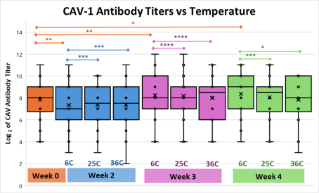
Figure 1. Box and whisker plot of CAV-1 SVN log2 antibody titers, with arithmetic mean indicated by X and range indicated by bars. Timepoints shown are week 0 baseline (orange), and week 2 (blue), week 3 (purple) and week 4 (green). Geometric means for samples held at experimental temperatures at weeks 2-4 were compared with those of refrigerated controls and baseline values. The Bonferroni corrected significance of p values indicating equivalence between each paired TOST are represented by * for p < 0.0056, ** for p < 0.0011, *** for p<0.00011, and **** for p< 0.000011.
3.3. CAV-1 SVN Assay Coefficient of Variance
This SVN assay had an intra assay CV of 20% and inter assay CV of 24.6%.
3.4. Equivalence Testing Results - CPV-2 Hemagglutination Inhibition
Results of equivalence testing for CPV-2 antibody titers are shown (Table 3) with normality (d’Agostino and Pearson), mean of differences (95% CI), correlation coefficient, P value for r, Cohen’s d for effect size, post hoc power, and P value for equivalence with Bonferroni correction (alpha = 0.00556). TOST test showed significant equivalence for weeks 3 and 4 experimental groups. Week 4 comparison for refrigerated control versus timepoint 0 was not significant.
Table 3. CPV-2 equivalence testing with normality, mean of differences (95% CI), correlation coefficient, P value for r, Cohen’s d, post hoc power, and P value for equivalence (Bonferroni corrected alpha = 0.00556). F = frige/6°C; 0 = Baseline/Week 0; RT = room temperature/25°C; Inc = incubator/36°C.

Comparison CPV-2

Normality

Mean of differences (95% CI)

Correlation coefficient

P value for r

Cohen’s d

Power

P value for equivalence

Significance (Bonferroni corrected alpha = 0.00556)

Week 2

F v 0

No

0.227

-0.0916, 0.546

0.887

<.0001

0.132

0.996

0.00022

**

F v RT

Yes

0.182

-0.151, 0.515

0.876

<.0001

0.101

0.993

0.00019

**

F v Inc

Yes

0.182

-0.131, 0.4947

0.898

<.0001

0.0965

0.997

0.000098

***

Week 3

F v 0

Yes

0.273

0.0708, 0.475

0.937

<.0001

0.176

1.0

0.0000019

****

F v RT

Yes

0.273

0.0412, 0.504

0.916

<.0001

0.178

1.0

0.000012

***

F v Inc

Yes

0.273

-0.0088, 0.554

0.890

<.0001

0.169

0.996

0.00011

***

Week 4

F v 0

Yes

1.682

1.396, 1.968

0.887

<.0001

1.251

NA

1.0 ns

ns

F v RT

Yes

0.182

-0.00201, 0.366

0.898

<.0001

0.165

1.0

0.000000082

****

F v Inc

No

0.182

0.037, 0.327

0.933

<.0001

0.177

1.0

1.6E-09

****

Note: power could not be determined for the week 4 pairing of 6°C and baseline because the effect size was greater than 1.
TOST testing (with Bonferroni correction) found the CPV-2 antibody titers for experimental groups were statistically equivalent to the refrigerated control through week 4 with p < 0.001 for each pairing. Week 4 results for the CPV-2 experimental groups and refrigerated control were not statistically equivalent to the results from time point 0. (Figure 2).
Figure 2. Box and whisker plot of CPV-2 HI log2 antibody titers, with arithmetic mean indicated by X and range indicated by vertical bars. Timepoints shown are week 0 baseline (orange), and week 2 (blue), week 3 (purple) and week 4 (green). Geometric means for samples held at experimental temperatures at weeks 2-4 were compared with refrigerated control and baseline values. The Bonferroni corrected significance of p values indicating equivalence between each paired TOST are represented by * for p < 0.0056, ** for p < 0.0011, *** for p<0.00011, and **** for p< 0.000011.
3.5. CPV-2 HI Assay Coefficient of Variance
This HI assay had a CV% of 16.67% (intra-assay) and 24.1% (inter-assay).
4. Discussion
4.1. Summary of Key Findings
This study determined that canine vaccinal antibodies against CPV-2 and CAV-1 maintain stability for 3 weeks when stored at temperatures up to 36°C, as measured by two gold-standard serological assays. Statistical equivalence testing found no significant differences between experimental temperature groups and refrigerated controls for the majority of comparisons, supporting the hypothesis that current shipping restrictions may be unnecessarily conservative. These findings have important implications for veterinary diagnostic practices and could lead to significant cost reductions for practitioners and pet owners.
4.2. Comparison with Existing Literature
Scientific literature contains a few publications regarding expected temperature excursions during transport . As part of a study to determine the effect of high temperature shipment on a human allograft product , investigators added thermal trackers to 72 shipments to 5 warm climate US cities during the months of August-September. That study found overall average package internal temperatures of 26.2°C ± 2.3°C. However, temperature spikes >40°C were seen, most frequently during truck transport in Phoenix, AZ, the warmest of the sites tested. These data support the choice of 36°C for high temperature storage in the current study, but suggest that thermal cycling effects should be examined in the future.
It is important to note that the experimental high temperature used in the current study was below clinical parameters for normal canine body temperature (37.5-39.2°C, Merck Veterinary Manual) . In general, protein denaturation occurs at temperatures greater than 80°C although studies of thermal instability of monoclonal antibody products use lower temperatures (25-40°C) .
While the majority of studies in the human medical literature focus on monoclonal antibody, some studies of temperature effects on native human antibody for diagnostic testing have shown favorable results .
Veterinary medical literature includes multiple studies of impact of heat treatments on colostral bovine antibody, and have included temperatures ranging from <60°C to 76°C . Heat treatment of stored colostrum is a common practice to decrease potential pathogen load before feeding to newborn calves. A meta-analysis of 26 publications showed good antibody resistance to thermal degradation at colostrum heat treatments below 60°C as evidenced by comparative passive antibody levels in calves fed heat treated versus stored raw colostrum. However, these studies generally examined total antibody levels, and were not specific for any particular antigen or diagnostic assay.
4.3. Biological Mechanisms
Canine antibodies are specifically folded proteins held by disulfide bonds which are susceptible to fragmentation from peptide bond cleavage. In addition, Nowak et al suggest mechanisms of thermal instability include degradation pathways, formation of aggregates, deamidation and oxidation . While beyond the scope of the current manuscript, mechanisms of antibody thermal stability found in the current study can be presumed to reflect the presence of stabilizing factors in canine serum, including albumin and other proteins.
Differential stability observed between CPV-2 and CAV-1 antibodies was not significant and presumably reflects differences in assay methodologies. Differences in antibody isotype (IgG versus IgM) may play a role; however, it is important to note that both HI and SVN detect both classes of antibody without distinguishing between IgG and IgM. Future studies may investigate differential thermal stability between antibody classes.
4.4. Application of Test Methods
The assays used in this study reflect the two most commonly used “gold standard” laboratory-based tests for canine vaccinal antibody testing. Hemagglutination inhibition (HI) assay is the standard laboratory assay for determination of CPV-2 antibody levels, and serum virus neutralization (SVN) is the standard method for determination of antibody levels against CAV-1,2 and CDV. These tests were chosen to reflect the most common scenario encountered for canine sera that are “shipped out” to a reference laboratory. The inter-assay CV% which were found in this study are similar to those reported for similar serologic applications, including HI quantification of human antibody against influenza virus and an SVN for rabies antibody .
Because laboratory based canine vaccinal antibody tests determine biologic functionality, these diagnostic tools should not be viewed with the same lens as that applied to chemical analyses. Multiple aspects of serologic tests are difficult to standardize, and thus an inherent level of variability is unavoidable . Inter-assay variability can be especially problematic for longitudinal studies . The current study encountered “session effect” for week 4 HI results when a new lot of reference CPV-2 virus was required. While the 4-week HI timepoint groups varied from time point 0 with lower geometric mean titers, there was statistically no difference in CPV-2 antibody titers between groups tested at this session. However, this finding highlights the importance of assay standardization and quality control in longitudinal studies.
4.5. Clinical Importance Versus Statistical Significance
The current study used a high level of power to detect a small effect size, and applied a conservative significance threshold. However, it is important to note that significant p values simply indicate that equivalence between controls and experimental groups was unlikely to be due to chance alone. After Bonferroni correction, these data have only 0.5% probability of false equivalence. While p values are compelling, it is important to interpret these findings with an understanding of clinical importance.
The concept of “clinical importance” stems from the realization that proof of statistical significance may or may not indicate an actual benefit to stakeholders . For all medical decisions, it is essential to fully consider potential harms and benefits of a particular course of action. Canine antibody testing itself is not invasive and carries minimal direct risk to the patient, however incorrect test results have important clinical consequences.
Samples mistakenly reported as having antibody below protective threshold or undetectable (aka false negative) will lead to unnecessary vaccination of a dog that is actually already immune. On the other hand, determination of antibody titer above protective threshold when it is not (aka false positive) would result in a susceptible dog remaining unprotected. In general, a false negative result is most problematic for dogs which have had adverse vaccine reactions in the past , whereas a false positive test could result in poor clinical outcome for a wider population of dogs. If antibody titers are indeed adversely impacted by high shipping temperatures (the null hypothesis), the most likely outcome would be increased false negative results. Therefore, the statistical significance and clinical importance of this study are aligned. Accessible shipping requirements reduce overall potential harm (decreased cost without increased false positives), while retaining the benefit of discovering unprotected individuals (true negatives).
4.6. Practical Implications
Because expensive shipping requirements negatively impact all stakeholders, the current study findings have significant practical implications for veterinary diagnostic laboratories and practitioners. Cost-benefit analysis suggests that less restrictive requirements could reduce shipping costs by 60-80% (source FedEx, https://www.fedex.com/en-us/online/rating.html), and overall costs by 48-74% (depending on laboratory). Removing requirements for overnight shipping and/or heavy ice packs would make titer testing more accessible to a broad range of pet owners. Implementation of revised shipping protocols should include careful quality control monitoring. Sample acceptance criteria must continue to include lack of visible contamination, severe hemolysis, and other factors that can interfere with testing independently of antibody stability.
While this study found antibody was highly stable over transport conditions, veterinary clinicians must determine the impact of potential false negative result for their patient, and ship samples accordingly. For example, a sample from a dog that has experienced systemic allergic reaction after vaccination may warrant sample shipping on ice packs. Timely result turnaround is another important consideration which may impact clinical decisions about shipping choices. For this example, samples from a shelter experiencing an outbreak of CPV-2 may warrant overnight shipping. On the other hand, sera from a group of healthy dogs for routine vaccinal antibody testing could be shipped through a standard postal service at a much-reduced cost.
4.7. Study Strengths and Limitations
Strengths of the current study include the fact that all samples tested originated from the field, and thus represented a variety of initial collection and handling techniques. The sample set included a variety of breeds, ages, and vaccines administered, and thus was highly representative of typical submissions to canine serology reference laboratories. Post hoc power was >92% for all test groupings.
This study was limited in that the only variable examined was the effect of static elevated temperature over time. The effects of agitation, thermal cycling, and repeated freeze/thaw were not included in this study. Limited resources also restricted the number of timepoints tested. While these results suggest approaching endpoint at 4 weeks, sample delay beyond this timeframe is infrequent. The possibility of microbial contamination after 4 weeks of transport is high enough to reject such samples. Future studies should include more extensive quality control measures and extend over a longer observation period to more fully characterize antibody stability.
4.8. Future Research
The current study focused specifically on a single species, sample type, and test situation (canine serum for vaccinal antibody testing). Larger questions regarding antibody stability beyond the scope of this manuscript include: differential stability across classes of antibody, comparative differences in antibody stability between species, stability of antibody in samples other than serum, and comparative antibody stability between different assays other than SVN and HI.
5. Conclusion
This novel study demonstrates statistical equivalence between samples stored at elevated temperatures (up to 36°C) and refrigerated controls using two gold-standard serologic assays. These findings provide evidence that canine vaccinal antibodies in separated serum maintain stable titers for up to 4 weeks at temperatures encountered during commercial shipping. The practical implications of these findings are substantial, with potential for significant cost reductions and improved accessibility to important diagnostic testing. Reference laboratories may consider adjustments to acceptance criteria to allow shipment of serum without refrigeration if other quality indicators remain acceptable.
Abbreviations

HI

Hemagglutination Inhibition

SVN

Serum Virus Neutralization

CPV-2

Canine Parvovirus Type 2

CAV-1

Canine Adenovirus Type 1

TOST

Two One-Sided T-test

CDV

Canine Distemper Virus

CAVIDS

Companion Animal Vaccines and ImmunoDiagnostic Service laboratory

MEM

Minimal Essential Medium

IgG

Immunoglobulin Type G

IgM

Immunoglobulin Type M

Acknowledgments
The authors express gratitude for the contributions of Ms. Xiao-jun Wu and Mr. William Kingsbury for technical and administrative support respectively.
Author Contributions
Paige Hamilton: Conceptualization, Data curation, Formal Analysis, Resources, Writing – original draft
Laurie Larson: Conceptualization, Formal Analysis Funding acquisition, Project administration, Supervision, Writing – original draft, Writing – review & editing
Funding
This work is not supported by any external funding.
Data Availability Statement
The data supporting the outcome of this research work has been reported in this manuscript.
Conflicts of Interest
The authors declare no conflicts of interest.
References
[1] Jensen WA, Totten JS, Lappin MR, Schultz RD. Use of serologic tests to predict resistance to Canine distemper virus-induced disease in vaccinated dogs. J Vet Diagn Invest. 2015 Sep; 27(5): 576-80.
[2] Mila H, Grellet A, Desario C, Feugier A, Decaro N, Buonavoglia C, Chastant-Maillard S. Protection against canine parvovirus type 2 infection in puppies by colostrum-derived antibodies. J Nutr Sci. 2014 Nov 13; 3: e54.
[3] Schultz RD, Thiel B, Mukhtar E, Sharp P, Larson LJ. Age and long-term protective immunity in dogs and cats. J Comp Pathol. 2010 Jan; 142 Suppl 1: S102-8.
[4] Spibey N, Greenwood NM, Sutton D, Chalmers WS, Tarpey I. Canine parvovirus type 2 vaccine protects against virulent challenge with type 2c virus. Vet Microbiol. 2008 Apr 1; 128(1-2): 48-55.
[5] Abdelmagid OY, Larson LJ, Payne L, Tubbs A, Wasmoen T, and Schultz RD. Evaluation of the efficacy and duration of immunity of a canine combination vaccine against virulent parvovirus, infectious canine hepatitis virus, and distemper virus experimental challenges. Veterinary Therapeutics: Research in Applied Veterinary Medicine 5, no. 3 (2004): 173-86.
[6] Larson LJ and Schultz RD. Comparison of selected canine vaccines for their ability to induce protective immunity against canine parvovirus infection. AJVR, Vol. 58, No. 4, April 1997, pp 360- 363. PMID: 9099379.
[7] Chalmers WS, Baxendale W. A comparison of canine distemper vaccine and measles vaccine for the prevention of canine distemper in young puppies. Vet Rec. 1994 Oct 8; 135(15): 349-53.
[8] Macartney L, Thompson H, McCandlish IA, Cornwell HJ. Canine parvovirus: interaction between passive immunity and virulent challenge. Vet Rec. 1988 Jun 11; 122(24): 573-6.
[9] (PDF) 2017 AAHA Canine Vaccination Guidelines.” ResearchGate. Accessed July 28, 2025. Ford RB, Larson LJ, McClure KD, Schultz RD, Welborn LV. 2017 AAHA Canine Vaccination Guidelines. J Am Anim Hosp Assoc. 2017 Sep/Oct; 53(5): 243-251.
[10] Larson, LJ, Thiel B, Santana V, and Schultz RD. “Canine nomograph evaluation improves puppy immunization.” Clinical Theriogenology 12, no. 3 (September 1, 2020): 216-21.
[11] Archer, SW. “Serum and Plasma Collection & Sample Shipping and Handling Instructions,” n.d.
[12] “Serum Sample Collection & Shipping Procedures » National Scleroderma Core Centers | Boston University.” Accessed May 6, 2025.
[13] “Collection, Storage and Shipment of Specimens for Laboratory Diagnosis and Interpretation of Results.” In Surveillance Guidelines for Measles, Rubella and Congenital Rubella Syndrome in the WHO European Region. World Health Organization, 2012.
[14] “CDC-DPDx-Serum/PlasmaSpecimens,”January 9, 2019.
[15] Henriksen LO, Faber NR, Moller MF, Nexo E, Hansen AB. Stability of 35 biochemical and immunological routine tests after 10 hours storage and transport of human whole blood at 21 degrees C. Scand J Clin Lab Invest 2014; 74: 603
[16] Li SQ, Bomser JA, Zhang QH. Effects of pulsed electric fields and heat treatment on stability and secondary structure of bovine immunoglobulin G. J Agric Food Chem. 2005 Feb 9; 53(3): 663-70.
[17] Felding P, Petersen PH, Hørder M. The stability of blood, plasma and serum constituents during simulated transport. Scand J Clin Lab Invest 1981; 41: 35-40.
[18] National Weather Service;
[19] Appel, M, and Robson DS. “A microneutralization test for canine distemper virus.” AJVR 34, no. 11 (November 1973): 1459-63. PMID: 4201293.
[20] Schultz RD, Adams LS. Immunologie methods for the detection of humoral and cellular immunity. Veterinary Clinics of North America, Vol 8, Issue 4, 1978, Pages 721-753, ISSN 0091-0279,
[21] Carmichael LE, Joubert JC, Pollock RV. “Hemagglutination by canine parvovirus: serologic studies and diagnostic applications.” AJVR 41, no. 5 (May 1980): 784-91. PMID: 6250432.
[22] Faul F, Erdfelder E, Lang A-G, and Buchner A. “G*Power 3: A flexible statistical power analysis program for the social, behavioral, and biomedical sciences.” Behavior Research Methods 39, no. 2 (May 2007): 175-91.
[23] Lakens D. Equivalence Tests: A practical primer for t tests, correlations, and meta-analyses. Soc Psychol Personal Sci. 2017 May; 8(4): 355-362.
[24] Claudia A, J Ji, Krug E, Liu J, McCaig L, Rozaieski B, Santos C, Sloan J, St. Charles AM, Wiegeshoff F. Industry perspective on temperature cycling studies tom regulatory temperature excursion support requirements: survey outcome and recommendations. Journal of Pharmaceutical Sciences, Volume 112, Issue 12, 2023, Pages 2981-2990, ISSN 0022-3549,
[25] Nybo M, Cadamuro J, Cornes MP, Gómez Rioja R, Grankvist K. Sample transportation - an overview. Diagnosis (Berl). 2019 Mar 26; 6(1): 39-43.
[26] Jørgensen P, Jacobsen P, Poulsen JH. Identifying the potential of changes to blood sample logistics using simulation. Scand J Clin Lab Invest 2013; 73: 279-85.
[27] Schallenberger M, Lovick H, Locke J, Meyer T, Juda G. The effect of temperature exposure during shipment on a commercially available demineralized bone matrix putty. Cell Tissue Bank (2016) 17: 677-687.
[28] Merck Veterinary Manual.
[29] Matsuura Y, Takehira M, Joti Y. et al. Thermodynamics of protein denaturation at temperatures over 100 °C: Cut A1 mutant proteins substituted with hydrophobic and charged residues. Sci Rep 5, 15545 (2015).
[30] de Wit JN, Swinkels GA. A differential scanning calorimetric study of the thermal denaturation of bovine beta-lactoglobulin. Thermal behaviour at temperatures up to 100 degrees C. Biochim Biophys Acta. 1980 Jul 24; 624(1): 40-50.
[31] Ma H, Ó’Fágáin C, O’Kennedy R. Antibody stability: A key to performance - Analysis, influences and improvement. Biochimie, Volume 177, 2020, Pages 213-225, ISSN 0300-9084,
[32] McConnell AD, Zhang X, Macomber JL, Chau B, Sheffer JC, Rahmanian S, Hare E, Spasojevic V, Horlick RA, King DJ, Bowers PM. A general approach to antibody thermostabilization. MAbs. 2014; 6(5): 1274-82.
[33] McAllister G, Shepherd S, Templeton K, Aitken C, Gunson R. Long term stability of HBsAg, anti-HBc and anti-HCV in dried blood spot samples and eluates. J Clin Virol. 2015 Oct; 71: 10-7.
[34] Wang TQ, Wang TF, Jia YQ, Cao ZP, Zhu BL. Stability of IgE in postmortem plasma and hemolyzed samples. Fa Yi Xue Za Zhi. 2021 Feb; 37(1): 11-14. English, Chinese.
[35] Correia IR. Stability of IgG isotypes in serum. MAbs. 2010 May-Jun; 2(3): 221-32.
[36] Gelsinger SL, Jones CM, Heinrichs AJ. Effect of colostrum heat treatment and bacterial population on immunoglobulin G absorption and health of neonatal calves. J Dairy Sci. 2015 Jul; 98(7): 4640-5.
[37] Malik MI, Rashid MA, Raboisson D. Heat treatment of colostrum at 60°C decreases colostrum immunoglobulins but increases serum immunoglobulins and serum total protein: A meta-analysis. J Dairy Sci. 2022 Apr; 105(4): 3453-3467.
[38] Nowak C, Katiyar A, Bhat R, Sun J, et al. Forced degradation of recombinant monoclonal antibodies: a practical guide. MAbs. 2017; 9(8): 1217-30.
[39] Waldock J, Zheng L, Remarque EJ, Civet A, Hu B, Jalloh SL, Cox RJ, Ho S, Hoschler K, Ollinger T, Trombetta CM, Engelhardt OG, Caillet C. 2021. Assay harmonization and use of biological standards to improve the reproducibility of the hemagglutination inhibition assay: a FLUCOP collaborative study. mSphere 6: e00567-21.
[40] Smith TG, Gilbert AT. Comparison of a micro-neutralization test with the rapid fluorescent focus inhibition test for measuring rabies virus neutralizing antibodies. Trop Med Infect Dis. 2017; 2(3): 24.
[41] Dimech W. The standardization and control of serology and nucleic acid testing for infectious diseases. Clin Microbiol Rev. 2021 Dec 15; 34(4): e0003521.
[42] Caruana EJ, Roman M, Hernández-Sánchez J, Solli P. Longitudinal studies. J Thorac Dis. 2015 Nov; 7(11): E537-40.
[43] Ranganathan P, Pramesh CS, Buyse M. Common pitfalls in statistical analysis: Clinical versus statistical significance. Perspect Clin Res. 2015 Jul-Sep; 6(3): 169-70.
[44] Tizard IR. Adverse consequences of vaccination. Vaccines for Veterinarians. 2021: 115-130. e1.
Cite This Article
  • APA Style

    Hamilton, P., Larson, L. (2025). Canine Vaccinal Antibody Remains Stable for 4 Weeks at Simulated Shipping Temperatures. Animal and Veterinary Sciences, 13(5), 125-134. https://doi.org/10.11648/j.avs.20251305.12

    Copy | Download

    ACS Style

    Hamilton, P.; Larson, L. Canine Vaccinal Antibody Remains Stable for 4 Weeks at Simulated Shipping Temperatures. Anim. Vet. Sci. 2025, 13(5), 125-134. doi: 10.11648/j.avs.20251305.12

    Copy | Download

    AMA Style

    Hamilton P, Larson L. Canine Vaccinal Antibody Remains Stable for 4 Weeks at Simulated Shipping Temperatures. Anim Vet Sci. 2025;13(5):125-134. doi: 10.11648/j.avs.20251305.12

    Copy | Download

  • @article{10.11648/j.avs.20251305.12,
      author = {Paige Hamilton and Laurie Larson},
      title = {Canine Vaccinal Antibody Remains Stable for 4 Weeks at Simulated Shipping Temperatures
    },
      journal = {Animal and Veterinary Sciences},
      volume = {13},
      number = {5},
      pages = {125-134},
      doi = {10.11648/j.avs.20251305.12},
      url = {https://doi.org/10.11648/j.avs.20251305.12},
      eprint = {https://article.sciencepublishinggroup.com/pdf/10.11648.j.avs.20251305.12},
      abstract = {To establish evidence-based shipping requirements for sera submitted to reference laboratories for vaccinal antibody testing, the stability of canine antibody was determined over four weeks at temperatures simulating ground transport conditions. Known positive canine serum samples (n = 22) were tested to determine quantitative antibody titers using two gold-standard serologic assays. Antibody titer against canine parvovirus (CPV-2) via hemagglutination inhibition (HI) assay and against canine adenovirus (CAV-1) via serum virus neutralization (SVN) assay. Samples were aliquoted and held at static temperatures: in a refrigerator (6°C), at room temperature (25°C), and in an incubator (36°C) Samples were randomized and repeat tested at weeks 2, 3, and 4. Statistical equivalence was determined using paired two one-sided t-test (TOST), with zone of indifference of ± 1 dilution. For both antibody assays, experimental groups demonstrated statistical equivalence to refrigerated controls through week 4 (p < 0.05 for all comparisons.) These results demonstrate that canine vaccinal antibodies remain stable for four weeks at continuous elevated temperatures that might be encountered during ground shipment. This finding supports the implementation of less restrictive shipping requirements for canine vaccinal antibody testing, potentially reducing costs for veterinary practitioners and pet owners, and ultimately allowing greater access to important diagnostic testing.
    },
     year = {2025}
    }
    

    Copy | Download

  • TY  - JOUR
    T1  - Canine Vaccinal Antibody Remains Stable for 4 Weeks at Simulated Shipping Temperatures
    
    AU  - Paige Hamilton
    AU  - Laurie Larson
    Y1  - 2025/09/11
    PY  - 2025
    N1  - https://doi.org/10.11648/j.avs.20251305.12
    DO  - 10.11648/j.avs.20251305.12
    T2  - Animal and Veterinary Sciences
    JF  - Animal and Veterinary Sciences
    JO  - Animal and Veterinary Sciences
    SP  - 125
    EP  - 134
    PB  - Science Publishing Group
    SN  - 2328-5850
    UR  - https://doi.org/10.11648/j.avs.20251305.12
    AB  - To establish evidence-based shipping requirements for sera submitted to reference laboratories for vaccinal antibody testing, the stability of canine antibody was determined over four weeks at temperatures simulating ground transport conditions. Known positive canine serum samples (n = 22) were tested to determine quantitative antibody titers using two gold-standard serologic assays. Antibody titer against canine parvovirus (CPV-2) via hemagglutination inhibition (HI) assay and against canine adenovirus (CAV-1) via serum virus neutralization (SVN) assay. Samples were aliquoted and held at static temperatures: in a refrigerator (6°C), at room temperature (25°C), and in an incubator (36°C) Samples were randomized and repeat tested at weeks 2, 3, and 4. Statistical equivalence was determined using paired two one-sided t-test (TOST), with zone of indifference of ± 1 dilution. For both antibody assays, experimental groups demonstrated statistical equivalence to refrigerated controls through week 4 (p < 0.05 for all comparisons.) These results demonstrate that canine vaccinal antibodies remain stable for four weeks at continuous elevated temperatures that might be encountered during ground shipment. This finding supports the implementation of less restrictive shipping requirements for canine vaccinal antibody testing, potentially reducing costs for veterinary practitioners and pet owners, and ultimately allowing greater access to important diagnostic testing.
    
    VL  - 13
    IS  - 5
    ER  - 

    Copy | Download

Author Information
  • Department of Pathobiological Sciences, School of Veterinary Medicine, University of Wisconsin-Madison, Madison, the United States

    Biography: Paige Hamilton is a third-year veterinary medical student at the University of Wisconsin - Madison School of Veterinary Medicine, and a current employee of the Companion Animal Vaccines and ImmunoDiagnostic Service (CAVIDS) laboratory. Paige’s aspirations involve working within the field of laboratory animal medicine with a focus on infectious disease etiology and experimental treatments.

  • Department of Pathobiological Sciences, School of Veterinary Medicine, University of Wisconsin-Madison, Madison, the United States

    Biography: Laurie Larson is a veterinarian, senior scientist, veterinary immunology instructor, and director of the Companion Animal Vaccines and ImmunoDiagnostic Service (CAVIDS) laboratory at the University of Wisconsin-Madison School of Veterinary Medicine. Dr. Larson received her DVM degree from Iowa State University in 1987. After several years in small animal clinical practice, she has worked in the field of veterinary vaccinology for 34 years. She owes much to her long-time mentor, Dr. Ronald Schultz.

  • Abstract
  • Keywords
  • Document Sections

    1. 1. Introduction
    2. 2. Materials and Methods
    3. 3. Results
    4. 4. Discussion
    5. 5. Conclusion
    Show Full Outline
  • Abbreviations
  • Acknowledgments
  • Author Contributions
  • Funding
  • Data Availability Statement
  • Conflicts of Interest
  • References
  • Cite This Article
  • Author Information