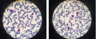
Background: Acute leukemia has a particularly bad prognosis in the newborn era. Its prognosis is significantly poorer than in older youngsters. The occurrence of leukaemia in the neonatal period can present diagnostic challenges due to the rarity of the condition and its clinical presentation, which may be misleading or inconspicuous. Case report: A case of neonatal acute myeloblastic leukaemia (AML) in a newborn with trisomy 21 is presented herein. The patient was admitted with respiratory distress and a clinical examination revealed a facial dysmorphic syndrome, pallor, hepatosplenomegaly, and a bone marrow failure syndrome with bone marrow invasion by 36% myeloblasts, confirming the diagnosis of AML. The immunophenotyping results indicated that the patient had AML0, with a low CD13+ and CD33+ MPO myeloid population. During the patient's hospitalisation, a multi-resistant Klebsiella pneumoniae urinary tract infection was diagnosed, and septic shock was diagnosed after four days. Conclusion: Acute neonatal leukaemia is a rare and complex condition that requires the expertise of both neonatologists and paediatric haematologists.
| Published in | American Journal of Laboratory Medicine (Volume 9, Issue 2) |
| DOI | 10.11648/j.ajlm.20240902.11 |
| Page(s) | 14-17 |
| Creative Commons |
This is an Open Access article, distributed under the terms of the Creative Commons Attribution 4.0 International License (http://creativecommons.org/licenses/by/4.0/), which permits unrestricted use, distribution and reproduction in any medium or format, provided the original work is properly cited. |
| Copyright |
Copyright © The Author(s), 2024. Published by Science Publishing Group |
Leukaemia, Neonatal, Trisomy 21, Blasts
ALL | Acute Lymphoblastic Leukaemia |
AML | Acute Myeloid Leukaemia |
M4 | Acute Myelomonocytic |
M5 | Monocytic |
CNS | Central Nervous System |
KMT2A | Lysine Methyltransferase 2A |
| [1] | Roberts I, Fordham NJ, Rao A, Bain BJ. Neonatal leukaemia. Br J Haematol. 2018; 182(2): 170–84. https://doi.org/10.1111/bjh.15246 |
| [2] | Ishii E, Oda M, Kinugawa N, et al. Features and outcome of neonatal leukemia in Japan: experience of the Japan Infant Leukemia Study Group. Pediatr Blood Cancer. 2006; 47(3): 268-72. |
| [3] | Bader JL, Miller RW. US cancer incidence and mortality in the first year of life. Am J Dis Child. 1979; 133(2): 157–59. |
| [4] | B. Brethon et al. Infant acute leukemia Bull Cancer (Paris) (2016). |
| [5] | Pierce MI. Leukemia in the newborn infant. J Pediatr. 1959; 54(5): 691–706. |
| [6] | Resnik KS, Brod BB. Leukemia cutis in congenital leukemia. Analysis and review of the world literature with report of an additional case. Arch Dermatol. 1993; 129(10): 1301–1306. |
| [7] | Zhang IH, Zane LT, Braun BS, Maize J, Zoger S, Loh ML. Congenital leukemia cutis with subsequent development of leukemia. J Am Acad Dermatol 2006; 54: S22-7. |
| [8] | Ariffin, E., Jones, H. & Bhatnagar, N. Congenital acute myeloid leukaemia with KMT2A rearrangement. British Journal of Haematology (2018), 182, 169. |
| [9] | Bain, B. J., Chakravorty, S. & Ancliff, P. Congenital acute megakaryoblastic leukemia. American Journal of Hematology (2015), 90, 963. |
| [10] | Debord, C., Grain, A., Theisen, O., Boutault, R., Rialland, F. & Eveillard, M. (2018) A blueberry muffin rash at birth. British Journal of Haematology, 182, 168. |
| [11] | Gamis, A. S. & Smith, F. O. (2012) Transient myeloproliferative disorder in children with Down syndrome: clarity to this enigmatic disorder. British Journal of Haematology, (2012), 159, 277–287. |
| [12] | Coenen EA, Zwaan CM, Reinhardt D, et al. Pediatric acute myeloid leukemia with t(8;16) (p11; p13), a distinct clinical and biological entity: A collaborative study by the International-Berlin-Frankfurt-Münster AML-study group. Blood. 2013; 122(15): 2704–13. |
| [13] | Oliver R, Juergens AL, 2nd, Hatch D, et al. Neonatal acute lymphoblastic leukemia. Pediatr Emerg Care. 2020; 36(2): e102–3. |
| [14] | Hanada, T., Kanamitsu, et al A Long-term survivor after congenital acute myeloid leukemia with t(8;16) (p11; p13). Acta Medica Okayama, (2016), 70, 31–35. |
| [15] | Gy-arf-as, T., Wintgens, J., Biskup, W., Oschlies, I., Klapper, W., R. Siebert, S. Bens, C. Haferlach, R. Meisel, M. Kuhlen et A. Borkhardt. Rémission spontanée transitoire dans la leucémie myéloïde aiguë congénitale réarrangée MLL-AF10 présentant avec insuffisance cardiorespiratoire et iléus méconial. Pédiatrie Moléculaire et Cellulaire, (2016), 3, 30. TTT. |
| [16] | Wong TN, Ramsingh G, Young AL, Miller CA, Touma W, Welch JS, Lamprecht TL, Shen D, HundalJ, Fulton RS, et al: Role of TP53 mutations in the origin and evolution of therapy‑related acute myeloid leukaemia. Nature 518: 552‑555, 2015. |
| [17] | H. Ishida, A. Iguchi Panel-based next-generation sequencing facilitates the characterization of childhood acute myeloid leukemia in clinical settings Published in Biomedical Reports 28 August 2020. |
| [18] | Quek L, Ferguson P, Metzner M, Ahmed I, Kennedy A, Garnett C, Jeffries S, Walter C, Piechocki K, Timbs A, et al: Mutational analysis of disease relapse in patients allografted for acute myeloid leukemia. Blood Adv 1: 193‑204, 2016. |
| [19] | Papaemmanuil E, Gerstung M, Bullinger L, Gaidzik VI, Paschka P, Roberts ND, Potter NE, Heuser M, Thol F, Bolli N, et al: Genomic classification and prognosis in acute myeloid leukemia. N Engl J Med 374: 2209‑2221, 2016. |
| [20] | GreifPA, HartmannL, VosbergS, StiefSM, MattesR, Hellmann I, Metzeler KH, Herold T, Bamopoulos SA, Kerbs P, et al: Evolution of cytogenetically normal acute myeloid leukemia during therapy and relapse: An exome sequencing study of 50 patients. Clin Cancer Res 24: 1716‑1726, 2018. |
| [21] | Shiba N, Yoshida K, Shiraishi Y, Okuno Y, Yamato G, Hara Y, Nagata Y, Chiba K, Tanaka H, Terui K, et al: Whole‑exome sequencing reveals the spectrum of gene mutations and the clonal evolution patterns in paediatric acute myeloid leukaemia. Br J Haematol 175: 476‑489, 2016. |
| [22] | Bolouri H, Farrar JE, Triche T Jr, Ries RE, Lim EL, Alonzo TA, Ma Y, Moore R, Mungall AJ, Marra MA, et al: The molecular landscape of pediatric acute myeloid leukemia reveals recurrent structural alterations and age‑specific mutational interactions. Nat Med 24: 103‑112, 2018. |
APA Style
Cherrafi, F., Boumaazi, H., Yahyaoui, H., Ameur, M. A., Chakour, M. (2024). Neonatal Leukaemia: A Case Report and Review of the Literature. American Journal of Laboratory Medicine, 9(2), 14-17. https://doi.org/10.11648/j.ajlm.20240902.11
ACS Style
Cherrafi, F.; Boumaazi, H.; Yahyaoui, H.; Ameur, M. A.; Chakour, M. Neonatal Leukaemia: A Case Report and Review of the Literature. Am. J. Lab. Med. 2024, 9(2), 14-17. doi: 10.11648/j.ajlm.20240902.11
AMA Style
Cherrafi F, Boumaazi H, Yahyaoui H, Ameur MA, Chakour M. Neonatal Leukaemia: A Case Report and Review of the Literature. Am J Lab Med. 2024;9(2):14-17. doi: 10.11648/j.ajlm.20240902.11
@article{10.11648/j.ajlm.20240902.11,
  author = {Fedwa Cherrafi and Hiba Boumaazi and Hicham Yahyaoui and Mustapha Ait Ameur and Mohammed Chakour},
  title = {Neonatal Leukaemia: A Case Report and Review of the Literature
},
  journal = {American Journal of Laboratory Medicine},
  volume = {9},
  number = {2},
  pages = {14-17},
  doi = {10.11648/j.ajlm.20240902.11},
  url = {https://doi.org/10.11648/j.ajlm.20240902.11},
  eprint = {https://article.sciencepublishinggroup.com/pdf/10.11648.j.ajlm.20240902.11},
  abstract = {Background: Acute leukemia has a particularly bad prognosis in the newborn era. Its prognosis is significantly poorer than in older youngsters. The occurrence of leukaemia in the neonatal period can present diagnostic challenges due to the rarity of the condition and its clinical presentation, which may be misleading or inconspicuous. Case report: A case of neonatal acute myeloblastic leukaemia (AML) in a newborn with trisomy 21 is presented herein. The patient was admitted with respiratory distress and a clinical examination revealed a facial dysmorphic syndrome, pallor, hepatosplenomegaly, and a bone marrow failure syndrome with bone marrow invasion by 36% myeloblasts, confirming the diagnosis of AML. The immunophenotyping results indicated that the patient had AML0, with a low CD13+ and CD33+ MPO myeloid population. During the patient's hospitalisation, a multi-resistant Klebsiella pneumoniae urinary tract infection was diagnosed, and septic shock was diagnosed after four days. Conclusion: Acute neonatal leukaemia is a rare and complex condition that requires the expertise of both neonatologists and paediatric haematologists.
},
 year = {2024}
}
TY  - JOUR
T1  - Neonatal Leukaemia: A Case Report and Review of the Literature

AU  - Fedwa Cherrafi
AU  - Hiba Boumaazi
AU  - Hicham Yahyaoui
AU  - Mustapha Ait Ameur
AU  - Mohammed Chakour
Y1  - 2024/07/15
PY  - 2024
N1  - https://doi.org/10.11648/j.ajlm.20240902.11
DO  - 10.11648/j.ajlm.20240902.11
T2  - American Journal of Laboratory Medicine
JF  - American Journal of Laboratory Medicine
JO  - American Journal of Laboratory Medicine
SP  - 14
EP  - 17
PB  - Science Publishing Group
SN  - 2575-386X
UR  - https://doi.org/10.11648/j.ajlm.20240902.11
AB  - Background: Acute leukemia has a particularly bad prognosis in the newborn era. Its prognosis is significantly poorer than in older youngsters. The occurrence of leukaemia in the neonatal period can present diagnostic challenges due to the rarity of the condition and its clinical presentation, which may be misleading or inconspicuous. Case report: A case of neonatal acute myeloblastic leukaemia (AML) in a newborn with trisomy 21 is presented herein. The patient was admitted with respiratory distress and a clinical examination revealed a facial dysmorphic syndrome, pallor, hepatosplenomegaly, and a bone marrow failure syndrome with bone marrow invasion by 36% myeloblasts, confirming the diagnosis of AML. The immunophenotyping results indicated that the patient had AML0, with a low CD13+ and CD33+ MPO myeloid population. During the patient's hospitalisation, a multi-resistant Klebsiella pneumoniae urinary tract infection was diagnosed, and septic shock was diagnosed after four days. Conclusion: Acute neonatal leukaemia is a rare and complex condition that requires the expertise of both neonatologists and paediatric haematologists.

VL  - 9
IS  - 2
ER  -
Faculty of Medicine and Pharmacy, Cadi Aayad University, Marrakech, Morocco; Hematology Laboratory, Avicenna Military Hospital, Marrakech, Morocco
Information