The green synthesis of metal oxide nanoparticles via plant extracts is an environmentally friendly, simple, inexpensive, and rapid, method for synthesizing nanoparticles for biological applications. In this study, Zanthoxylum chalybeum bark extracts were used to explore the biosynthesis of copper oxide nanoparticles (CuO NPs). The CuO NPs were successfully synthesized from an aqueous extract of Zanthoxylum chalybeum stem bark and characterized. The morphological, optical, and structural characteristics of the nanoparticles were assessed via scanning electron microscopy (SEM), X-ray diffraction (XRD), UV‒visible spectrophotometer, and Fourier transform infrared (FTIR) spectroscopy. Nanocrystalline CuO NPs, with an average crystalline size of 18.26 nm and a band gap energy of 1.45 eV, were confirmed via XRD and UV‒vis spectrophotometry, respectively. The SPR (surface plasmon resonance) peak was identified at wavelength of 529 nm in the UV-Vis spectrum. FT-IR analysis confirmed the presence of various functional groups that trigger the synthesis of CuO NPs. Morphological studies via SEM revealed spherical nanoparticles. Antifungal evaluation of the Candida albicans fungal strain and antibacterial evaluation of four bacterial strains (Staphylococcus aureus, Bacillus subtilis, Escherichia coli, and Pseudomonas aeruginosa) revealed greater potency of the green-synthesized CuO NPs than the Zanthoxylum chlamydium extracts and erythromycin (positive control). The green-synthesized CuO NPs obtained may be used as an antibacterial and antifungal agent for various therapeutic uses in medicine.
| Published in | Advances in Biochemistry (Volume 14, Issue 2) |
| DOI | 10.11648/j.ab.20261402.16 |
| Page(s) | 57-65 |
| Creative Commons |
This is an Open Access article, distributed under the terms of the Creative Commons Attribution 4.0 International License (http://creativecommons.org/licenses/by/4.0/), which permits unrestricted use, distribution and reproduction in any medium or format, provided the original work is properly cited. |
| Copyright |
Copyright © The Author(s), 2026. Published by Science Publishing Group |
Biosynthesis, Nanoparticles, Antifungal, Antibacterial Activity
Peak NO | 2θ values (°) | FWHM | Particle size (0.9*0.154)/(nm) |
|---|---|---|---|
peak 1 | 31.87 | 0.46 | 17.95 |
peak 2 | 34.53 | 0.33 | 25.20 |
peak 3 | 36.37 | 0.46 | 18.17 |
peak 4 | 47.66 | 0.58 | 14.97 |
peak 5 | 56.73 | 0.5 | 18.05 |
peak 6 | 62.99 | 0.55 | 16.93 |
peak 7 | 68.08 | 0.58 | 16.52 |
Average Particle size (nm) | 18.26 |
Sample Name Microbial Strains | CuO NPs (S1) | Zanthoxylum chalybeum extract (S5) | Positive Control (erythromycin) |
|---|---|---|---|
Bacillus subtilis | 13.33±0.47 | 7.67±0.47 | 10.67±0.47 |
Pseudomonas aeruginosa | 11.33±0.94 | 7.50±0.41 | 7.67±0.47 |
Escherichia coli | 11.33±0.47 | 5.83±0.24 | 7.83±0.24 |
Staphylococcus aureus | 6.67±0.47 | 5.33±0.47 | 7.33±0.47 |
Candida albicans | 15.33±1.25 | 9.67±0.94 | 9.83±0.24 |
Anova: Single Factor | ||||||
|---|---|---|---|---|---|---|
Groups | Count | Sum | Average | Variance | ||
Cuo NPs | 5 | 57.99 | 11.598 | 10.33912 | ||
Plant extract | 5 | 36 | 7.2 | 2.9464 | ||
Positive control | 5 | 43.33 | 8.666 | 2.21168 | ||
ANOVA | ||||||
Source of Variation | SS | df | MS | F | P value | F crit |
Between Groups | 50.14697 | 2 | 25.07349 | 4.85381 | 0.028537 | 3.885294 |
Within Groups | 61.9888 | 12 | 5.165733 | |||
Total | 112.1358 | 14 | ||||
CuO NPs | Copper Oxide Nanoparticles |
HCl | Hydrochloric Acid |
XRD | X-ray Diffraction |
SEM | Scanning Electron Microscopy |
FTIR | Fourier Transform Infrared Spectroscopy |
UV−Vis | Ultraviolet-Visible Spectroscopy |
SPR | Surface Plasmon Resonance |
| [1] | J. Gu, A. Aidy, and S. Goorani, “Antioxidant potentials of copper nanoparticles green-synthesized by Calendula officinalis,” J. Exp. Nanosci., vol. 17, no. 1, pp. 285–296, 2022, |
| [2] | A. I. Osman, Y. Zhang, M. Farghali, A. K. Rashwan, and A. S. Eltaweil, Synthesis of green nanoparticles for energy, biomedical, environmental, agricultural, and food applications: A review, vol. 22, no. 2. Springer International Publishing, 2024. |
| [3] | Mali, S. C., Dhaka, A., Githala, C. K., & Trivedi, R. (2020). Green synthesis of copper nanoparticles using Celastrus paniculatus Willd. leaf extract and their photocatalytic and antifungal properties. Biotechnology Reports, 27. |
| [4] | D. M. Nzilu, E. S. Madivoli, D. S. Makhanu, S. I. Wanakai, G. K. Kiprono, and P. G. Kareru, “Green synthesis of copper oxide nanoparticles and its efficiency in degradation of rifampicin antibiotic,” Sci. Rep., pp. 1–18, 2023, |
| [5] | A. H. Abbas and N. Y. Fairouz, “Materials Today : Proceedings Characterization, biosynthesis of copper nanoparticles using ginger roots extract and investigation of its antibacterial activity,” Mater. Today, Proc., vol. 61, pp. 908–913, 2022, |
| [6] | K. M. Doman et al., “Synthesis of silver and copper nanoparticles using Spirulina platensis and evaluation of their anticancer activity,” Int. J. Environ. Health Res., vol. 34, no. 2, pp. 661–673, 2024, |
| [7] | S. Vijayaram, H. Razafindralambo, Y. Zhang, and S. Seerangaraj, “Applications of Green Synthesized Metal Nanoparticles — a Review,” Biol. Trace Elem. Res., vol. 202, no. 1, pp. 360–386, 2024, |
| [8] | R. Radhakrishnan, F. Liakath, A. Khan, and A. Muthu, “Green Synthesis of Copper Oxide Nanoparticles Mediated by Aqueous Leaf Extracts of Leucas aspera and Morinda tinctoria,” vol. 10, no. 4, pp. 2706–2714, 2021. |
| [9] | M. V. D. Prakash et al., “Eco-friendly green synthesis of copper nanoparticles from Tinospora cordifolia leaves: optical properties with biological evaluation of applications,” Mater. Technol., vol. 38, no. 1, 2023, |
| [10] | D. Singh, D. Jain, and D. Rajpurohit, “Bacteria-assisted green synthesis of copper oxide nanoparticles and their potential applications as antimicrobial agents and plant growth stimulants,” no. April, pp. 1–10, 2023, |
| [11] | N. M. Kimani, C. O. Ochieng, M. D. Ogutu, K. O. Yamo, J. O. Onyango, and C. B. R. Santos, “Inhibition Kinetics and Theoretical Studies on Zanthoxylum chalybeum Engl. Dual Inhibitors of α-Glucosidase and α-Amylase,” J. Xenobiotics, vol. 13, no. 1, pp. 102–120, 2023, |
| [12] | S. D. Mbinile, L. K. Munishi, I. B. Ngondya, and P. A. Ndakidemi, “Spatial distribution and anthropogenic threats facing medicinal plant Zanthoxylum chalybeum in Simanjiro Area, Northern Tanzania,” Sci. African, vol. 10, p. e00562, 2020, |
| [13] | N. Quadri, M. M. Setty, A. Awasthi, U. Nayak, M. Singh, and S. Sharma, “Synthesis, characterization, genotoxicity assessment, and antibacterial applications of Zanthoxylum armatum silver nanoparticles (ZASNPs) with antibiotic efficacy enhancement potential,” Nanoscale, vol. 17, no. 3, pp. 1555–1567, 2024, |
| [14] | T. Khairy et al., “Antibacterial activity of green synthesized copper oxide nanoparticles against multidrug-resistant bacteria,” pp. 1–21, 2024. |
| [15] | M. Priya et al., “Green synthesis, characterization, antibacterial, and antifungal activity of copper oxide nanoparticles derived from Morinda citrifolia leaf extract,” Sci. Rep., pp. 1–13, 2023, |
| [16] | O. Article and S. Erkan, “Evaluation of surface properties of modified Ti6Al4V alloy with copper nanoparticles organic nanostructure for biomedical applications : dependency on anticorrosive, antibacterial, and biocompatibility,” vol. 43, no. 3, pp. 361–380, 2025, |
| [17] | E. A. Mohamed, “Green synthesis of copper & copper oxide nanoparticles using the extract of seedless dates,” Heliyon, vol. 6, no. 1, p. e03123, 2020, |
| [18] | J. Singh, S. Kaur, G. Kaur, S. Basu, and M. Rawat, “Biogenic ZnO nanoparticles : a study of blueshift of optical band gap and photocatalytic degradation of reactive yellow 186 dye under direct sunlight,” pp. 272–280, 2019. |
| [19] | I. JAHAN and İ. IŞILDAK, “Microwave Irradiation System for a Rapid Synthesis of Non-Toxic Metallic Copper Nanoparticles from Green Tea,” Trak. Univ. J. Nat. Sci., no. August 2020, |
| [20] | M. C. Ogwuegbu, A. S. Ayangbenro, D. M. N. Mthiyane, O. O. Babalola, and D. C. Onwudiwe, “Green synthesis of CuO nanoparticles using Ligustrum lucidum extract, and the antioxidant and antifungal evaluation”. |
| [21] | N. Sedefoglu, S. Er, K. Veryer, Y. Zalaoglu, and F. Bozok, “Green synthesized CuO nanoparticles using macrofungi extracts: Characterization, nanofertilizer and antibacterial effects,” Mater. Chem. Phys., vol. 309, p. 128393, Nov. 2023, |
| [22] | S. P. Lakshmanan, S. T. Jostar, G. J. Arputhavalli, S. Jebasingh, and C. M. R. Josephine, “Role of Green Synthesized CuO Nanoparticles of Trigonella Foenum-Graecum L. Leaves and their Impact on Structural, Optical and Antimicrobial Activity,” Int. J. Nanosci. Nanotechnol., vol. 17, no. 2, pp. 109–121, 2021. |
| [23] | G. Thirumoorthy et al., “Phytofabricated bimetallic synthesis of silver-copper nanoparticles using Aerva lanata extract to evaluate their potential cytotoxic and antimicrobial activities,” Sci. Rep., pp. 1–15, 2024, |
| [24] | F. Faridatul, W. Ode, S. Rizki, A. Luqman, I. Astuti, and R. Hertadi, “Heliyon Green synthesis of copper ions nanoparticles functionalized with rhamnolipid as potential antibacterial agent for pathogenic bacteria,” Heliyon, vol. 10, no. 1, p. e24242, 2024, |
| [25] | A. N. Labaran et al., “Biosynthesis of copper nanoparticles using Alstonia scholaris leaves and its antimicrobial studies,” Sci. Rep., pp. 1–10, 2024, |
| [26] | O. H. Sahib, L. Z. Mohammed, and M. A. Mahdi, “Biosynthesis of Copper Nanoparticles Using Passiflora Flower Extract and Study their Antimicrobial, Antioxidant and Anticancer Activity,” vol. 26, pp. 2953–2963, 2025, |
| [27] | S. Jabeen et al., “Biogenic Synthesis of Copper Oxide Nanoparticles from Aloe vera: Antibacterial Activity, Molecular Docking, and Photocatalytic Dye Degradation,” 2024, |
| [28] | I. L. Ikhioya et al., “The Green Synthesis of Copper Oxide Nanoparticles Using the Moringa oleifera Plant and Its Subsequent Characterization for Use in Energy Storage Applications,” vol. 172, pp. 162–172, 2023, |
| [29] | S. G. Ali et al., “Green Synthesis of Copper Oxide Nanoparticles from the Leaves of Aegle marmelos and Their Antimicrobial Activity and Photocatalytic Activities,” 2023. |
| [30] | J. L. Lopez-Miranda et al., “Antibacterial and Anti-Inflammatory Properties of ZnO Nanoparticles Synthesized by a Green Method Using Sargassum Extracts,” 2023. |
| [31] | M. A. Khan, N. B. Moghul, M. A. Butt, M. M. Kiyani, I. Zafar, and A. I. Bukhari, “Assessment of antibacterial and antifungal potential of Curcuma longa and synthesized nanoparticles: A comparative study,” J. Basic Microbiol., vol. 61, no. 7, pp. 603–611, 2021, |
| [32] | M. Bayat, M. Zargar, E. Chudinova, T. Astarkhanova, and E. Pakina, “In vitro evaluation of antibacterial and antifungal activity of biogenic silver and copper nanoparticles: The first report of applying biogenic nanoparticles against Pilidium concavum and Pestalotia sp. fungi,” Molecules, vol. 26, no. 17, 2021, |
APA Style
Chelang’a, P., Ayabei, K., Tarus, P., Njenga, L. (2026). Green Synthesized CuO Nanoparticles Using Zanthoxylum chalybeum Extracts: Characterization and Antibacterial Effects. Advances in Biochemistry, 14(2), 57-65. https://doi.org/10.11648/j.ab.20261402.16
ACS Style
Chelang’a, P.; Ayabei, K.; Tarus, P.; Njenga, L. Green Synthesized CuO Nanoparticles Using Zanthoxylum chalybeum Extracts: Characterization and Antibacterial Effects. Adv. Biochem. 2026, 14(2), 57-65. doi: 10.11648/j.ab.20261402.16
@article{10.11648/j.ab.20261402.16,
  author = {Posla Chelang’a and Kiplagat Ayabei and Paul Tarus and Lemeitaron Njenga},
  title = {Green Synthesized CuO Nanoparticles Using Zanthoxylum chalybeum Extracts: Characterization and Antibacterial Effects},
  journal = {Advances in Biochemistry},
  volume = {14},
  number = {2},
  pages = {57-65},
  doi = {10.11648/j.ab.20261402.16},
  url = {https://doi.org/10.11648/j.ab.20261402.16},
  eprint = {https://article.sciencepublishinggroup.com/pdf/10.11648.j.ab.20261402.16},
  abstract = {The green synthesis of metal oxide nanoparticles via plant extracts is an environmentally friendly, simple, inexpensive, and rapid, method for synthesizing nanoparticles for biological applications. In this study, Zanthoxylum chalybeum bark extracts were used to explore the biosynthesis of copper oxide nanoparticles (CuO NPs). The CuO NPs were successfully synthesized from an aqueous extract of Zanthoxylum chalybeum stem bark and characterized. The morphological, optical, and structural characteristics of the nanoparticles were assessed via scanning electron microscopy (SEM), X-ray diffraction (XRD), UV‒visible spectrophotometer, and Fourier transform infrared (FTIR) spectroscopy. Nanocrystalline CuO NPs, with an average crystalline size of 18.26 nm and a band gap energy of 1.45 eV, were confirmed via XRD and UV‒vis spectrophotometry, respectively. The SPR (surface plasmon resonance) peak was identified at wavelength of 529 nm in the UV-Vis spectrum. FT-IR analysis confirmed the presence of various functional groups that trigger the synthesis of CuO NPs. Morphological studies via SEM revealed spherical nanoparticles. Antifungal evaluation of the Candida albicans fungal strain and antibacterial evaluation of four bacterial strains (Staphylococcus aureus, Bacillus subtilis, Escherichia coli, and Pseudomonas aeruginosa) revealed greater potency of the green-synthesized CuO NPs than the Zanthoxylum chlamydium extracts and erythromycin (positive control). The green-synthesized CuO NPs obtained may be used as an antibacterial and antifungal agent for various therapeutic uses in medicine.},
 year = {2026}
}
TY  - JOUR
T1  - Green Synthesized CuO Nanoparticles Using Zanthoxylum chalybeum Extracts: Characterization and Antibacterial Effects
AU  - Posla Chelang’a
AU  - Kiplagat Ayabei
AU  - Paul Tarus
AU  - Lemeitaron Njenga
Y1  - 2026/05/19
PY  - 2026
N1  - https://doi.org/10.11648/j.ab.20261402.16
DO  - 10.11648/j.ab.20261402.16
T2  - Advances in Biochemistry
JF  - Advances in Biochemistry
JO  - Advances in Biochemistry
SP  - 57
EP  - 65
PB  - Science Publishing Group
SN  - 2329-0862
UR  - https://doi.org/10.11648/j.ab.20261402.16
AB  - The green synthesis of metal oxide nanoparticles via plant extracts is an environmentally friendly, simple, inexpensive, and rapid, method for synthesizing nanoparticles for biological applications. In this study, Zanthoxylum chalybeum bark extracts were used to explore the biosynthesis of copper oxide nanoparticles (CuO NPs). The CuO NPs were successfully synthesized from an aqueous extract of Zanthoxylum chalybeum stem bark and characterized. The morphological, optical, and structural characteristics of the nanoparticles were assessed via scanning electron microscopy (SEM), X-ray diffraction (XRD), UV‒visible spectrophotometer, and Fourier transform infrared (FTIR) spectroscopy. Nanocrystalline CuO NPs, with an average crystalline size of 18.26 nm and a band gap energy of 1.45 eV, were confirmed via XRD and UV‒vis spectrophotometry, respectively. The SPR (surface plasmon resonance) peak was identified at wavelength of 529 nm in the UV-Vis spectrum. FT-IR analysis confirmed the presence of various functional groups that trigger the synthesis of CuO NPs. Morphological studies via SEM revealed spherical nanoparticles. Antifungal evaluation of the Candida albicans fungal strain and antibacterial evaluation of four bacterial strains (Staphylococcus aureus, Bacillus subtilis, Escherichia coli, and Pseudomonas aeruginosa) revealed greater potency of the green-synthesized CuO NPs than the Zanthoxylum chlamydium extracts and erythromycin (positive control). The green-synthesized CuO NPs obtained may be used as an antibacterial and antifungal agent for various therapeutic uses in medicine.
VL  - 14
IS  - 2
ER  -
Department of Chemistry and Biochemistry, University of Eldoret, Eldoret, Kenya
Department of Chemistry and Biochemistry, University of Eldoret, Eldoret, Kenya
Department of Chemistry and Biochemistry, University of Eldoret, Eldoret, Kenya
Department of Chemistry, Jomo Kenyatta University of Agriculture and Technology, Nairobi, Kenya
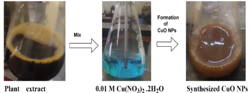
Figure 1. The graphical flowchart for the green synthesis of CuO NPs using Zanthoxylum chalybeum stem bark extract.
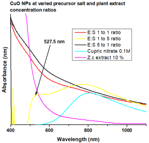
Figure 2. Biosynthesis of CuO NPs at various concentration ratios of precursor salt to plant extract.
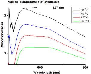
Figure 3. Biosynthesis of CuO NPs at various temperatures.
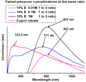
Figure 4. Biosynthesis of CuO NPs at various precursor molar concentrations.
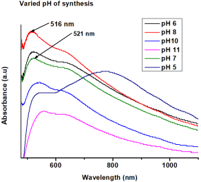
Figure 5. Biosynthesis of CuO NPs at various pH values.
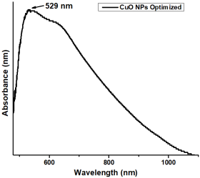
Figure 6. Optimized CuO NPs.
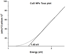
Figure 7. Tauc plot of CuO NPs.
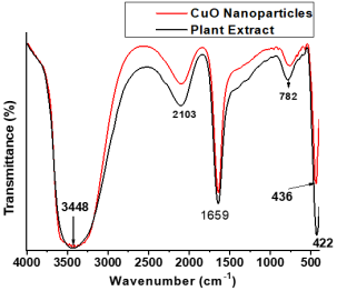
Figure 8. FTIR spectrum of CuO NPs.
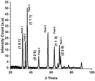
Figure 9. XRD spectrum of CuO NPs.
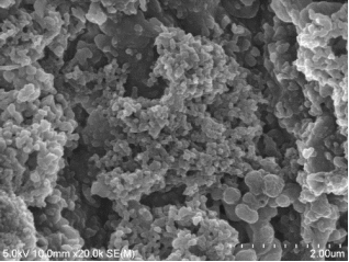
Figure 10. SEM image of CuO NPs.
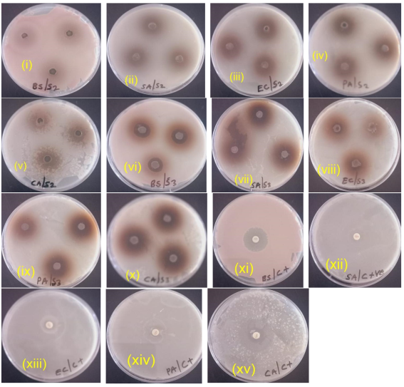
Figure 11. Zones of inhibition of (a) plant extract against (i) Bacillus subtilis; (ii) Staphylococcus aureus; (iii) Escherichia coli; (iv) Pseudomonas aeruginosa; (v) Candida albicans (Fungus), and (b) AgNPs against (vi) Bacillus subtilis; (vii) Staphylococcus aureus; (viii) Escherichia coli; (ix) Pseudomonas aeruginosa; (x) Candida albicans (Fungus); and (c) erythromycin against (xi) Bacillus subtilis; (xii) Staphylococcus aureus; (xiii) Escherichia coli; (xiv) Pseudomonas aeruginosa; and (xv) Candida albicans (Fungus).
Information

