Background: Arteriovenous malformation (AVM) is a congenital vascular anomaly in which there are abnormal connections between arteries and veins in the form of fistula or nidus without an intervening capillary bed. External ear AVMs can cause swelling, redness, bleeding, and pain. Timely diagnosis and treatment are crucial to prevent complications. According to literature, there are different techniques to treat auricular AVM which include surgical excision followed by ear reconstruction, sclerotherapy and embolization. Stereotactic radiosurgery (SRS) has an established role in treating intracranial AVM with excellent obliteration rates but limited literature exists on its efficacy in treating auricular AVM in terms of obliteration rates and cosmetic outcome. Case Presentation: We report a rare case of right external ear AVM which was post embolization and post excision followed by reconstruction but had persistent residual nidus in right pinna treated by stereotactic radiosurgery. He was treated to a dose of 21 Gy in 3 fractions at 7 Gy per fraction for 3 consecutive days. He had completed treatment without any complications. Follow up after 2 weeks of treatment, clinically, patient had redness, swelling and persistent pulsations but no episode of bleeding. At the six-month follow-up, redness, swelling, and pulsations had decreased. Follow up after 1 year post treatment, clinically, redness, swelling and pulsations were not present. Dynamic Brain MRI with angiography showed complete obliteration of residual AVM nidus in right pinna. Patient was satisfied with the cosmetic outcome. Conclusion: We conclude that stereotactic radiosurgery can be used as an effective treatment modality for auricular AVMs. In our case, radiosurgery provided an excellent control and obliteration of the nidus and good cosmetic result eliminating the need for surgical reconstruction at the site of the nidus.
| Published in | International Journal of Neurosurgery (Volume 9, Issue 1) |
| DOI | 10.11648/j.ijn.20250901.12 |
| Page(s) | 10-16 |
| Creative Commons |
This is an Open Access article, distributed under the terms of the Creative Commons Attribution 4.0 International License (http://creativecommons.org/licenses/by/4.0/), which permits unrestricted use, distribution and reproduction in any medium or format, provided the original work is properly cited. |
| Copyright |
Copyright © The Author(s), 2025. Published by Science Publishing Group |
Arteriovenous Malformation, Digital Substraction Angiography, External Ear, Nidus, Dynamic Contrast Enhanced Mri, Onyx, Embolization, Excision
Schobinger stage | Features |
|---|---|
I (quiescence) | Cutaneous blush or warmth |
II (expansion) | Bruit, audible pulsation, and enlarging lesion |
III (destruction) | Pain, ulceration, bleeding, and infection |
IV (decompensation) | Cardiac failure |
Structures | Target coverage and OAR constraints | |
|---|---|---|
GTV ( residual nidus) | V100 | 98 % |
Dmax | 107% | |
PTV | V100 | 95 % |
Dmax | 107% | |
BRAINSTEM | Dmax | 6.38 Gy |
RIGHT COCHLEA | Dmean | 10.73 GY |
Mandible | Dmax | 20 Gy |
AVM | Arteriovenous Malformation |
SRS | Stereotactic Radiosurgery |
MRI | Magnetic Resonance Imaging |
DSA | Digital Substraction Angiography |
ECA | External Carotid Artery |
GTV | Gross Tumor Volume |
PTV | Planning Target Volume |
V100 | Volume Receiving 100% of Prescription Dose |
OAR’s | Organs at Risk |
QA | Quality Assurance |
IMRT | Intensity Modulated Radiotherapy |
CBCT | Cone Beam Computed Tomography |
Dmax | Maximum Dose |
Dmean | Mean Dose |
| [1] | Liu R, Chen J, Jia L, Pan B, Jiang H. Surgical management of auricular arteriovenous malformations: A literature review. Laryngoscope Investig. Otolaryngol. 2022 Apr; 7(2): 604-13. |
| [2] | Kim JB, Lee JW, Choi KY, et al. Clinical characteristics of arteriovenous malformations of the head and neck. Dermatol. Surg. 2017 Apr 1; 43(4): 526-33. |
| [3] | Kohout MP, Hansen M, Pribaz JJ, Mulliken JB. Arteriovenous malformations of the head and neck: natural history and management. Plast. Reconst. Surg. 1998 Sep 1; 102(3): 643-54. |
| [4] | Zou Y, Qiao C, Lin X, et al. Clinical course of extracranial arteriovenous malformations. J CraniofacSurg. 2020 Mar 1; 31(2): 372-6. |
| [5] | Kansy K, Bodem J, Engel M, et al. Interdisciplinary treatment algorithm for facial high-flow arteriovenous malformations, and review of the literature. J. Craniomaxillofac. Surg. 2018 May 1; 46(5): 765-72. |
| [6] | Fernández-Alvarez V, Suárez C, de Bree R, et al. Management of extracranial arteriovenous malformations of the head and neck. AurisNasus Larynx. 2020 Apr 1; 47(2): 181-90. |
| [7] | Vilela Chagas Ferreira M, Goldenberg DC, Kharmandayan V, Gemperli R. Management of arteriovenous malformation of the ear: a protocol for resection and reconstruction. Laryngoscope. 2020 May; 130(5): 1322-6. |
| [8] | Wu JK, Bisdorff A, Gelbert F, Enjolras O, Burrows PE, Mulliken JB. Auricular arteriovenous malformation: evaluation, management, and outcome. Plast. Reconst. Surg. 2005 Apr 1; 115(4): 985-95. |
| [9] | Bulstrode NW, Lese I, Aldabbas M, Glover M, Robertson F, Rennie A. Arterio-venous malformations of the ear: description of distinct anatomical presentation and multidisciplinary management approach. J PlastReconstrAesthet Surg. 2021 Jul 1; 74(7): 1574-81. |
| [10] | Wang D, Su L, Han Y, Fan X. Ethanol embolotherapy of high-flow auricular arteriovenous malformations with electrolytically detachable coil-assisted dominant outflow vein occlusion. Eur J VascEndovasc Surg. 2014 Nov 1; 48(5): 576-84. |
| [11] | Hua C, Jin Y, Yang X, et al. Midterm and long-term results of ethanol embolization of auricular arteriovenous malformations as first-line therapy. J VascSurg: Venous and Lymphatic Disorders. 2018 Sep 1; 6(5): 626-35. |
| [12] | Pham TH, Wong BJ, Allison G. A large arteriovenous malformation of the external ear in an adult: report of a case and approach to management. Laryngoscope. 2001; 111(8): 1390-4. |
| [13] | Jin Y, Lin X, Chen H, et al. Auricular arteriovenous malformations: potential success of superselective ethanol embolotherapy. J. Vasc. Interv. Radiol. 2009 Jun 1; 20(6): 736-43. |
| [14] | Zheng LZ, Fan XD, Zheng JW, Su LX. Ethanol embolization of auricular arteriovenous malformations: preliminary results of 17 cases. Am. J. Neuroradiol. 2009 Oct 1; 30(9): 1679-84. |
| [15] | Yang F, Yang B, Qu Z, Tan Y, Lu F, Liao Z. Percutaneous ethanol sclerotherapy for auricular arteriovenous malformation: Our experience with 11 patients. Clin. Otolaryngol. 2020 Sep; 45(5): 811-7. |
| [16] | Whitty LA, Murray JD, Null WE, Elwood ET, Jones GE. An arteriovenous malformation of the external ear in the pediatric population: a case report and review of the literature. Can. J. Plast. Surg. 2009 Dec; 17(4): 45-7. |
| [17] | Kim SH, Han SH, Song Y, Park CS, Song JJ. Arteriovenous malformation of the external ear: a clinical assessment with a scoping review of the literature. Braz. J. Otorhinolaryngol. 2017 Nov; 83: 683-90. |
| [18] | Gupta A, Gupta S, Kumar A, Bhattacharaya S, Jha M, Tiwari V. High-flow vascular malformation of ear: a case report. World J. Plast. Surg. 2018 May; 7(2): 256. |
APA Style
Deputy, M., Hunugundmath, S., Nirhali, A., Naik, V., Gadhave, S. (2025). A Rare Case of External Ear Arteriovenous Malformation Treated with Linac Based Radiosurgery. International Journal of Neurosurgery, 9(1), 10-16. https://doi.org/10.11648/j.ijn.20250901.12
ACS Style
Deputy, M.; Hunugundmath, S.; Nirhali, A.; Naik, V.; Gadhave, S. A Rare Case of External Ear Arteriovenous Malformation Treated with Linac Based Radiosurgery. Int. J. Neurosurg. 2025, 9(1), 10-16. doi: 10.11648/j.ijn.20250901.12
@article{10.11648/j.ijn.20250901.12,
  author = {Mariya Deputy and Sanjay Hunugundmath and Amit Nirhali and Vishram Naik and Sharad Gadhave},
  title = {A Rare Case of External Ear Arteriovenous Malformation Treated with Linac Based Radiosurgery
},
  journal = {International Journal of Neurosurgery},
  volume = {9},
  number = {1},
  pages = {10-16},
  doi = {10.11648/j.ijn.20250901.12},
  url = {https://doi.org/10.11648/j.ijn.20250901.12},
  eprint = {https://article.sciencepublishinggroup.com/pdf/10.11648.j.ijn.20250901.12},
  abstract = {Background: Arteriovenous malformation (AVM) is a congenital vascular anomaly in which there are abnormal connections between arteries and veins in the form of fistula or nidus without an intervening capillary bed. External ear AVMs can cause swelling, redness, bleeding, and pain. Timely diagnosis and treatment are crucial to prevent complications. According to literature, there are different techniques to treat auricular AVM which include surgical excision followed by ear reconstruction, sclerotherapy and embolization. Stereotactic radiosurgery (SRS) has an established role in treating intracranial AVM with excellent obliteration rates but limited literature exists on its efficacy in treating auricular AVM in terms of obliteration rates and cosmetic outcome. Case Presentation: We report a rare case of right external ear AVM which was post embolization and post excision followed by reconstruction but had persistent residual nidus in right pinna treated by stereotactic radiosurgery. He was treated to a dose of 21 Gy in 3 fractions at 7 Gy per fraction for 3 consecutive days. He had completed treatment without any complications. Follow up after 2 weeks of treatment, clinically, patient had redness, swelling and persistent pulsations but no episode of bleeding. At the six-month follow-up, redness, swelling, and pulsations had decreased. Follow up after 1 year post treatment, clinically, redness, swelling and pulsations were not present. Dynamic Brain MRI with angiography showed complete obliteration of residual AVM nidus in right pinna. Patient was satisfied with the cosmetic outcome. Conclusion: We conclude that stereotactic radiosurgery can be used as an effective treatment modality for auricular AVMs. In our case, radiosurgery provided an excellent control and obliteration of the nidus and good cosmetic result eliminating the need for surgical reconstruction at the site of the nidus.
},
 year = {2025}
}
TY  - JOUR
T1  - A Rare Case of External Ear Arteriovenous Malformation Treated with Linac Based Radiosurgery

AU  - Mariya Deputy
AU  - Sanjay Hunugundmath
AU  - Amit Nirhali
AU  - Vishram Naik
AU  - Sharad Gadhave
Y1  - 2025/02/24
PY  - 2025
N1  - https://doi.org/10.11648/j.ijn.20250901.12
DO  - 10.11648/j.ijn.20250901.12
T2  - International Journal of Neurosurgery
JF  - International Journal of Neurosurgery
JO  - International Journal of Neurosurgery
SP  - 10
EP  - 16
PB  - Science Publishing Group
SN  - 2640-1959
UR  - https://doi.org/10.11648/j.ijn.20250901.12
AB  - Background: Arteriovenous malformation (AVM) is a congenital vascular anomaly in which there are abnormal connections between arteries and veins in the form of fistula or nidus without an intervening capillary bed. External ear AVMs can cause swelling, redness, bleeding, and pain. Timely diagnosis and treatment are crucial to prevent complications. According to literature, there are different techniques to treat auricular AVM which include surgical excision followed by ear reconstruction, sclerotherapy and embolization. Stereotactic radiosurgery (SRS) has an established role in treating intracranial AVM with excellent obliteration rates but limited literature exists on its efficacy in treating auricular AVM in terms of obliteration rates and cosmetic outcome. Case Presentation: We report a rare case of right external ear AVM which was post embolization and post excision followed by reconstruction but had persistent residual nidus in right pinna treated by stereotactic radiosurgery. He was treated to a dose of 21 Gy in 3 fractions at 7 Gy per fraction for 3 consecutive days. He had completed treatment without any complications. Follow up after 2 weeks of treatment, clinically, patient had redness, swelling and persistent pulsations but no episode of bleeding. At the six-month follow-up, redness, swelling, and pulsations had decreased. Follow up after 1 year post treatment, clinically, redness, swelling and pulsations were not present. Dynamic Brain MRI with angiography showed complete obliteration of residual AVM nidus in right pinna. Patient was satisfied with the cosmetic outcome. Conclusion: We conclude that stereotactic radiosurgery can be used as an effective treatment modality for auricular AVMs. In our case, radiosurgery provided an excellent control and obliteration of the nidus and good cosmetic result eliminating the need for surgical reconstruction at the site of the nidus.

VL  - 9
IS  - 1
ER  -
Department of Radiation Oncology, Sahyadri Super Speciality Hospitals, Pune, India
Biography: Mariya Deputy: Junior Consultant in department of Radiation Oncology at Sahyadrisuperspeciality hospitals, Pune, India. Special interest in Brain tumors, Breast cancer, Gynaecological cancers, SRS and SBRT.
Research Fields: Oncology, Radiation Oncology, Molecular oncology.

Department of Radiation Oncology, Sahyadri Super Speciality Hospitals, Pune, India
Biography: Sanjay Hunugundmath: Lead Consultant in department of Radiation Oncology at Sahyadrisuperspeciality hospitals, Pune, India. He did his Masters in Radiation Oncology from Amrita Institute of medical sciences, Kochi. He completed his fellowship in stereotactic radiation drom National Cancer Centre, Singapore. He had extensive training in Clinical Oncology from TMC Kolkata.
Research Fields: Oncology, Radiation Oncology, Molecular oncology.

Department of Radiation Oncology, Sahyadri Super Speciality Hospitals, Pune, India
Biography: Amit Nirhali: Chief Medical Physicist and RSO in department of Radiation Oncology at SahyadriSuperspeciality Hospitals, Pune, India. He has 15 years of extensive experience in commissioning Linac, Brachytherapy, Tomotherapy machines, treatment planning, quality assur-ance of RT machines, setting up and designing radiotherapy departments. Key interest in intergrating machine learning, radiomics and AI in Radiation Oncology.
Research Fields: Medical Physics, Radiation Oncology, Artificial Intelligence in Radiation Oncology.

Department of Radiation Oncology, Sahyadri Super Speciality Hospitals, Pune, India
Biography: Vishram Naik: Senior Medical physicist in department of Radiation Oncology at Sahyadrisuperspeciality hospitals, Pune, India. 7 years of experience as a Medical Physicist with wide experience in treatment planning and quality assurance of RT. Key interest areas are Dosimetery and Advanced algorithm in Radiation Oncology.
Research Fields: Medical Physics, Radiation Oncology.

Department of Radiation Oncology, Sahyadri Super Speciality Hospitals, Pune, India
Biography: Sharad Gadhave: Senior Radiation Therapist in department of Radiation Oncology at Sahyadrisuperspeciality hospitals, Pune, India. He did his advance diploma in Radiotherapy Technology from Tata Memorial Hospital, Mumbai. He ha wide experience in treating special pro-cedure such as SRS, SBRT, DIBH, IGRT etc.
Research Fields: Radiation Oncology.

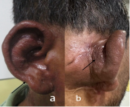
Figure 1. Pretreatment clinical photograph of patient. Anterior aspect (a) and posterior aspect (b) pointed with arrows.
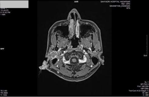
Figure 2. Pretreatment Dynamic Brain MRI with contrast. Residual nidus is pointed with arrow.
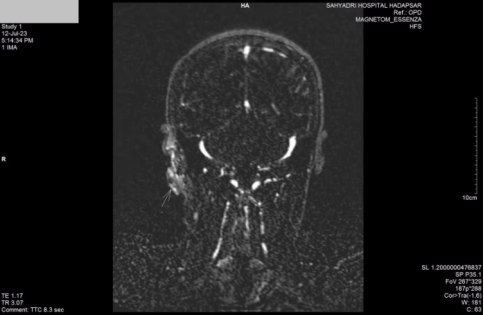
Figure 3. Pretreatment MR Angiography. Residual nidus is pointed with arrow.
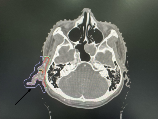
Figure 4. Planning CT scan with contrast. It shows red, gross tumor volume and green, planning target volumepointed with arrow.
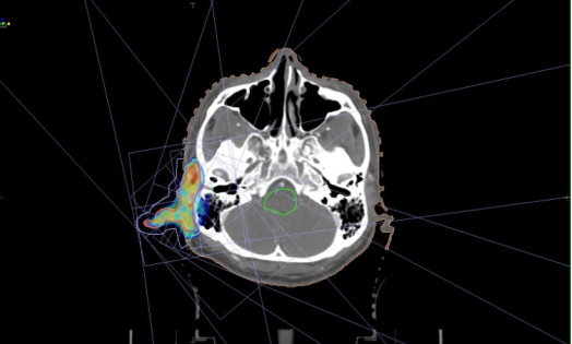
Figure 5. IMRT plan on planning CT scan. It shows 9 field IMRT plan, virtual bolus and 100 % dose wash.
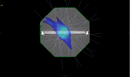
Figure 6. Pretreatment patient specific quality assurance done with PTW OCTAVIOUS 1500 Phantom on which gamma analysis was done.
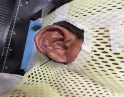
Figure 7. Immobilization of patient on treatment couch with head and neck orfit and head rest pointed with arrow.
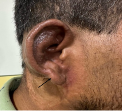
Figure 8. Clinical photograph of patient 2 weeks post treatment.
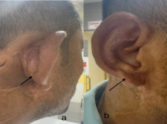
Figure 9. 1 year post treatment clinical photograph. Posterior aspect (a) and anterior aspect (b) pointed with arrows.
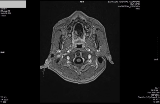
Figure 10. 1 year post treatment Dynamic Brain MRI with contrast shows complete resolution of residual nidus pointed with arrow.
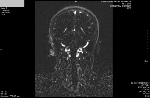
Figure 11. 1 year post treatment MR Angiography shows complete resolution of residual nidus pointed with arrow.
Information

